Coronary heart disease is considered one of the leading causes of death worldwide. This is a pathology that leads to disruption of the heart and is accompanied by narrowing of the coronary arteries. Coronary angiography is used to diagnose coronary artery disease. The study shows the condition of the heart vessels, blood circulation in them and places of blockage. To carry out such a procedure, there are rules for preparing the patient, contraindications and possible complications.
What it is
The coronary arteries supply blood to the heart, carrying nutrients and oxygen. This is necessary for the normal and uninterrupted functioning of the heart muscle. But sometimes, due to congenital abnormalities, atherosclerotic plaques or spasms, the lumen in the arteries narrows, this is called stenosis. The most dangerous is occlusion, that is, cessation of blood flow. This picture leads to coronary artery disease or myocardial infarction, and in severe cases, death.
Content:
- What it is
- When is coronary angiography prescribed?
- How to prepare
- How it's made
- Contraindications for the procedure
- Is this examination dangerous?
- Where can I get it and how much does it cost?
The condition and functioning of the coronary arteries and vessels can be checked using coronary angiography. This is a type of angiography and fluoroscopy. The study is carried out by filling the arteries with a special substance that does not transmit x-rays - contrast. A catheter delivers contrast to the heart.
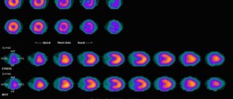
When the substance is distributed throughout the vessels, a series of X-rays are taken. In such images, the blood network, arterial patency, and stages of the disease are clearly visible. Based on such a study, the doctor makes a conclusion about treatment methods.
There are the following types of examination:
- CT coronary angiography. The procedure is carried out using computed tomography, contrast is injected into a vein without catheterization of the coronary vessels of the heart. During the examination, the radiologist sees places of narrowing, the thickness of the walls of blood vessels, and the detailed structure of the circulatory system in this area.
- Interventional selective coronary angiography. The catheter is moved through the vein to the coronary artery itself, and contrast is injected. The procedure is carried out under X-ray control, the doctor sees the result in real time on the screen.
- Ultrasound coronary angiography. An unpopular technique, but used for research. It happens in the same way as the previous method, but an ultrasound sensor is attached to the end of the catheter. This is done to assess the condition of the vascular walls.
- The most common method of examination in the CIS is interventional. Computed tomography is increasingly being carried out in private clinics, as this method provides more information. Other examination methods are prescribed in isolated cases.
When is coronary angiography prescribed?
The purpose of this procedure is to examine in detail the condition of the arteries of the heart, looking for narrowing or blockage. Coronary angiography of blood vessels is usually used by cardiologists and cardiac surgeons. It is needed before complex operations to make the correct diagnosis and choose the right treatment. The patient is referred for coronary angiography in the following cases:
- ineffectiveness of drug therapy in patients with coronary heart disease;
- positive stress test (stress echocardiography, bicycle/treadmill testing);
- before any heart surgery in patients over 40 years of age;
- angina pectoris after a heart attack, stenting or coronary bypass surgery;
- all emergency situations when there is a suspicion of a cardiovascular accident - acute myocardial infarction (in the first 6 hours);
- unstable angina;
- lack of information about other diagnostic methods.
It is also prescribed to evaluate the effectiveness of therapy for the heart and blood vessels. Checking is needed for atypical pain in the chest area without a clear cause. It is often used before coronary artery bypass surgery and other interventions.
Description of the diagnostic technique
Coronary angiography is a visualization technique that is carried out using ionizing radiation. A classic X-ray apparatus is used as a scanning unit. To add contrast to the area being studied, a staining iodide solution is used, which is administered intravenously at the beginning of the procedure, in the middle of the screening, or throughout the entire session in the form of injection injections to measure the intensity and pressure of blood flow in the veins.
The study is used to assess the functionality of the heart arteries, and also as a concomitant procedure for stenting. The technique is the most informative in studying the consequences of ischemic pathology, the state of the system as a whole, detection of pathological processes, their complexity and further planning of therapeutic and restorative manipulations. During screening, the function of the coronary bed is assessed, places of abnormal narrowing, their size and extent, and atherosclerotic anomalies are identified. Indications for scanning are determined only by the treating specialist after excluding possible contraindications.
How to prepare
Coronary fluoroscopy is performed after preparing the patient. The procedure can be emergency or planned. Emergency diagnostics are carried out with minimal analyzes and tests; the type and number of such checks is selected by the attending physician depending on the situation. For routine examination the following is prescribed:
- consultation of specialists: neurologist, cardiologist, surgeon;
- blood tests: coagulability, Rh factor and group, biochemical analysis;
- fluorography;
- allergy tests;
- Analysis of urine;
- electrocardiogram.
A full list of examinations is prescribed based on the clinical picture. The procedure is performed on an empty stomach to avoid nausea and the gag reflex. If the contrast agent will be injected into the thigh, you will need to remove hair from that area the day before.
A few days before the scheduled examination, the patient is shown a large amount of water, alcohol is prohibited. You cannot eat on the day of the event.
Before the examination itself, you need to warn the doctor about what medications you took and how you feel. You also need to take a change of clothes and personal hygiene items for your stay in the hospital.
Advantages and disadvantages of ultrasound and MRI of the heart
Today, high-quality cardiac MRI requires very expensive equipment - a tomograph with a magnetic field power of at least 3 Tesla. Research using such an ultra-high-field device will be expensive, so the main disadvantage of cardiac MRI is its high price. In addition, MRI of the coronary vessels and heart cannot measure the speed of blood flow and its actual volume. Cardiac ultrasound can better cope with this task.
How it's made
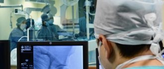
Angiography of the heart vessels is performed in a hospital; the patient remains in the hospital for several days for careful preparation and supervision after the procedure. All manipulations are carried out in the sterile conditions of the X-ray operating room - this is an office equipped specifically for such purposes. Before the invasion, the patient signs consent documents.
The patient then changes into hospital gowns. He is placed on the operating table and the position is fixed so that sudden movement does not disrupt the procedure. A cardiac monitor is connected to monitor the heartbeat and blood pressure.
The anesthesiologist administers local anesthesia; the patient will not feel pain, but will remain conscious. A catheter is inserted into a vein in the thigh or arm.
The puncture site is selected depending on the clinical picture. More often the radial artery in the arm is punctured - this is the most painless and safe, less often - the femoral artery in the groin area.
A conductor (thin wire) is inserted through the needle into the lumen of the vessel, and the needle is removed. A catheter (hollow tube) is passed through the guidewire. The catheter is advanced through the vessels to the coronary arteries themselves. There are no nerve endings in the vessels, so the patient will not feel anything. Contrast is fed into the catheter, which fills the vessels and is clearly visualized by fluoroscopy. On the screen, the doctor sees the advancement of the catheter.
When the substance fills the arteries, they become clearly visible on the monitor. To obtain high-quality images, the radiologist may ask you to hold your breath. Data from the screen can be recorded on a digital medium so that doctors can then assess the patient’s condition.
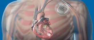
If the patency of the vessels is impaired, a mesh stent can be installed or balloon dilatation can be performed. This is done directly during coronary angiography; such manipulations restore the lumen of the vessel and normal blood flow. After the procedure, the instruments are removed, the puncture site is disinfected and a tight bandage is applied.
The patient remains on bed rest for another day. Some private clinics offer this service on an outpatient basis, the patient rests for a few hours and goes home. The whole process will take no more than an hour.
Reviews from patients confirm that fluoroscopy of blood vessels is quick and without severe pain. When the catheter is inserted, discomfort is felt, and when the contrast enters, a feeling of warmth is felt in the chest.
Contact your doctor immediately if...
- There is bleeding, new bruising, or swelling at the access site.
- Increased pain or discomfort in the area where the catheter was placed.
- Signs of infection develop, such as redness, discharge from the access site, and fever.
- Change in temperature or color of the leg or arm where the cut was made.
- Feeling of severe weakness.
- Have chest pain or shortness of breath.
If there is severe bleeding from the access site, apply a pressure bandage above the site and call emergency medical help immediately!
Contraindications for the procedure
Coronary angiography has only relative contraindications. There is a list of diseases that increase the risk of complications. But even against the background of such diseases, fluoroscopy of blood vessels can be performed if the patient’s life depends on it. Coronary angiography is performed with extreme caution when:
- impaired blood clotting;
- decompensated diabetes mellitus;
- allergies to contrast agent;
- arterial hypertension;
- severe renal failure - the contrast agent has a bad effect on diseased kidneys;
- internal bleeding - postponed until this problem is resolved.
All possible contraindications must be taken into account when prescribing coronary angiography. Absolute contraindications include only a severe allergic reaction to contrast. In most cases, the study is simply postponed.
Rotablationplasty
This method has been used since 1989. A diamond bur is introduced into the vessel, capable of rotating at a frequency of 190,000 rpm. in a minute. It is used to remove tissue at the site of restenosis. The size of the crushed particles in this way is 5–10 micrometers (the length of a red blood cell is 7.5 micrometers). The resulting channel is very small in diameter, but sufficient for performing PTCA. As a rule, the method is used in case of formation of calcifications at the site of restenosis. The diameter of the bur head varies from 1.25 to 2.5 mm. Because the rotablation bur cable does not penetrate the stenosis site well, the success rate of the operation is 80–95%. Complications include vascular spasms, which occur more often (5%) than with PTCA.
Is this examination dangerous?
Everyone who is about to undergo this procedure is interested in what the consequences and complications may be. This type of diagnosis is considered a safe manipulation; the process is controlled using an X-ray or computed tomograph. Nevertheless, the risk of complications is still present; the most dangerous consequence is damage to blood vessels by the catheter.
The likelihood of such a complication is very low; it can occur from sudden movements or carelessness of the doctor. If the patient has not undergone proper preparation, an allergic reaction or bleeding may occur (with poor clotting). All these risks can be reduced if you carefully prepare and relax before the procedure.
If you are overcome by fear and anxiety in the X-ray operating room, you can warn the doctor about this and he will prescribe sedatives.