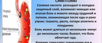
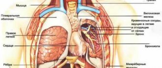
If a person experiences pain in the area of the left side of the hypochondrium, the first suspicions immediately fall on the pancreas or stomach. However, another, neighboring organ may also be disturbing.
The spleen is one of the most mysterious organs, the functions of which are not fully understood. This is the most important organ that helps the immune system: it is the one that copes with filtering blood cells infected with viruses. The spleen is a reservoir organ, a blood depot.
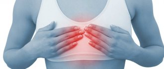
Pain syndrome in the spleen without the influence of external factors occurs only in one case - when it is significantly enlarged. Often, patients simply complain of discomfort, a pulling sensation in the left half of the abdomen.
Diseases that cause pain in the spleen:
- Infectious diseases (typhoid fever, hepatitis, anthrax, infectious mononucleosis, syphilis, tuberculosis) Many infectious diseases affect the spleen, causing pain and starting a destructive process.
- Traumatic conditions of the spleen They are divided into two types: open and closed type. The first includes stab and gunshot wounds of the chest area on the left or the upper part of the abdominal cavity. Closed injuries - bruises and fractures. In some cases, the spleen may rupture, which is accompanied by internal bleeding. Damage to an organ is usually characterized by acute pain radiating to any part of the body, as well as a general deterioration of the condition.
- Organ infarction Occurs as a result of thromboembolism, as well as thrombosis. The condition of a person with a splenic infarction is directly related to the magnitude of the pathology. If it is small, then it is probably asymptomatic. With a large heart attack, pain occurs in the left hypochondrium with irradiation to the back area, which responds to breathing.
- Spleen abscess The pathology picture is fever, pain in the abdomen and chest, muscle tension.
- Parasitic diseases (most often echinococcosis)
- Benign and malignant neoplasms
- An enlarged spleen is often combined with an enlarged liver (hepatolienal syndrome), which is observed in liver cirrhosis, portal hypertension, and blood diseases.
Any pain in the body is a signal for help.
Do not blindly ignore it and take painkillers indiscriminately. Pain and discomfort in the left hypochondrium may indicate several problems.
Only a doctor can identify the exact cause.
If you experience pain of an unclear nature in the left hypochondrium, you can contact our clinic. To make a correct diagnosis, specialists will conduct an examination and interview, and also prescribe the necessary tests. During the consultation, you can discuss all the symptoms in order to choose the most effective and safe therapy.
Diagnosis and treatment
Diagnosis of spleen diseases begins with a thorough examination by a doctor. After palpation and history taking, additional studies will be required - ultrasound, radiography, MRI or puncture. Laboratory diagnostics are also required.
The primary task is to correctly diagnose in order to prescribe effective treatment. It should only occur under the supervision of an experienced specialist.
Preventive measures to maintain the organ in a healthy state are very simple: proper nutrition and a healthy lifestyle. The spleen begins to work better with regular physical activity, as well as with special breathing exercises.
Pain in the spleen occurs when the organ has significantly increased in size. This may indicate various diseases, which can only be diagnosed by a qualified doctor. It is highly recommended not to make a diagnosis on your own, much less select a treatment.
After all, the spleen is one of the important organs responsible for supporting human immunity.
Timely contact with specialists will help you solve the problem at an early stage with minimal damage.
For early diagnosis of health problems, you can undergo a comprehensive diagnostics of the body at our center.
The best diagnosis of pain in the spleen is MRI of the spleen
You can sign up for a consultation right now: online or by phone
In the 21st century, trauma has become a pandemic. A systematic review of the literature [63] found that 5.8 million people die from injury every year, and by 2021 this figure could rise to 8.4 million. Road traffic accidents are the most common cause of death in older people from 15 to 44 years old.
Among closed injuries of abdominal organs, splenic injury, according to various authors, ranges from 16 to 50%, ranking 1st-2nd in frequency [11, 14, 27, 57, 77].
Surgery for spleen injuries has gone through several stages. Splenectomy was performed in China in the second century AD [64], but was first described in the medical literature in 1549 [43]. In 1581, Viard removed part of the spleen that had prolapsed through a stab wound in the abdominal wall [24]. In both cases there was recovery. Until the 1980s, splenectomy was the only operation for splenic injury. Thus, in the manual on operative surgery it is written: “In connection with this circumstance characterizing the parenchymal organ, except for splenectomy, that is, complete removal of the spleen, no other interventions on it are recommended” [15]. Increased knowledge about the numerous functions of the spleen [18], including the immune function [7, 16, 17], led to the development of organ-sparing operations [2, 8, 10] and autotransplantation of spleen tissue [1, 19]. After the successful use of conservative therapy for solid organ injuries in children, this tactic was extended to adults [68].
Morphological aspects of splenic injury, including two-stage rupture, were studied in the works of A.P. Vilka et al., M.A. Sapozhnikova and I. Riezzo et al. [9, 20, 69].
To damage the spleen, especially a pathologically altered one, great force is not required. Cases of its spontaneous rupture in various diseases are well known [25, 42, 46, 51, 74, 87]. The tensile strength of the spleen is 4/5 provided by the intact capsule [73].
Currently, the generally accepted classification of injuries (Organ Injury Scaling), The American Association for the Surgery of Trauma [58]. The section on splenic injuries was published in 1989 and revised in 1994 ( see table
).
Damage to the spleen can be assumed based on the mechanism of injury (a blow to the left half of the body), in the presence of fractures of the VIII-XII ribs on the left. K. Boris et al. [30] showed that the likelihood of splenic injury in victims with fractures of five or more ribs on the left side exceeds that in victims with fractures of one to four ribs, but the severity of splenic injury does not correlate with the number of broken ribs. The clinical picture of splenic rupture is characterized by signs of intra-abdominal bleeding. The symptoms of Kehr (phrenicus symptom, i.e. pain in the area of the left shoulder girdle and left shoulder joint) and Vanka-vstanka (increased pain in the horizontal position of the patient) are considered pathognomonic for spleen injury, although they are very rare [3]. It should be remembered that in a victim with impaired consciousness and with a combined injury with dominant injuries in other areas of the body, the clinical picture may be veiled [79]. Therefore, any patient with abdominal trauma is advised to undergo instrumental examination and clinical observation.
The capabilities and limitations of various diagnostic methods (ultrasound, CT, laparocentesis and laparoscopy) were discussed in the previous lecture. According to a survey, more than 80% of Swiss surgeons begin instrumental examination for abdominal trauma with ultrasound [72], and the author of the lecture supports this point of view. Since the FAST technique has low sensitivity in detecting splenic injury [35], in hemodynamically stable victims, ultrasound should examine the abdominal and retroperitoneal organs. Suspicion of abdominal trauma based on ultrasound findings (including fluid in the abdominal cavity and retroperitoneal hemorrhage) is an indication for CT scanning.
CT is the most accurate diagnostic tool to determine the severity of splenic injury and detect signs of ongoing bleeding [70]. The use of CT in trauma, according to F. Swaid et al. [77], contributed to a significant reduction in the number of diagnostic laparotomies. Intravenous contrast enhancement and scanning in the arterial, portal venous and delayed phases are mandatory [31]. In the arterial phase, arterial damage is better identified [80], while in the venous and delayed phase, ongoing bleeding and parenchymal rupture are detected. A false arterial aneurysm, the presence of a zone of increased accumulation of contrast agent in the parenchyma (blush), and a large amount of fluid in the abdominal cavity are signs of ongoing bleeding [32]. Considering the risk of rebleeding in patients treated conservatively, many researchers recommend performing a repeat CT scan within 48 hours [55] to 7 days [60] after injury.
Laparocentesis and diagnostic peritoneal lavage have been used less frequently in recent years than non-invasive diagnostic methods. The indication for the use of laparocentesis and peritoneal lavage is the uninformativeness of ultrasound and the inability to perform CT. We consider it necessary to remind you that laparocentesis and laparoscopy do not allow diagnosing central splenic hematoma and, therefore, carrying out therapy adequate for this condition. In such situations, intense intra-abdominal bleeding with a two-stage rupture of the spleen comes as a complete surprise to both the victim and the surgeon.
All researchers consider hemodynamic parameters to be the main factor influencing the choice of tactics for splenic injury [48, 49, 53, 86, 89].
Victims with unstable hemodynamics and a large amount of fluid in the abdominal cavity, according to ultrasound or a positive laparocentesis result, require immediate laparotomy [75].
Surgery for intense intra-abdominal bleeding is aimed at saving lives, and therefore should be performed as quickly as possible. The author of the lecture, who has 25 years of experience in emergency surgery, mainly in trauma surgery, considers it necessary to recall the strict sequence of actions during laparotomy in a patient with abdominal trauma. As already mentioned in the previous lecture, the universal access for abdominal trauma is the upper mid-median laparotomy.
Laparotomy, removing the tamponing effect, can lead to hypotension. If the decrease in pressure is pronounced or critical, and inspection of the abdominal organs is difficult or impossible due to large hemoperitoneum, it is advisable to press the aorta with a fist to the spine immediately below the diaphragm. This technique allows you to achieve some hemodynamic stabilization, provides additional time for infusion therapy and blood aspiration, which is advisable to perform using reinfusion devices.
After evacuation of blood from the abdominal cavity, a full and rapid inspection is performed. If necessary, a nasogastric tube is inserted to empty the stomach. The small intestine is ventrated from the abdominal cavity, and the transverse colon is pulled caudally - this improves the view of the upper abdominal cavity. The abdominal organs are examined and palpated in the following order: the diaphragmatic surface of the right lobe of the liver and the right half of the diaphragm; visceral surface of the right lobe of the liver and gallbladder; diaphragmatic and visceral surfaces of the left lobe of the liver; left subphrenic space (spleen and left half of the diaphragm). Each stage of the examination ends with tamponade of the right subdiaphragmatic, subhepatic and left subdiaphragmatic spaces. Then the abdominal part of the esophagus, the anterior wall of the stomach with the lesser omentum, and the part of the duodenum visible through the peritoneum are examined. After this, the colon is inspected in the direction from the cecum to the sigmoid with simultaneous examination of the peritoneum of the lateral canals and palpation of the kidneys. Having moved the transverse colon caudally, sequentially, from the ligament of Treitz to the ileocecal junction, the small intestine, its mesentery and both mesenteric sinuses are examined. Billroth or vascular clamps are applied to the damaged arteries - depending on the need for reconstructive vascular surgery; through ruptures of the gastrointestinal tract are isolated with napkins moistened with an antiseptic.
After the revision of the abdominal cavity is completed, the tampons are removed. Blotting the swab with blood indicates ongoing bleeding and indicates its possible source. It should be emphasized that a full revision of the abdominal organs precedes the restorative stage of the operation. A typical mistake of young surgeons is that they first suture the damage that first caught their eye, then continue the inspection and interrupt it again to repair another discovered damage, etc. With such an incorrect sequence of actions, injury to the aorta or inferior vena cava will be the last to be detected with all the ensuing consequences. Indications for revision of the retroperitoneal space and methods for its implementation will be discussed in one of the following lectures.
So, a spleen injury was discovered. What to do? The key to any operation on the spleen is to mobilize it and bring it to the level of the laparotomy wound. Unlike patients with hematological diseases, this can be done in the majority of victims. Attempts to perform surgery on the spleen deep in the left subphrenic space are fraught with additional blood loss, poor hemostasis, and injury to the tail of the pancreas. Two methods of mobilization of the spleen have been proposed; they can be conventionally called “anterior” and “posterior”.
In the first method, the gastrosplenic ligament is dissected in the avascular zone, then the ligament incision is expanded with mandatory hemostasis from short gastric arteries and veins, providing sufficient access to the omental bursa. Complete dissection of the gastrosplenic ligament is not required at this stage of the operation. After visualization of the splenic artery along the upper edge of the pancreas, the parietal peritoneum above it is dissected over a short distance. The artery is carefully “bypassed” from all sides (for this, a long dissector with short sharp tips bent at a right angle is most convenient), after which a non-absorbable ligature of USP size 0 or 1 is placed under it. After tying the ligature, the blood flow to the spleen is significantly reduced and its size, blood loss is reduced. The second stage completes the division of the gastrosplenic ligament, ligating and crossing the branches of the left gastroepiploic artery and vein in the caudal direction, and the short gastric vessels in the cranial direction. The distance between the stomach and the spleen is minimal in the area of the upper pole of the spleen; the ligatures applied here can slip off during postoperative dilatation of the stomach, so ligation of the short gastric vessels in this place is best done by suturing the serous and muscular membranes of the stomach wall with 8-shaped sutures. Then the surgeon with his left hand covers the spleen from the diaphragmatic side and moves it towards the laparotomy wound. This technique allows you to stretch the splenorenal ligament, which does not contain blood vessels. The splenorenal ligament is cut with scissors and the spleen is brought out into the laparotomy wound. The next step is to cross and ligate the splenocolic ligament between the clamps. The author draws the attention of readers to the fact that in the currently used international anatomical nomenclature, the spleen has only two ligaments - the gastrosplenic and splenorenal, which was previously called the diaphragmatic-splenic [13]. Surgeons also distinguish the splenocolic and pancreasplenic ligaments [15, 90]. The authors of a well-known anatomy textbook [21] call the gastrosplenic and splenorenal ligaments large, and the rest small.
The “posterior” method is used for a mobile spleen; it is easier to perform in thin patients and in the presence of a wide costal angle. Mobilization begins with displacement of the spleen towards the laparotomy wound and intersection of the splenorenal ligament. Having penetrated the fatty tissue with the index finger of the right hand and then the hand, the surgeon dissects it, thereby mobilizing the spleen with the tail of the pancreas [5]. It should be emphasized that during this maneuver it is necessary to feel the anterior surface of the kidney with the back surface of the finger or hand; this indicates that it is “in the layer”, which eliminates additional injury to the spleen and pancreas. Rotation of the spleen and its displacement towards the laparotomy wound make it possible to quickly apply clamps and stop bleeding. Since with this method of mobilization, clamps are often applied simultaneously to the “leg” of the spleen and the gastrosplenic ligament, you need to be very careful not to catch the tail of the pancreas and the greater curvature of the stomach in the clamp. The operation is completed by ligating and cutting the splenocolic and gastrosplenic ligaments (if this was not performed at the previous stage).
Some surgeons [4, 22] believe that ligation of the splenic artery can be combined with organ-sparing surgery on the spleen and this does not lead to an increase in postoperative complications such as pancreatitis and splenic ischemia. Other authors [86] write: “...those spleens that we could previously save during surgery, now we treat without surgery,” so if surgery is indicated for a patient with a splenic injury, it is always a splenectomy [90]. This is also supported by the fact that with a closed injury, unlike a stab wound, the volume of intraorgan damage is unknown and is most often greater than its external manifestations.
In our practical activities we act as follows. If, based on the results of the examination, a decision is made about splenectomy, the bleeding is not intense, and the anatomical conditions are complex, we perform mobilization using the “anterior” method. If the bleeding is intense and access to the spleen is technically simple, we choose the “posterior” method. If an organ-preserving operation is planned, then we do not ligate the splenic artery, but put it on a tourniquet, which we remove after completing the surgical procedure on the spleen.
For victims with stable hemodynamics and signs of injury to the abdominal organs according to ultrasound, a CT scan is performed and the possibility of conservative therapy is clarified. In first-level trauma centers, in 50-75% of cases with splenic injury, conservative therapy is started [55, 61, 66]. Necessary organizational conditions for conservative therapy are repeated examinations, laboratory and instrumental examination methods, constant availability of the operating room and anesthesiology service [76].
Endovascular embolization is an important component of conservative therapy [47, 59, 65, 75], although it is used in less than 10% of cases [38]. Indications for angiography and its effectiveness remain the subject of debate. Most authors believe that angiography is indicated for grade IV-V splenic damage, ongoing bleeding (blush) [29], and vascular damage [56]. Some authors consider hypotension that cannot be corrected by infusion therapy and the need for repeated blood transfusions to be additional indications for angiography [34].
The indication for endovascular surgery is the detection during angiography of extravasation of a contrast agent (into the abdominal cavity or intraparenchymal), a false arterial or arteriovenous aneurysm. Indirect indications are “breakage”, spasm or thrombosis of a branch of the splenic artery. There are three embolization methods. During proximal embolization, the trunk of the splenic artery is occluded after its branches branch off to the pancreas (dorsal pancreatic artery). Distal (superselective) embolization involves identification and occlusion of only the damaged vessel. The combination of two methods is called combined embolization. Each method has its own indications, but the format of the lecture for surgeons does not allow discussing this issue. Proximal embolization is more reliable and faster, but complications occur more often after it, although J. Frandon et al. [44], B. Schnüriger et al. [71] disagree with this. “Major” complications of embolization include rebleeding [88], abscess [78] of the spleen and sepsis, pancreatitis [62], total splenic infarction, and “minor” complications include infarction of part of the spleen [50]. General complications of angiography (vessel damage, nephropathy, etc.) are also not discussed in this lecture.
According to a meta-analysis by J. Requarth et al. [66], who included 10,157 victims, conservative therapy was successful in 91.7% of cases. Obviously, the stricter the criteria for selecting victims for conservative therapy, the better its results. Conservative treatment of splenic injuries is more successful in first-level trauma centers, university clinics, and hospitals that regularly use angiography [28].
Predictors of failure of conservative therapy F. Carvalho et al. [36] consider the overall severity of the injury and the degree of damage to the spleen. In 2000, G. Velmahos et al. [81, 82] noted that conservative therapy for splenic injury of grade III or more and the need for transfusion of 1 liter of blood is less successful; by 2010, they somewhat changed their opinion [83] and called such factors already grade V injury to the spleen and cranial -brain injury.
How long to observe a patient with a splenic injury who is treated without surgery? There are known cases of late splenic rupture [38, 67]. In a retrospective study by R. Crawford et al. [39] found that of 691 patients with splenic injury, 499 (72%) received conservative therapy, which was unsuccessful in 36 (7%). In 26 victims, bleeding occurred in the first 3 days after admission, in 7 - on the 4-8th day, in 3 - on the 12-22nd day. In all patients with late bleeding, except one, it occurred during their hospital stay, the cause of which was other injuries. There were no deaths. The authors conclude that observation of a victim with an isolated splenic injury in a hospital for more than 3 days is impractical.
What to do if conservative therapy fails? If the clinical picture of bleeding is obvious, there is no alternative to open surgery. If ongoing bleeding is manifested by an increase in the amount of fluid in the abdominal cavity according to ultrasound, and hemodynamics continue to remain stable, then it is advisable to perform laparoscopic splenectomy [23, 26].
Stopping bleeding from a splenic rupture is carried out using sutures, chemical (Tachocomb, SURGICEL, 3% sodium tetradecyl sulfate solution) or physical (ultrasonic, electrical, argon plasma) effects, however, due to the extreme rarity of such observations, we do not include them in this lecture We are considering.
Autotransplantation of fragments [19, 45] or tissue [6, 12, 54] of the spleen, quite common in the 90s of the 20th century, is now used much less frequently, since its practical benefit has not been proven.
Postoperative complications and complications during conservative treatment of a victim with splenic injury include re-bleeding, traumatic pancreatitis, infiltration and abscess of the left subdiaphragmatic space. Iatrogenic trauma to the greater curvature of the stomach can lead to necrosis of its wall, the formation of an abscess and gastric fistula. Treatment of these complications is carried out according to the general principles of surgery. The author of the lecture is convinced that careful adherence to all stages of the operation reduces the occurrence of such complications to a minimum.
A specific complication of splenectomy, overwhelming postsplenectomy infection (OPSI), or overwhelming postsplenectomy infection, was described by H. King and H. Shumaker [52] in children operated on for various hematological diseases. The overwhelming postsplenectomy infection is caused by encapsulated microorganisms (pneumococci and meningococci), the infection develops very quickly and is fatal in 50% of cases. The overwhelming postsplenectomy infection in adult patients operated on for trauma, in contrast to the pediatric population, is a casuistry [37]. J. Wang et al. [85] conducted a telephone survey of 889 patients who had undergone splenectomy and found that overwhelming postsplenectomy infection occurred in one of them, accounting for 0.1%.
Many countries have recommendations for patients who have undergone splenectomy or have a nonfunctioning spleen, which include mandatory vaccinations, patient education and education, and antibiotic therapy, either on demand or ongoing [40], but audits show that these recommendations are poorly adhered to by both doctors and patients [ 33, 41, 84].
To summarize, we consider it necessary to say that careful adherence to the stated principles of diagnosis and treatment of victims with closed splenic trauma allows us to reduce mortality and the number of complications to a minimum. So, over the past 25 years at the Research Institute of Emergency Medicine named after. N.V. Sklifosovsky, the death of a patient with a closed isolated injury to the spleen is casuistry.