The structure of the eye has a complex structure, but it is not necessary to study it all in detail to understand what intraocular pressure is and why it suddenly deviates from the norm. It is enough to imagine the eye as a liquid surrounded by multiple membranes. The outer one is the sclera, behind it is a network of blood vessels, and even deeper is the ciliary body. When the muscles contract, the shape of the lens changes and the person can see something up close. But this is not the only function of the ciliary body.
Another important task assigned to it is the secretion of intraocular fluid. By circulating between the various chambers of the eye, it ensures normal metabolism and maintains a certain level of intraocular pressure (IOP). In other words, the fluid secreted by the ciliary body constantly puts pressure on the eye, setting the parameters of its normal size and shape. As soon as the amount of this fluid increases excessively or there are problems with its outflow, the pressure will jump. This will entail distortion of the shape, and, consequently, of the entire optical system of the eye.
Important! Intraocular pressure directly affects the quality and acuity of vision. When it deviates from the norm, the shape of the eyeball changes and the mechanisms of accommodation that allow close vision are disrupted.
What is ophthalmotonus?
Intraocular pressure is normal when approximately 2 mm³ of fluid enters and flows out of the eye every minute. It is with this ratio that it has an indicator of 12 to 22 mmHg. The value may depend on age, physical activity, even the pressure is different in the morning and evening. However, so far it does not exceed 22 mm Hg. and does not fall below 12 mmHg, then it is considered normal. If the process of inflow and outflow is unstable, then the pressure is not normal. There are three types of such violations.
- Transient - there is a short-term deviation from the usual indicator and a return to the limits.
- Labile - pressure increases occur on a regular basis.
- Stably elevated blood pressure is constantly observed, and this poses a great danger to the health of the visual organs.
However, IOP varies depending on age. Until the age of 40, it is 12-22 mm Hg; at 50-60 years, a value of 23-25 mm Hg is considered normal, and after 70 years, this figure increases to 23-26 mm Hg. These values do not indicate a pathological condition.
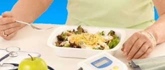
Nutrition
Some adjustments are made to the diet. The amount of foods that contribute to the rise of insulin is reduced. This applies to potatoes, pasta, sugar, bakery products, and cereals. It is recommended to consume more dark-colored berries (blackberries, blueberries), as well as vegetables that contain lutein (spinach, Brussels sprouts, broccoli).
Omega-3 fatty acids should be included in the menu. They can be consumed in the form of dietary supplements or natural products - seafood (salmon, tuna, salmon, herring).
Causes and symptoms of high blood pressure
Increased IOP is much more common than hypotonicity. Its increase may be due to various factors:
- increased production of intraocular fluid;
- significant physical activity;
- frequent consumption of coffee or alcohol;
- severe stress, etc.
An additional risk factor is also the presence of chronic diseases that affect the condition of the eyes: diabetes mellitus, cardiovascular pathologies, hereditary factors. If you have relatives suffering from glaucoma, then you should definitely visit an ophthalmologist for a vision test. With minor deviations from the IOP norm, a person does not even notice discomfort - there are no symptoms of the disorder. They appear only during labile surges or with constantly elevated IOP, when the values already reach 40-50 mmHg. With an average intraocular pressure of 22 to 40 mm Hg. nothing hurts, my eyes don’t turn red, and it doesn’t affect my vision. It is not for nothing that glaucoma, which is inevitable with a persistently increased IOP, is called the “silent thief of vision.”
In old age, you also need to carefully monitor the state of ophthalmotonus. According to medical statistics, 20% of people over 65 years of age are diagnosed with glaucoma, after 70 years of age this figure is 30%, and among people over 80 years of age - 40%. This is explained simply: over time, the outflow of fluid from the eye worsens.
Exercise for the eyes
Purpose of the exerciseExecution techniqueTime costsRelaxing muscles, relieving tension. Close your eyes with your palm. After relaxing, blink a couple of times. Repeat blinking at 5 second intervals. 2 minutes. Increasing the flexibility of the eye muscles. Draw an infinity sign with your eyes, imagining it mentally. Movements are performed exclusively by the pupils. 2 min. Strengthening muscles, improving vision. Select 2 objects in the room: close (30 cm) and distant. Focus your gaze on one and the other object alternately. For 1-1.5 minutes. on every subject. Change the position of the eyes 10-15 times. Improving focus Focus your gaze on your arm extended forward. Bring the phalanges closer to the nose, not reaching 8 cm. Move your hand back. For 2-3 minutes. keep your finger on your finger.Eye exercises improve visual function, normalize the outflow of tear fluid, and reduce stress on the optic nerve.
Symptoms of increased intraocular pressure
Here are some manifestations and signs that may indicate that ophthalmotonus is increased:
- headaches in the temples, dizziness;
- painful sensations in the eyes with sudden movements of the head, raising the eyebrows;
- decreased peripheral vision;
- blurred vision after waking up;
- redness of the eyes due to a pronounced capillary network.
All of these signs can directly indicate the presence of increased intraocular pressure. If they persist, you should visit an ophthalmologist for a comprehensive examination of your vision organs. The most dangerous consequence of increased IOP is glaucoma, which is also called the eye “plague”.
Glaucoma is an ophthalmological disease in which intraocular pressure is constantly elevated or there are sudden changes. If it is not reduced to normal in time, the optic nerve dies, and this inevitably leads to blindness.
According to statistics, approximately 14-15% of blind people in the world have lost their vision due to glaucoma. The overwhelming majority of people with this pathology are over 50 years old. However, it can also occur in infants (congenital) and young adults (juvenile form). The pressure increases due to poor outflow of fluid from the angle of the anterior segment of the eye, which causes its excess to form. As a result, the eyeball begins to put pressure on the optic nerve, causing its damage and, as a result, vision impairment. Then peripheral vision begins to decrease, the visibility area is limited. If the optic nerve dies, the person goes blind. These changes are irreversible. Loss of vision can also occur suddenly as a result of an acute attack.
People at risk (we listed them above) should be especially attentive to the state of their vision. If there are signs of deterioration, you should definitely visit an ophthalmologist for a full diagnosis. Glaucoma cannot be completely cured, but medications can slow the progression of the disease by maintaining stable IOP. In some cases, operations are prescribed to normalize intraocular pressure. They are aimed at increasing the outflow of aqueous humor or reducing its production.
Measurement according to Maklakov
- The patient lies down on the couch, and the doctor administers anesthesia by instilling several drops of dicaine into each eye in turn.
- Then the head is fixed and asked to look at one point.
- A small weight treated with special marking paint is carefully lowered onto the open eye, under the pressure of which the eyeball should be slightly deformed.
- Now the weight is lowered onto a sheet of paper to see how much paint is left on it. Intraocular pressure is determined by the intensity of the imprint.
- The procedure is repeated again in both eyes to avoid the possibility of misinterpretation.
Naturally, some amount of paint from the load will remain on the surface of the eyeball, but it will quickly be washed away with tears. Instead of weights, ophthalmologists sometimes use a portable device that looks like a ballpoint pen. They also apply pressure on the eye, having previously treated the eyeball with an anesthetic.
This method also has an alternative – non-contact tonometry. No weights are placed on the eye, but instead a controlled air flow is used. Many patients find this method more acceptable, but in reality it is rarely used - it is not as accurate.
Important! In patients with glaucoma, eye pressure fluctuates much more noticeably throughout the day than in healthy people. Having such suspicions, the doctor may ask the patient to come to the clinic several times throughout the day. To ensure the accuracy of the diagnosis, you need to measure your blood pressure at least three times before lunch, and the same number in the evening.
How to measure the pressure inside the eye yourself?
Without the help of a doctor and special equipment, it is impossible to determine with what force the fluid secreted by the ciliary body presses on the eye. Nevertheless, it is possible to understand that blood pressure is greatly increased, and ophthalmologists recommend that everyone master this simple technique.
Close your eyes and relax. Now gently press your index finger on the eyeball. You should feel an elastic ball that gives in to your pressure - this is normal. If the eye is very hard and practically does not deform, the IOP level is most likely elevated and it is better to consult a specialist to make sure that its value is not critical
What is low ophthalmotonus
In addition to increased IOP, low blood pressure also occurs, although much less frequently. This condition may be caused by the following reasons:
- retinal detachment;
- inflammatory processes in the eyes;
- eye injuries;
- low blood pressure;
- dehydration and some others.
With reduced ophthalmotonus, the following symptoms are observed: dry eyes, painful sensations when blinking, pop-up dots, burning, blurred vision. Low IOP is no less dangerous than high IOP. Clinical examination of this condition reveals papilledema, venous congestion, maculopathy, papillar atrophy, vitreous opacities, and keratopathy. Visual functions deteriorate significantly.
Folk remedies
Alternative medicine can be used to supplement the treatment prescribed by your doctor. There are simple but effective recipes that have stood the test of time.
Aloe decoction
Used to wash eyes. Aloe (about 5 leaves) is poured with boiling water (250-300 ml). Boil the product for 5 minutes. After cooling, the decoction is ready for use.
Decoction of nettle and lily of the valley
Used for lotions. Nettle and lily of the valley (2 tsp each) are poured with boiling water (200 ml). Cover the container with a lid and let the product brew for 9-10 hours. Cotton pads soaked in liquid are applied to closed eyes (for 5-7 minutes).
Potato compresses
The raw root vegetable is grated on a fine grater. Combine it with apple cider vinegar (5 ml). Let it brew for half an hour. The pulp is applied to gauze napkins and placed on closed eyes. You need to keep the compress for 10 minutes.
Regardless of which prescription is chosen to stabilize eye pressure, the course duration is at least 1 month. Regularity of procedures – 2-3 rubles. in Week.
How can you measure intraocular pressure?
To measure IOP, contact and non-contact tonometers of various modifications are used, including pneumotonometers. The ophthalmologist decides which method to use for diagnosis, depending on the patient’s age, existing pathology, etc.
At the Center for Contact Vision Correction on Tverskaya, intraocular pressure can be checked using various methods. You can make an appointment by calling +7, +7 (800) 100 95 96. Our specialists will carry out other diagnostic procedures if necessary. We are glad to see residents of Moscow and the Moscow region, as well as guests of the capital, in our Center. The Central Clinical Hospital on Tverskaya is open every day, without days off or breaks, from 9.00 to 21.00 (doctors are seen until 20.00 ).
Cost of services for glaucoma
| № | Service name | Price in rubles | Make an appointment |
| 2010025 | Set of disposable consumables for antiglaucomatous surgery | 36000 | Sign up |
| 2010024 | Molteno valve implantation | 54000 | Sign up |
| 2010023 | Implantation of the EX-Pres shunt valve | 54000 | Sign up |
| 2010022 | Ahmed valve implantation | 54000 | Sign up |
| 2010021 | Summing collagen or silicone drainage | 9000 | Sign up |
| 2010020 | Pupilloplasty | 30000 | Sign up |
| 2010019 | Iridoplasty | 22200 | Sign up |
| 2010018 | Basal iridotomy | 10800 | Sign up |
| 2010001 | Sinutrabeculectomy (STE) | 42000 | Sign up |
| 2010002 | Non-penetrating deep sclerectomy (NPDS) | 46200 | Sign up |
| 2010004 | Antiglaucomatous surgery for primary glaucoma of the first category of complexity | 23400 | Sign up |
| 2010005 | Antiglaucomatous surgery for primary glaucoma of the second category of complexity | 30600 | Sign up |
| 2010006 | Antiglaucomatous surgery for primary glaucoma of the third category of complexity | 37200 | Sign up |
| 2010007 | Antiglaucomatous surgery for secondary or refractory glaucoma of the first category of complexity | 29500 | Sign up |
| 2010008 | Antiglaucomatous surgery for secondary or refractory glaucoma of the second category of complexity | 42000 | Sign up |
| 2010009 | Antiglaucomatous surgery for secondary or refractory glaucoma of the third category of complexity | 48000 | Sign up |
| 2010010 | Antiglaucomatous surgery with drainage of the anterior chamber angle for primary glaucoma of the first category of complexity | 28200 | Sign up |
| 2010011 | Antiglaucomatous surgery with drainage of the anterior chamber angle for primary glaucoma of the second category of complexity | 39300 | Sign up |
| 2010012 | Antiglaucomatous surgery with drainage of the anterior chamber angle for primary glaucoma of the third category of complexity | 45600 | Sign up |
| 2010013 | Antiglaucomatous surgery with drainage of the anterior chamber angle for secondary or refractory glaucoma of the first category of complexity | 33360 | Sign up |
| 2010014 | Antiglaucomatous surgery with drainage of the anterior chamber angle for secondary or refractory glaucoma of the second category of complexity | 43800 | Sign up |
| 2010015 | Antiglaucomatous surgery with drainage of the anterior chamber angle for secondary or refractory glaucoma of the third category of complexity | 54000 | Sign up |
| 2010017 | Reconstruction of the anterior chamber angle in secondary glaucoma | 27000 | Sign up |
Making an appointment Today: 8 registered
Prevention measures
It is always easier to prevent diseases, so experts recommend adhering to the following rules in everyday life.
- Time for work and rest should be balanced. The body (including the visual system) needs at least 8 hours a day to rest.
- You need to take breaks throughout the day. At this time, you can allocate 3-5 minutes to perform eye exercises or massage.
- Before going to bed, it is recommended to massage the eyeballs with your fingertips, making circular movements clockwise and counterclockwise.
- Observe personal hygiene rules.
- Stick to a balanced diet. Include more fresh fruits, berries and vegetables, and foods containing Omega-3 acids in the menu.
- Use vitamin complexes, which include microelements beneficial for eye health.
- When performing exercises, you should not put too much pressure on your eyeballs. Heavy physical exercises involving heavy lifting and other overexertion are also not recommended. This automatically increases the IOP.
- Spend more time outdoors on weekends. Active games and hikes are the best cure for all diseases.
Periodically you need to visit an ophthalmologist for a preventive examination. This will help identify the problem at an early stage, which will simplify treatment.