Duration of bleeding according to Duque: time, norm, deviations
Duque's bleeding duration is an important diagnostic test that can be used to determine the rate at which capillary bleeding stops. Thanks to this, the condition of blood vessels, their ability to contract during traumatic injury and hemostasis are assessed.
Procedure and indications for use
To establish the duration of bleeding according to Duke, a special Frank needle is used. It has a specific design, thanks to which the laboratory assistant can control the puncture depth (no more than 4 mm). After all, the study requires minor damage to the soft tissue for blood to appear.
The puncture is made on the ring finger or on the earlobe. After the first drop of blood appears, the time is noted, and the biological fluid is blotted with filter paper every 10-15 seconds. This must be done very carefully so as not to damage the wound itself and thus increase the duration of bleeding. The countdown ends as soon as no traces of blood are detected during the next application of the filter.
Analysis for the duration of bleeding according to Duque is prescribed for the following diseases:
- varicose veins;
- routine examination before surgery;
- tendency to thrombus formation, the appearance of bruises with minor mechanical impact;
- severe liver pathologies;
- autoimmune systemic diseases;
- pregnancy planning.
What affects the duration of bleeding
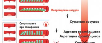
Stopping blood after damage to the skin or mucous membrane occurs due to the activation of the coagulation system. This process includes primary hemostasis, hemocoagulation, coagulation, plasma hemostasis and secondary hemostasis. As a result, strands of blood protein (fibrin) are formed, on which platelets accumulate, forming a blood clot.
This prevents the free flow of blood and helps stop bleeding.
In some cases, a disruption of the coagulation processes occurs, as a result of which the rate of stopping bleeding is significantly prolonged or, conversely, reduced.
If the results of the study reveal not the norm, but a change in indicators, this indicates abnormalities in the patient’s body.
The following pathologies are characterized by a long clotting time:
- thrombopenic Werlhoff disease;
- thrombopenic purpura;
- mourn;
- phosphorus poisoning;
- hemorrhagic diathesis;
- leukemia;
- splenomegalic cirrhosis of the liver;
- long-term use of antiplatelet agents, for example, ascorbic acid or drugs based on it;
- bleeding with hypofibrinogenemia;
- DIC syndrome;
- congenital malformations of blood vessels, accompanied by deterioration of precapillary contraction.
A decrease in the duration of hemostasis most often indicates a violation of the research methodology. Only in some cases does it manifest itself with increased spasticity of the blood vessels.
Interpretation of results
Duques bleeding time is an important diagnostic procedure. The duration of the study is from 2 to 4 minutes, which depends on the individual characteristics of the human body. An important role is also played by what medications the patient is taking at the time of the study. Before the procedure, you must notify your doctor about this so as not to get erroneous results.
Only a specialist can interpret the sample, just like answering the question of whether this is the norm or not. After all, assessing the condition of the circulatory system, as well as confirming or excluding the presence of certain pathologies, is possible only based on the results of a comprehensive examination.
If the Duke test is normal, but there are clinical manifestations of hemostasis disorders, it is imperative to look at the blood clotting time. The duration of bleeding may be within acceptable limits in hemophilia and hypoprothrombinemia.
But at the same time, blood clotting time is significantly prolonged.
To ensure that the results are correct, after the bleeding has completely stopped, you can apply a little pressure with your finger on the wound area. This will cause bleeding to resume. With healthy hemostasis it will stop very quickly, but with disturbances it will manifest itself in full force. The norm will remain far behind.
If you suspect a pathology of the circulatory system, you cannot ignore the doctor’s prescriptions. Only timely diagnosis is the key to a quick recovery.
Indications for use
The duration of bleeding according to Duque is one of the simplest methods, but its results can be a reason for a more detailed examination. The procedure does not require special expensive equipment or complex skills. It is prescribed at the initial stages of a general examination if various diseases are suspected, as well as during routine diagnostics.
Indications for the study may be:
Also read:Bleeding time and its norm
- phlebeurysm;
- examination before planned surgery;
- tendency to form blood clots;
- chronic liver diseases;
- autoimmune pathologies;
- a tendency to develop hematomas - this indicates weakness of the blood vessels;
- general examination during pregnancy planning.
The main purpose of the Duque bleeding time is to assess platelet activity. Normally, they are activated immediately as soon as a violation of the integrity of the vessel wall occurs. They clog the damaged area to prevent fluid from leaving the bloodstream. This process is called white thrombus formation. Any violations, either in the direction of increasing or decreasing the rate of thrombus formation, can be the result of dangerous diseases.
For analysis you will need a special needle that controls the depth of tissue puncture
Bleeding time
Bleeding time (Duke's test) is the duration of bleeding during an injection. To test, use a Frank needle with replaceable blades or special disposable lancets (if they are not available, then use syringe needles) to inject the fingertip to a depth of 4 mm, after which every 10-30 seconds. Using filter paper, without touching the wound, remove a drop of blood; The time is counted from the moment the first drop appears until the bleeding stops. Normally, bleeding stops after 1.5 - 2 minutes. The test reflects the process of stopping capillary bleeding, in which the main role is played by blood platelets, forming the so-called provisional thrombus. In addition, serotonin is released from the blood platelets, causing contraction of the capillary walls. Test results do not always accurately reflect the state of blood clotting. Thus, with thrombocytopathies, the bleeding time is usually sharply prolonged, but the blood clotting time is normal; with hemophilia, hypoprothrombinemia, the blood clotting time is prolonged, and the bleeding time is most often normal. More clearly, violations of bleeding time are detected when pressing or stretching the edges of the wound after the bleeding has stopped: in this case, normally, bleeding resumes for a short time, and in thrombocytopenia it resumes with the same intensity and for a long period (2-3 minutes or more).
Decoding the results
The normal duration of capillary bleeding is from 1 to 4 minutes. During this time, in a healthy person, a spasm of the damaged vessel occurs, and platelets are activated. Thanks to these processes, the section of the vascular wall in the area of the puncture is closed by a thrombus, which retains the fluid in the lumen of the capillary. The readings are deciphered on site, and the results are assessed by a doctor.
Duke's test is often performed in combination with other tests. Thus, it is equally important to assess the rate of blood clotting, count the number of platelets and determine their percentage with other formed elements. There are also specific tests that can help assess the degree of platelet aggregation with collagen and adrenaline.
IMPORTANT! To evaluate the correctness of the result, you can put a little pressure on the puncture area after the bleeding has completely stopped. Normally, its slight resumption is allowed, but the blood quickly stops.
Bleeding time: norm and criteria for blood clotting
Bleeding time is the interval between the time tissue damage occurs and the blood flow stops. This indicator is quite important for absolutely all people, as it indicates the level of blood clotting.
Normal and deviations in bleeding time
Bleeding time
After damage to the skin, it will be normal if the flow of blood stops within a couple of minutes from the moment of puncture. If a person’s puncture time is increased or decreased, this indicates the presence of violations.
During the study, not only platelet counting is performed, but also their adhesion. They stick to the damaged vessel wall.
If during a puncture the bleeding time increases, this indicates that the patient has:
- DIC syndrome
- Hereditary thrombocytopenia
- Vitamin deficiency C
Also, prolonged use of aspirin or anticoagulants can negatively affect bleeding time.
In some cases, patients may experience a decrease in bleeding time. This is most often observed when the patient has renal failure or thrombocytopathy. This phenomenon is also observed during the development of diseases such as von Willebrand disease or acute leukemia. The performance of platelets is their ability to connect. The rate of spontaneous aggregation ranges from 0 to 20 percent.
If a patient has atherosclerosis, diabetes mellitus, heart disease, or blood disease, this indicates an increase in aggregation capacity.
It may also decrease if the patient has diseases that are associated with impaired functioning of blood clots. The reaction of a blood clot is determined by the process of contraction, compaction and release of blood serum in the form of a clot. This action occurs after the protein contained in platelets is formed after protein formation. The normal retraction index is from 48 to 64 percent.
If there is an interruption in the duration of bleeding, the patient must immediately seek help from a doctor. Otherwise, this could have disastrous consequences.
Basic criteria for blood clotting
The duration of the bleeding process is directly affected by blood clotting. Thanks to this process, blood loss during wounds is prevented. Blood clotting is part of the work of hemostasis. Coagulation consists of primary hemostasis, hemocoagulation, coagulation, plasma hemostasis, secondary hemostasis.
Thanks to this process, the formation of strands of blood protein called fibrin is observed in the blood. It forms blood clots, which eliminates the possibility of blood flow and stops bleeding. Bleeding disorders are affected by a variety of causes. In order to avoid undesirable consequences, you need to know the normal blood clotting time and compare it with your indicators.
In order to find out as accurately as possible the duration of bleeding in your body, you need to take a blood test called a coagulogram and hemostasiogram.
Thanks to the results of this comprehensive analysis, the presence of certain diseases in the patient is determined. Initially, a normal bleeding time is required, which ranges from 1 to 3 minutes. It takes 10 minutes for the bleeding process to complete.
You can find out the duration of bleeding as accurately as possible using a special analysis. For this purpose, you must contact a medical center.
Indicators
Interpretation of a blood test for coagulation
Bleeding and the speed at which it stops directly depends on certain indicators.
The rate of blood clotting directly depends on:
- Thrombosed time
- Bleeding time
- Clotting time
- Antithrombin 3
- Fibrinogen
The main indicator of the characteristics of the blood clotting process is thrombin time. Normally it should last from 14 to 21 seconds. This indicator directly depends on what methods of determining it are used. Antithrombin 3 is an indicator that affects the formation of the smallest number of blood clots. This is a regulator of the coagulation circulatory system.
The fibrinogen level should be from 2 to 4 g/l.
Thanks to this criterion, it is possible to characterize the functions of the blood coagulation system and determine the possibility of inflammatory processes in the body. It may be influenced by several medical factors.
Bleeding in an adult should last between 2 and 4 minutes. The level of blood clotting directly depends on its characteristics. A blood clot should form within 2-5 minutes. The faster it is formed, the sooner the bleeding will stop.
Estimation of bleeding duration according to Duque
Bleeding time according to Duque
The bleeding time, the rate of which depends on the individual characteristics of the patient, can be estimated using a fairly simple method according to Duques. This is a special technique with which the condition of the circulatory system, namely the vessels, is assessed. From the start to the stop of bleeding, no more than 3 minutes should pass using this method.
Hemostasis is a biological complex with the help of which blood is stopped in a timely manner. The duration of bleeding using this method is an assessment of the state of platelets. In the absence of damage to the vascular walls, platelet activation should be carried out immediately.
When assessing the duration of bleeding according to Duque, platelet activity is assessed.
For this purpose, the number of platelets and a specific platelet formula are calculated. Also, in order to evaluate the indicators, factors such as the ability of platelets to aggregate with collagen, adenosine diphosphate and platelet aggregation, coagulability - von Willebrand factor activity are assessed.
More information about blood clotting can be found in the video.
The duration of bleeding using this method is most often determined using a special needle. Its design includes a hollow body and trigger, as well as a small tip and coupling for the spring. The needle is characterized by a high level of convenience, as it provides the ability to adjust the puncture needle.
In most cases, the piercing is done in places such as a finger or earlobe.
If the patient has normal hemostasis, then he can easily cope with bleeding. This will take him no more than two minutes. If blood clotting slows down, the duration of bleeding will be prolonged. This may indicate the presence of liver pathologies, hemophilia and various other diseases. The measurement of the fence directly depends on the area in which the puncture is made - in the earlobe or finger.
The Duque method for determining the duration of bleeding is not the only one. Very often, laboratory assistants use other parts of the human body to conduct research. In this case, difficulties in venous outflow are artificially provoked. To conduct the study, a puncture is made in the upper area of the forearm. Droplets of blood that form at the puncture site are removed with sterile wipes. After three minutes, only small spots should remain on the napkin. The duration of bleeding is a fairly important factor in human life. To determine it, it is necessary to undergo special studies.
Coagulogram No. 2 (prothrombin (according to Quick), INR, fibrinogen)
A coagulogram is a study of the hemostatic system that allows you to evaluate the external and general pathways of blood coagulation and identify the risk of hypercoagulation (excessive clotting) or hypocoagulation (bleeding).
Synonyms Russian
Hemostasiogram: prothrombin index (PI), prothrombin time (PT); international normalized ratio (INR); factor I (first) of the plasma coagulation system.
English synonyms
Coagulation studies (coagulation profile, coag panel, coagulogram): Prothrombin time (Pro Time, PT, Prothrombin time ratio, P/C ratio); International Normalized Ratio (INR); Fibrinogen (FG, Factor I).
Units
% (percent), sec. (second), g/l (grams per liter).
What biomaterial can be used for research?
Venous blood.
How to properly prepare for research?
- Do not eat for 12 hours before the test.
- Avoid physical and emotional stress and do not smoke for 30 minutes before the test.
General information about the study
The hemostasis system consists of many biological substances and biochemical mechanisms that ensure the preservation of the liquid state of the blood and prevent and stop bleeding. It maintains the balance between blood clotting and anti-clotting factors. Significant disturbances in the compensatory mechanisms of hemostasis are manifested by processes of hypercoagulation (excessive thrombus formation) or hypocoagulation (bleeding), which can threaten the patient’s life.
When tissues and blood vessels are damaged, plasma components (clotting factors) participate in a cascade of biochemical reactions, which results in the formation of a fibrin clot. There are internal and external pathways of blood coagulation, which differ in the mechanisms that trigger coagulation. The internal pathway is realized when blood components come into contact with the collagen of the subendothelium of the vessel wall. This process requires coagulation factors XII, XI, IX and VII. The extrinsic pathway is triggered by tissue thromboplastin (factor III) released from damaged tissues and the vascular wall. Both mechanisms are closely interrelated and from the moment of formation of active factor X they have common paths of implementation.
The study of indicators such as PTI (prothrombin index) and INR (international normalized ratio) allows us to assess the state of the external coagulation pathway. PTI is calculated as the ratio of the standard prothrombin time (the clotting time of control plasma after the addition of tissue thromboplastin) to the plasma clotting time, expressed as a percentage. INR is a prothrombin test standardized in accordance with international recommendations. It is calculated by the formula: INR = (patient prothrombin time / control prothrombin time) x MICH, where MICH (international sensitivity index) is the sensitivity coefficient of thromboplastin relative to the international standard. INR and PI are inversely proportional indicators, that is, an increase in INR corresponds to a decrease in the patient’s PTI and vice versa.
Reference PTI values depend on the set and characteristics of the reagents and differ in the activity of thromboplastin used in the test. The results of INR determination, thanks to standardization, make it possible to compare the results of different laboratories.
Tests for PTI (or a similar indicator - prothrombin according to Quick) and INR in a coagulogram help to identify disorders in the external and internal blood coagulation pathways associated with a deficiency or defect of fibrinogen (factor I), prothrombin (factor II), factors V (proaccelerin) , VII (proconvertin), X (Stewart-Prower factor). With a decrease in the concentration of these coagulation factors in the blood, the prothrombin time increases in relation to control laboratory parameters.
Plasma factors of the extrinsic coagulation pathway are synthesized in the liver. For the formation of prothrombin and some other coagulation factors, vitamin K is required, the deficiency of which leads to disturbances in the cascade of reactions and prevents the formation of a blood clot. This fact is used in the treatment of patients with an increased risk of thromboembolism and cardiovascular complications. Thanks to the prescription of the indirect anticoagulant warfarin, vitamin K-dependent protein synthesis is suppressed. PTI (or Quick prothrombin) and INR coagulation are used to monitor warfarin therapy in patients with factors that promote thrombosis (eg, deep vein thrombosis, presence of prosthetic valves, antiphospholipid syndrome).
Normally, a coagulogram in a healthy person has an INR in the range of 0.8-1.2; in patients being treated with indirect anticoagulants to prevent thromboembolic complications - 2.0-3.0, in patients with prosthetic valves and antiphospholipid syndrome - 2.5-3.5.
Simultaneous determination of fibrinogen in a coagulogram allows a comprehensive assessment of the state of the plasma hemostasis system.
Fibrinogen is blood clotting factor I, which is produced in the liver. Thanks to the action of the coagulation cascade and active plasma enzymes, it is converted into fibrin, which is involved in the formation of a blood clot and thrombus. Fibrinogen deficiency can be primary (due to genetic disorders) or secondary (due to excessive consumption in biochemical reactions), which is manifested by impaired formation of a stable blood clot and increased bleeding.
Fibrinogen is also an acute phase protein. Its concentration increases in the blood during diseases accompanied by tissue damage and inflammation. Determining the level of fibrinogen is important in the diagnosis of diseases with increased bleeding or thrombosis, as well as for assessing the synthetic function of the liver and the risk of cardiovascular diseases with complications.
What is the research used for?
- For a general assessment of the blood coagulation system.
- For the diagnosis of disorders of the external and general pathways of blood coagulation.
- To study the activity of coagulation factors I, II, V, VII, X.
- To monitor the patient's condition when prescribing anticoagulants.
- To assess the risk of cardiovascular complications.
- To assess the protein-synthesizing function of the liver (synthesis of blood clotting factors).
When is the study scheduled?
- With a comprehensive examination.
- When planning surgical interventions.
- When examining patients with nosebleeds, bleeding gums, blood in the stool or urine, hemorrhages under the skin and in large joints, chronic anemia, heavy menstrual flow, sudden loss of vision.
- When examining a patient with a history of episodes of thrombosis.
- With a hereditary predisposition to disorders of the hemostatic system.
- With a high risk of cardiovascular complications and thromboembolism.
- Before prescribing anticoagulants.
- When monitoring the hemostatic system while taking anticoagulants.
- For liver diseases.
What do the results mean?
Reference values (table of normal coagulogram indicators)
- Prothrombin according to Quick: 70 - 120%.
- INR
| Age | Reference values |
| Less than 3 days | 1,15 — 1,35 |
| 3 days – 1 month | 1,05 — 1,35 |
| 1 month – 1 year | 0,86 — 1,22 |
| 16 years | 0,92 — 1,14 |
| 6 – 11 years | 0,87 — 1,20 |
| 11 – 16 years | 0,97 — 1,30 |
| More than 16 years | 0,8 — 1,2 |
| Week of pregnancy | Reference values |
| 13-21st | 0,56 — 1,1 |
| 21st-29th | 0,5 — 1,13 |
| 29-35th | 0,58 — 1,17 |
| 35-42nd | 0,15 — 1,14 |
- Fibrinogen: 1.8 - 3.5 g/l.
| Week of pregnancy | Reference values |
| 1-13th | 2.12 - 4.33 g/l |
| 13-21st | 2.9 - 5.3 g/l |
| 21st-29th | 3 - 5.7 g/l |
| 29-35th | 3.2 - 5.7 g/l |
| 35-42nd | 3.5 - 6.5 g/l |
Reasons for an increase in INR and a decrease in the level of prothrombin according to Quick (indicates a possible deficiency of factors in the external hemostasis pathway and a tendency to increased bleeding):
- DIC syndrome (disseminated intravascular coagulation) during the period of hypocoagulation;
- hypofibrinogenemia (factor I deficiency);
- dysfibrinogenemia (synthesis of a defective protein that is unable to participate in the cascade of biochemical reactions);
- hereditary or acquired deficiency of factors II, V, VII;
- Factor X deficiency (eg, purpura due to amyloidosis);
- vitamin K deficiency;
- hemorrhagic disease of newborns;
- malabsorption with impaired fat absorption (due to celiac disease, chronic diarrhea);
- acute leukemia;
- antiphospholipid syndrome;
- congestive heart failure;
- liver pathology (hepatitis, cirrhosis, alcoholic liver disease);
- obstruction of the biliary tract, obstructive jaundice;
- pancreas cancer;
- Zollinger-Ellison syndrome (pancreatic adenoma);
- toxic shock syndrome;
- nephrotic syndrome (excessive urinary excretion of factors V and VII);
- oral anticoagulants (warfarin).
Causes of increased fibrinogen levels (indicates an increased risk of blood clots and cardiovascular complications):
- acute infection (eg pneumonia, tuberculosis);
- autoimmune diseases (rheumatoid arthritis, reactive arthritis);
- acute coronary syndrome, myocardial infarction;
- burns;
- cancer (breast, kidney, stomach);
- multiple myeloma;
- Hodgkin's disease (lymphogranulomatosis);
- glomerulonephritis, nephrotic syndrome, nephrosis;
- pregnancy;
- eclampsia;
- cerebrovascular disease, stroke;
- hepatitis;
- postoperative period;
- rheumatic fever;
- tissue damage.
Reasons for a decrease in INR and an increase in prothrombin according to Quick (indicates a tendency to form blood clots):
- DIC syndrome (period of hypercoagulation);
- deep vein thrombosis (initial stages);
- polycythemia;
- pregnancy (last months);
- increased activity of factor VII.
Reasons for decreased fibrinogen levels (may indicate an increased risk of bleeding):
- dysfibrinogenemia;
- hereditary afibrinogenemia;
- DIC syndrome;
- fibrinolysis;
- hemophilia A and B;
- liver pathology (hepatitis, cirrhosis);
- abortion;
- premature placental abruption;
- late stage of cancer;
- embolism (amniotic fluid, meconium, fat, tissue);
- anemia;
- eclampsia;
- leukemia;
- malabsorption;
- shock;
- sepsis;
- post-transfusion reactions.
What can influence the result?
- Factors that distort the result of the analysis: the presence of lupus anticoagulant in the blood (directly inhibits coagulation factors);
- transfusion of donor blood components in the last month (distorts the fibrinogen indicator).
- drinking alcohol, fatty foods;
- excess intake of vitamin K from food (found in beef or pork liver, green tea, broccoli, chickpeas, cabbage, turnips, soy, green leafy vegetables);
Important Notes
- Before prescribing anticoagulants, it is necessary to determine the initial PI and INR values. A PTI of less than 25% and an INR of more than 3.5 in patients taking anticoagulants are critical and require immediate medical attention. With an INR above 5.0 there is a high risk of bleeding, and with an INR below 0.5 there is a risk of blood clots.
- If fibrinogen decreases to a level of less than 1 g/l, urgent consultation with a specialist is necessary due to the high risk of bleeding.
Also recommended
- D-dimer
- Activated partial thromboplastin time (aPTT)
- Antithrombin III
- Thrombin time
- Vitamin K (phylloquinone)
- Complete blood count (without leukocyte formula and ESR)
- Erythrocyte sedimentation rate (ESR)
- Predisposition to increased blood clotting
- Genetic risk of thromboembolism
- Genetic risk of thromboembolism (advanced)
Who orders the study?
Cardiologist, surgeon, hematologist, hepatologist, therapist, obstetrician-gynecologist.
Literature
- Nazarenko G.I., Kishkun A. Clinical assessment of laboratory research results. – M.: Medicine, 2000. – 249-262.
- Fischbach FT, Dunning MB A Manual of Laboratory and Diagnostic Tests, 8th Ed. Lippincott Williams & Wilkins, 2008: 1344 p.
- Wilson D. McGraw-Hill Manual of Laboratory and Diagnostic Tests 1st Ed. Normal, Illinois, 2007: 666 p.
Duration of bleeding according to Duque - norm and pathology
The duration of bleeding according to Duques is an important indicator of the well-being of the body and its circulatory system. This analysis is a diagnostic procedure that allows us to identify various diseases and disorders of the hemostatic system.
All about the hemostasis system
The body has a special system called hemostasis. It ensures that blood remains in a liquid state while it moves through the vessels of the body, stops bleeding in case of damage to the vascular system or tissue integrity, and also dissolves blood clots that have already played a role.
In case of vascular damage, one of three mechanisms of the hemostasis system can be involved:
| Mechanism of hemostasis | Peculiarities |
| Coagulation | It is “turned on” by tissue factor released from damaged tissues and is regulated by blood clotting factors. The vessel affected by bleeding is blocked by a fibrin clot - a “red thrombus”, which includes red blood cells. |
| Vascular-platelet | This hemostasis uses vasospasm and blocking of its lumen with platelets. The resulting formation is called a “white thrombus.” |
| Fibrinolysis | This is the process of dissolving and eliminating a blood clot that has “spent” its time needed to restore the integrity of the vessel and surrounding tissues. |
Hemostasis is a complex biological system that involves the body controlling bleeding and eliminating its consequences. With any damage, platelets receive a signal from special proteins that are released at the time of injury.
Platelets form special projections on their surface that allow the cells to adhere tightly to each other and form a dense clot - a thrombus that covers the damaged area and stops bleeding. The rate at which this clot forms is the bleeding time.
What is bleeding time?
To check the condition of the hemostasis system, a simple test is used - tissue damage on the fingertip or earlobe.
Bleeding time is the moment from inflicting a wound until the last drop of blood stops flowing from the damaged area. This is an important indicator of platelet function and their effect on the condition of the wall of the affected vessel.
The test cannot reveal absolutely all disorders, but indicates the possible presence of thrombocytopenia, thrombocytopathy, problems with the elasticity of the walls of blood vessels and impaired contractility, von Willebrand disease.
If the analysis reveals a change in bleeding time, deeper and more extensive studies are required.
There are three main ways to determine bleeding time:
- Template method.
- According to Duque (Duke).
- According to Ivy.
The most commonly used is the Duque bleeding duration, the rate of which is a very important indicator, since it is the simplest and fastest diagnostic method. In medical practice, other methods of identifying important data can be used if the doctor considers it rational and necessary to prescribe them to the patient.
Useful video about blood clotting: