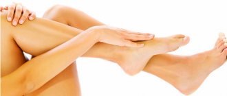
Why do spider veins appear on the legs?
Spider veins and spider veins on the legs are an expansion of small intradermal veins, as a result of which they become visible as “bluish veins.” The main reason for their appearance is hormonal status: female sex hormones help reduce the tone of the veins. Accordingly, in the overwhelming majority of cases they occur in the female part of our population. This may also explain their frequent occurrence during pregnancy. In addition, contributing factors include hereditary predisposition and prolonged static loads.
First signs and main symptoms
To avoid lengthy and complex treatment, it is important to identify the disease in the early stages. It makes no sense to say that only a specialist can do this, this is obvious. The patient’s main task is to distinguish the earliest symptoms in order to see a doctor before spider veins appear. The first signs include:
- itching, burning, regular pain in the legs after a long walk, swelling;
- the red-violet mesh is still absent at this stage, but by the end of the day muscle fatigue usually appears, the patient feels nervous tension due to severe pain inside the lower leg.
People with chronic diseases or family history should pay special attention to their body. The disease progresses at different rates, depending on the individual characteristics of the body and the presence of harmful factors. If it is not possible to avoid the appearance of capillaries, then timely treatment of spider veins is required. The main symptoms of telangiectasia are:
- red-violet dots appear on areas of the skin, which gradually turn into stripes;
- most often, the stripes spread quickly, intersecting each other, forming a pattern similar to a web;
- after a long walk, the legs quickly swell and get tired, swelling is observed in the shins;
- spider veins quickly progress, spread to new areas of the body, and the first signs of varicose veins appear;
- at a late stage of the disease, the patient experiences night cramps, a general deterioration in condition, incl. skin, bruising may occur due to cracked capillaries.
Patients should pay attention to the general condition of the body. Due to impaired blood circulation, a person experiences headaches and increased body temperature.
How to get rid of spider veins on your legs?
Conservative treatment (especially wearing compression stockings) is quite effective in preventing the occurrence of spider veins. However, if they have already appeared, there are two main treatment methods:
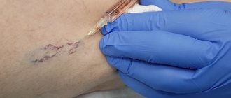
- Sclerotherapy. Using an injection, a sclerosant is injected into the main feeding vessel - a substance that causes damage and gluing of the vessel walls. Within 1-2 weeks, the vessel and the vascular network it feeds become fibrotic and disappear. The method is very effective for larger vessels.
- Percutaneous laser coagulation. Typically a neodymium laser is used. It passes through the transparent upper layers of the skin and is absorbed by the hemoglobin in the blood. The vessel heats up and it is “soldered”. This method is more effective for smaller vessels.
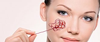
Telangiectasia - causes, symptoms, diagnosis, treatment
Vascular pathologies occupy a leading position among systemic diseases, and are becoming younger every year. If previously older people learned about varicose veins, in the modern world the disease manifests itself in people no older than 35 years of age, and sometimes affects teenagers and people under 25 years of age.
Varicose veins are accompanied by spider veins, in medical terminology – telangiectasia.
What it is
Telangiectasia is the dilation of capillaries, venules and arterioles visible to the naked eye.
Externally, the formations look like a network or an intricate star, hence the name “venous stars”.
The disease occurs at any age and sometimes occurs even in infants and newborns. The localization of the process is also different; telangiectasias occur on the trunk, limbs, mucous membranes and face.
The vessels increase in size, fill with blood, and appear in the subcutaneous fat layer. The color changes from red to bluish-violet, and when pressed, the area turns pale, but returns again.
Causes
For a long time it was believed that telangiectasia is associated with vascular pathologies. However, over time, studies were conducted that disproved the theory. Many experts are convinced of the root causes of vascular changes, but admit the influence of other provoking factors. For example, newborns do not have varicose veins, but a network of capillaries is formed, which means that there are other causes of the disease.
Causes:
- Varicose veins are the main cause of the disease. With this pathology, stagnation occurs, which impedes the movement of blood through small vessels.
- Hormonal imbalance - a deficiency or excess of certain hormones can cause the formation of vascular networks on the legs, arms, face and genitals.
- Mastocytosis or mast cell proliferation - systemic or cutaneous.
- Genetic predisposition. About 40% of daughters inherit the disease from their mother; in sons, the pathology is half as common.
- Dermatitis or epidermal pathological processes.
- Ataxia. It manifests itself especially clearly in the infancy and newborn period.
- Raynaud's disease is a disorder of the blood supply to the feet and hands.
- Prolonged exposure to ultraviolet rays - sunbathing or visiting a solarium.
- CS therapy, without medical supervision or in case of a peculiar allergic reaction.
- Vascular defect occurs in alcohol-dependent people, as well as in people leading a sedentary lifestyle.
- Sometimes stars develop in pregnant women with extreme weight gain, or due to hormonal changes.
Classification and types
Classification by type of vessels: – arterial; – venous; – capillary.
There are several types of shape:
- Stellate - many dilated capillaries of light red color.
- Spot-like - has the appearance of regular balls of a bright red hue, combined with other skin ailments.
- Linear – curved lines form on the delicate skin. The color ranges from red to purple, the location is the cheeks.
- Tree-like - from the name it is clear that outwardly telangiectasia resembles a tree or its root system. Such formations are more often found on the lower extremities and abdomen. They have a predominantly blue-violet hue.
Depending on the form of the disease, there are primary and secondary types. The first primary group includes the following species:
– nevoid TAE – affects the anatomical region of the cervical nerves; – generalized or essential TAE – first the affected area includes the feet, and then the disease spreads up the lower extremities; – hereditary – the stars are arranged randomly and asymmetrically, but do not affect the mucous membranes; – hereditary hemorrhagic TAE – affects the mucous membranes, as well as the endothelium of internal organs, areas of physiologically difficult blood supply (hands, feet), brain, liver and gastrointestinal tract; – telangiectatic marbled skin – the patient has a mesh on the face, which manifests itself after physical or mental stress; – ataxia-TAE – is considered a childhood form, since around 5 years old stars appear on the legs, knees, ears, conjunctiva, and soft palate.
The secondary form is combined with a severe underlying disease:
- keratosis solaris;
- basal cell carcinoma;
- imbalance of estrogen and collagen;
- a peculiar reaction to a transplanted donor organ - graft.
Why is it dangerous?
The disease itself is not a pathology, but a cosmetic defect.
The appearance of a characteristic capillary star is a sign of the disease, and may indicate a certain stage of the pathological process. TAEs indicate cirrhosis of the liver or internal gastric bleeding, appearing on the anterior wall of the enlarged abdominal cavity. The condition is dangerous and requires emergency hospitalization.
A similar process occurs in children, but indicates the accumulation of fluid in the abdominal cavity - ascites. Externally, the mesh diverges to the sides, resembling the head of a jellyfish.
Symptoms
Clinical manifestation of the disease in adults:
- a grid or winding lines, collected in bunches or located scattered;
- the color of the formations ranges from purple to red;
- absence of pain, burning or other discomfort at the site of mesh formation;
- periodic change in shade, under the influence of blood entering dilated vessels;
- Sometimes the site of TAE itches, but in this case we can talk about dermatological pathologies.
Symptoms in children of different ages:
- blurry mesh formations, mainly localized on the face;
- in newborn children, the disease is often found on the back of the head, but does not disappear over time;
- there is no pain, but sometimes swelling or swelling appears.
Based on the nature of the star, a knowledgeable specialist determines the form of the disease, the type and type of vessel. The localization of the pathological process plays an important role. Telangiectasia, collected in groups, can cause a clear cosmetic defect if they are formed on the prominent side of the face.
Localization
Interestingly, certain forms of the disease have favorite localization sites:
- Ataxia: soft palate, lower palate, conjunctiva, knees, elbows, ears.
- Hereditary hemorrhagic: oral cavity, mucous membranes, feet, fingers.
- Generalized: legs, toes, feet.
- Marbled skin - the skin of the face, cheeks, arms, torso and legs.
- The capillary pattern is diagnosed on the nose and bridge of the nose, found on the hands and abdomen, and sometimes occurs in the genital area, and even the perineum.
Which doctor should I contact?
The pathology is treated by a vascular surgeon and phlebologists.
Medical assistance is necessary if a cosmetic defect occurs.
If the skin around the star changes color and becomes faded, we are talking about a pathological process located in the deep veins or arteries.
Diagnostics
First, an external examination is carried out, during which the doctor notes such parameters as:
– individual characteristics of the patient; – form and nature of telangiectasia; – location; – type of vessel.
Only after a superficial examination are narrow-profile diagnostic studies prescribed:
- examination by a gynecologist and consultation with an endocrinologist;
- MRI;
- radiography;
- Ultrasound of internal organs;
- endoscopy;
- a number of laboratory tests, in particular, biochemistry, general analysis and determination of cholesterol concentration;
- urine test;
- daily monitoring.
Hardware methods are not used often, since sometimes it is enough for the doctor to correctly palpate the patient, collect an anamnesis and conduct a pinch test. The skin is pinched near the area of the capillary network or a tourniquet is applied.
By the nature of pinpoint hemorrhages, the degree of pathology and the possibility of a secondary form of the disease are judged.
If there are doubts about the diagnosis, the doctor refers the patient to additional diagnostic methods.
Treatment
The disease can only be treated with minimally invasive surgical interventions.
Drug therapy is used to prevent “stars”, as well as during the recovery period. Prescribed drugs:
- agents that normalize the biochemical composition of the blood and reduce cholesterol;
- having a liquefying effect;
- having immunostimulating properties.
You can completely get rid of the defect using several surgical techniques.
Surgical intervention
Surgical treatment is the main anti-thaleangiectasia therapy. The following techniques are used: – microsclerotherapy; – echosclerotherapy; – postoperative; – “foam-form” sclerotherapy.
The essence of the procedure: a special solution – sclerosant – is injected into the damaged vessel, identified by ultrasound.
The substance, once in the cavity, has a healing and anti-sticking effect. Thus, blood supply is restored, and under the action of pigment substances the defect is eliminated.
The operation is performed without general anesthesia, but at the request of the patient, the injection area is numbed.
In addition to sclerotherapy, laser coagulation is used, however, this procedure has many contraindications.
It is important to understand that the methods used to get rid of TAE do not eliminate the true cause of the disease. Only a specialist will prescribe the correct treatment, as well as the acceptable type of surgical intervention.
Which is better: sclerotherapy or laser coagulation?
These methods are not opposed, but rather complement each other. Larger vessels are better removed by sclerotherapy (it is easier to puncture them with a needle and inject the drug, there is less chance of rupture of the vein during injection, correctly administered sclerosant in the form of foam almost 100% leads to gluing of the vascular network). A small vascular network is difficult to treat with sclerotherapy due to technical reasons (the diameter of the vessels of the network can be comparable to the diameter of the needle). And for percutaneous laser coagulation, this is an advantage, since it is easier to heat and “seal” such a vessel with a laser without the possibility of burning the overlying skin. Thus, first, large vessels are removed using sclerotherapy, and then small ones are “reached” with a laser.
Multicolored legs
Thin and thick, red and blue... veins on the legs. Almost every woman studied them, intently examining her legs and wondering what they were? They appear out of the blue, and every year there are more and more of them. Some try not to notice these defects, others begin to worry when they are not even visible to others. However, the fact remains: this problem affects almost every person. Many patients are very surprised when they find out that they have dilated vessels, because this can mean anything: barely visible veins, or unaesthetic varicose veins. In fact, vessels are tubular organs (channels) of various sizes through which blood or lymph moves. In this article we will only touch on blood vessels, which in one way or another appear externally on the skin. On the skin, as a rule, you can see either thin red vessels with a diameter of less than a millimeter (usually capillaries or arterioles), or vessels of blue and purple shades up to several millimeters thick; quite often much larger vessels appear - the so-called varicose veins. If they begin to change and look like ugly subcutaneous nodes and tangles, then you should think about whether varicose veins have “fell” on your poor legs?
Is it possible to cure spider veins at home?
At home, you can and should prevent the appearance of spider veins. The most effective method is, of course, wearing compression hosiery (at least during pregnancy, on days of increased or prolonged static loads). Venotonic drugs have some effectiveness. A contrast shower, periodically throughout the day raising the legs above the level of the heart, and massage of the lower extremities (provided there are no varicose veins) will also be useful. However, it will not be possible to remove spider veins that have already appeared on the legs at home. This will require special procedures that can only be performed in a specialized clinic (sclerotherapy, percutaneous laser coagulation).
Be healthy!
Sign up for a free consultation with a phlebologist or ask any questions you have about phlebology and vein treatment by phone:
+7 (495) 255−50−11.
Basic questions about spider veins
- Is it painful to remove spider veins on the legs?
This question worries all women who decide to get rid of spider veins on the skin. The answer is very simple. For microsclerotherapy, the finest needles are used, so the injections are so painless that they surprise everyone who has undergone microsclerotherapy. In addition, our clinics use a cryosclerotherapy protocol. We always cool the skin with a special cooler, which makes injections absolutely painless. Treating intradermal vessels in the legs with laser or RFO is more difficult than with cryosclerotherapy and more expensive for the patient. Spider vein sclerotherapy is a painless and well-tolerated treatment procedure.
- What time of year is best for treatment?
It must be taken into account that the process of disappearance of the venous network after a course of sclerotherapy takes approximately two months. Therefore, if you are planning a vacation, then expect that you will be able to enjoy clear, star-free skin approximately 2-3 months after completing the course of treatment. Plan your visit to the phlebologist in advance so as not to be disappointed during your beach holiday. Treatment should not be done during pregnancy and breastfeeding, since the effect of the sclerosing drug on the child is unknown.
Advantages
Today, there are several methods for removing spider veins, but not all of them are equally safe. Often, after the intervention, scars and age spots remain on the skin. The removal of spider veins using laser coagulation is considered the most harmless and effective. This procedure is performed absolutely painlessly and has virtually no contraindications. When spider veins are removed in this way, laser radiation acts on the dilated vessel, heats it and “seals” it. As a result, the surrounding tissues are not affected, and the defects that bother the patient completely disappear. The Medicine and Beauty Clinic offers guaranteed removal of vascular spots and stars on the skin without complications.
Microsclerotherapy techniques used in the Moscow City
At the Moscow City Phlebology Center we use only the most modern types of microsclerotherapy (scleroobliteration of veins).
Sclerosants for microsclerotherapy
- Telangiectasia (spider veins), diameter up to 1 mm, are removed using a liquid form of the drug: polidocanol (0.25 - 0.75%), tetradecyl sulfate (0.1 - 0.2%).
- To remove venulectasias (diameter 1-2 mm) and reticular veins (2-4 mm), both microsclerotherapy solution and microfoam sclerotherapy (foam-form) are used. Concentration of active substance: polidocanol - 0.5-1%, sodium tetradecyl sulfate - 0.25-0.4%.
Types of modern sclerosants
All modern sclerosants can belong to one of the following groups:
- Detergent preparations. The chemical structure of these substances is based on fatty acids or alcohols. The mechanism of their action is based on the extraction of protein molecules from the surface membrane of the endothelial cell of venous vessels, the cell is irreversibly damaged and dies. This effect works only in the range of a certain sclerosant concentration, below which there is no effect on vascular cells. This group is represented by the following drugs:
- Ethoxysclerol (Polidocanol).
- Sodium tetradecyl sulfate (fiberwein).
Everything is ready for microsclerotherapy
In addition to these drugs, the following detergents are also used in the world: sodium morruate, ethanolamine oleate, glycerin.
- Cell toxin preparations are used by some specialists for sclerosis of vascular formations.
- Hypertonic and ionic solutions. Modern European phlebologists also use the following sclerosants of this group:
- Hypertonic sodium chloride solution (20 and 23.4% solutions).
- Sclerodex (a combination of a solution of 25% glucose and 10% sodium chloride).
- Ionized iodine preparations (variglobin, sclerodin).
- 75% glucose solution.
In Russia today only drugs from the detergent group are approved for use: polidocanol (ethoxysclerol) and tetradecyl sulfate (fiberwein). The issue of admitting 75% glucose solution into medical phlebological practice is being actively resolved.
Cosmetic microsclerotherapy (microscleroobliteration) at the Moscow Phlebology Center
The microsclerotherapy method is deservedly called the “gold standard” for the removal of telangiectasia. Due to its minimal invasiveness, good patient tolerance and good clinical effect, the microsclerotherapy technique has taken its rightful place in modern phlebological practice. Cosmetic microsclerotherapy at the Moscow City Phlebological Center is performed by some of the best phlebologists in Moscow and Russia according to the most modern European standards.
Microsclerotherapy performed by phlebologist of the MIFC Raskin V.V.
We use the most advanced technologies to achieve not only a good clinical effect, but also the best aesthetic result.
A good modern sclerosant, as it should be
A good modern sclerosant should have the following properties:
- The substance must not be toxic to the human body
- The sclerosing effect should be caused only by a strictly defined concentration of the drug, subject to prolonged contact with the vein wall
- No allergic reactions to sclerosant
- The drug must have the necessary strength to obliterate sufficiently large vessels, and be safe in case of extravasation (going beyond the boundaries of the vessel)
- The drug should not cause scarring or skin pigmentation effects
- No pain effect on drug administration
- The sclerosant must have good solubility in physiological solution.
Complications and side effects after a microsclerotherapy session
Minor ecchymosis (bruising) at the injection sites after the procedure is almost all that worries patients. These phenomena disappear within 1-2 weeks. Complications and side effects of sclerotherapy are quite rare, but you still need to know about them:
- Skin hyperpigmentation is a fairly rare phenomenon and goes away on its own within 6-12 months.
- Local tissue swelling. It resolves on its own and does not require separate treatment.
- Neurological complications: migraine-like headache, temporary impairment of visual fields, extremely rare complications that arise when working with the foam form of sclerosant in patients with congenital heart disease (patent foramen ovale). These phenomena disappear on their own within 10-15 minutes.
- Metting is the appearance of the finest red telangiectasia in the injection area. These manifestations disappear within a few months.
- Skin necrosis occurs when sclerosant enters the perivascular tissue in high concentrations. This is a very rare occurrence and is most often associated with improper manipulation technique.
- Allergic reactions to the drug. Modern sclerosants rarely cause sensitization of the body, but you should always remember this phenomenon.
Glue or cut?
Vein sclerotherapy
- Cost: 6,500 rub.
- Duration: 20-60 minutes
- Hospitalization: No hospitalization required
More details
The pages of modern magazines are full of advertisements about the non-surgical treatment of varicose veins, the so-called “sclerotherapy”. However, unfortunately, only rare forms of this pathology are eliminated non-surgically. Surgery remains the radical method. Sclerotherapy (introduction of “gluing” substances into dilated veins) only complements, but does not replace the treatment of varicose veins.
Fear of pain and large postoperative scars sometimes makes patients refuse surgery. Of course, surgery is a certain risk, but the risk of complications if you refuse surgery is much higher. There is an opinion that surgery does not lead to a lasting positive result, and varicose veins reappear even after it is performed. This is not entirely true. The further course of the disease depends on the type of structure of the venous system. There is a group of patients who, after surgery, “forget” about their veins. Another notices the appearance of new dilated ducts. There is no need to be afraid of this. Regular visits to a phlebologist will allow you to get rid of these troubles. And in this case, sclerotherapy takes a leading place.
How is the microsclerotherapy procedure performed in a modern phlebological center in Moscow?
In our phlebological center, we use some of the best thinnest needles with laser sharpening of 27-30 G. Using these tools, skin punctures are made that are practically not felt by patients. The procedure is performed by an experienced phlebologist, who has not only thousands of procedures under his belt, but also a significant number of master classes on this technology.
Master class performed by phlebologist Semenov A.Yu. on microsclerotherapy
Sessions are carried out with the patient in a horizontal position. After puncturing the skin, the tip of the needle enters the vessel, the specialist slowly and carefully injects the drug, making sure that the vein does not overstretch and rupture.
After the session, sterile bandages are applied and compression stockings are put on. The patient is recommended to take a half-hour walk.
What is microsclerotherapy (removal) of telangiectasia (spider veins)?
Microsclerotherapy of telangiectasia (spider veins) is a modern cosmetic procedure aimed at removing small-diameter venous vessels by introducing substances (sclerosants) specially developed for this purpose into their lumen. The technique is based on the effect of a chemical burn of the inner wall of the asterisk with subsequent fibrosis of the vessel.
Microsclerotherapy procedure for telangiectasis
The microsclerotherapy procedure for telangiectasia is the “gold standard” for the removal of these vascular formations that arise from persistent dilatation of the smallest veins. This state of affairs is associated with a high aesthetic effect, safety and good tolerability of microsclerotherapy for telangiectasia.
Contraindications
| Absolute | Relative |
|
|
If there are absolute contraindications, the procedure will have to be temporarily abandoned. Relative contraindications allow phototherapy, but only after consultation with the attending physician.
Let's join hands, friends.
If you find dilated venous nodules, you need to be examined by a phlebologist. A visual examination by a doctor is usually supplemented by the necessary examination - ultrasound duplex angioscanning, based on the results of which the phlebologist will give you the correct diagnosis and will be able to determine further treatment tactics.
Unfortunately, neither drug therapy, nor wearing elastic bandages or compression garments, nor physical therapy can lead to a cure, but only to a temporary slowdown in the progression of the disease, because It is impossible to eliminate the causes of its occurrence in a conservative way. However, conservative measures are indicated and effective in preventing complications when radical treatment is not possible.
Microfoam sclerotherapy
Another effective way to combat asterisks is microfoam sclerotherapy, which promotes the complete disappearance of diseased vessels.
It consists of introducing a special drug (sclerosant) into the vessel, which, by filling the vessel, promotes its “gluing”. Gradually the vessel disappears, turning into connective tissue. The treatment does not require special preparation and is not accompanied by rehabilitation, except for the need to wear compression stockings for 5 days. Along with the removal of the mesh, the doctor, based on the diagnostic conclusion, will prescribe treatment that will promote a deeper solution to the problem. Here everyone is treated with due attention, and the professionalism of the medical staff contributes to success in getting rid of the most difficult problems in the field of phlebology.
All reviews
"Spider" entangling your legs
Varicose veins are the most common vascular pathology, affecting up to 40% of the working population, it is a manifestation of congenital weakness of the walls and valves of the veins and in most cases is hereditary.
Aggravating factors, in addition to hereditary predisposition, are prolonged sedentary standing, pregnancy, and obesity.
Signs of varicose veins are varied and depend on the form and stage of development of the pathological process. External manifestations may include tortuous and dilated subcutaneous venous trunks and their tributaries, swelling of the feet and legs, darkening and thickening of the skin in the ankle area, fragility of blood vessels and a tendency to hemorrhage, trophic ulcers and bleeding from varicose nodes.
The main complaints with varicose veins include a feeling of fatigue and heaviness in the legs, cramps of the calf muscles at night, dull arching pain and swelling, which intensifies with a prolonged vertical position.
What is telangiectasia (spider veins)
Telangiectasia (spider veins) is a persistent dilation of the smallest vessels of the skin: venules, arterioles and capillaries. The dilations of the smallest arteries rarely exceed a diameter of 0.2 mm and are predominantly red in color. These are red telangiectasia. To remove them, methods based on physical influence are used: radiofrequency and laser percutaneous exposure.
Types of spider veins
The clinical significance of telangiectasia is still discussed by leading European phlebologists. However, the contribution of telangiectasia to the development of symptoms of chronic venous insufficiency is rather exaggerated. The leading clinical significance of telangiectasia is a pronounced cosmetic defect. The so-called “blue” telangiectasias are dilated venules. It is these formations that are the leading reason for turning to an aesthetic phlebologist.