Author:
Yakovleva Yulia Valerievna 5/5 (1 rating)
Med.
portal: Causes Symptoms Diagnostics Treatment Advantages of treatment at MGK Prices
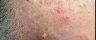
Xanthelasmas of the eyelids look like small single or multiple flat yellowish plaques located at the inner corner of the upper (usually) or lower eyelid. Xanthelasmas are benign formations that are not prone to malignancy; their appearance is associated with general disorders of lipid metabolism.
General information
Xanthelasma (xanthoma) of the skin of the eyelids is a benign formation in the form of a flat, slightly raised yellowish plaque on the skin.
The predominant localization of the pathological formation is the inner corner of the upper eyelid. Formations can be either multiple or single, symmetrical, varying in consistency and shape - soft/hard, even flat or nodular. Eyelid xanthelasmas are a type of xanthomas that appear on the eyelids when they are absent elsewhere on the body. Histologically, eyelid skin xanthoma consists of xanthoma cells (histiocytes), which contain intracellular fat deposits, that is, they contain vacuoles that are filled with partially esterified cholesterol . They are located predominantly in the upper reticular layer of the dermis/perivascular region, less often in the area of the appendages. These formations are not prone to malignant degeneration and do not pose a threat to life.
Xanthelasma of the eyelids (a type of xanthoma) is the most common manifestation of xanthomatosis. It occurs more often in females (1.1%) and much less frequently in males (0.3%) aged 15-75 years with a peak at the age of 30-50 years. Xanthomatosis is a disease characterized by multiple deposits of lipid substances, mainly in the skin (elbows, buttocks, thighs), less often in tendons, bones, dura mater), caused by hyperlipidemia .
How to get rid of xanthelasma
You should not try to remove plaques on your eyelids at home. This is fraught with inflammation of the eyelids (blepharitis) and the formation of rough, deforming scars while maintaining areas of xanthelasma. Such consequences of self-medication are much more difficult to eliminate than the formations themselves. Therefore, if you want to remove xanthoma plaques on your eyelids, you should consult a doctor.
Currently, surgical treatment of xanthelasma may include:
- classical surgical intervention, when the doctor uses instruments to excise and separate the formation from the surrounding tissues;
- electrocoagulation (diathermocoagulation), in which plaques are subjected to local destructive effects of high-frequency electric current;
- cryodestruction – cauterization of formations with liquid nitrogen, when the treated tissues die under the influence of ultra-low temperatures;
- chemical destruction, for this xanthelasmas are treated with trichloroacetic acid;
- laser therapy based on layer-by-layer controlled evaporation of tissue under the influence of high-frequency waves, also called a laser scalpel.
To decide which method is best to remove xanthelasmas, you need to evaluate the possible cosmetic consequences of each method. Excision of plaques with a scalpel under local anesthesia may be justified if they are large. In addition, this method of treatment is the cheapest. Its disadvantages are a fairly long period of healing under the scab, the often need for suturing the edges of the wound and the subsequent formation of scars on the eyelids.
The main disadvantage of cryodestruction is the low predictability of the depth of tissue necrosis that occurs. In most cases, one procedure does not lead to complete removal of xanthelasma, so repeated sessions of treating plaques with liquid nitrogen are required. If the surrounding healthy tissue is damaged too deeply, the scar formed after cryodestruction can itself become a cosmetic defect. Chemical destruction has the same disadvantages.
Electrocoagulation gives good and predictable results when removing small xanthelasmas. This method also often complements classical surgery if the surgical wound is large and prone to bleeding. Healing after diathermocoagulation occurs within 1.5-2 weeks under a crust with the formation of an inconspicuous atrophic scar. But depending on individual characteristics, the formation of keloid scars is possible.
Laser removal of xanthelasma is considered the most modern and minimally invasive method of local treatment. At the same time, the doctor clearly controls the depth and width of the impact, gradually removing all pathological areas. Healing after the procedure occurs quickly and without scarring.
“Do stains remain after xanthel laser removal?” - a very pressing question. Most often, patients fear the appearance of hyperpigmentation. Indeed, this problem can arise if the rules of skin care after laser therapy are not followed. In place of the falling off dark crust, delicate pink skin is visible, which differs in color from the surrounding tissues. This is a temporary phenomenon, usually within 3 weeks the shades gradually level out, and after 3-4 months the area of xanthelasma removal becomes invisible. But before that, regenerating skin requires protection from ultraviolet radiation. To do this, it is recommended to regularly apply sunscreen to your eyelids (even in cloudy weather) and use high-quality sunglasses.
After removal, xanthelasmas are not prone to recurrence, although if predisposing factors persist, new plaques may appear on adjacent areas of the skin.
Causes
The development of xanthelasmas is based on a disruption of the process of lipid metabolism with the gradual development of a state of hyperlipidemia, in which the content of lipoproteins of various fractions in the blood significantly increases. They are associated both with genetically determined (primary) disorders - primary hyperlipidemia types 2 and 4, and with fat metabolism disorders, which are based on various types of disorders/diseases - increased blood cholesterol ; hypothyroidism , diabetes mellitus , pancreatitis , cirrhosis , lipoid nephrosis , severe hepatitis .
In some cases, the cause of xanthelasma is age-related changes in the body, taking certain medications ( estrogens , corticosteroids , anabolic steroids , cyclosporine , antihypertensives , retinoids ), as well as abuse of cholesterol/fat-rich foods and alcohol-containing drinks.
Primary forms of xanthomatosis occur predominantly in persons under 40 years of age, including children, while secondary forms occur mainly in older/elderly people. However, xanthomas and xanthelasmas can also appear against the background of a normal lipid profile due to an inflammatory process on the skin, allergic contact dermatitis and erythroderma .
Contraindications
If you have xanthelasma of the eyelids, you should consult a specialist before removing them with a laser. Laser therapy should absolutely not be carried out if the following disorders are detected in the patient:
- herpetic type skin lesions;
- infectious diseases;
- diabetes mellitus;
- diseases of the cardiovascular system;
- dysfunction of the thyroid gland;
- oncological diseases;
- neurological disorders;
- diseases of the hematopoietic system.
It is also not recommended to carry out the procedure during pregnancy and lactation.
During the initial examination, the doctor will reliably assess the patient’s physical capabilities and make a decision on the possibility or impossibility of the operation.
Symptoms
Xanthelasma is a light orange/yellow plaque that slightly protrudes above the unchanged surface of the skin, with a predominant localization on the upper eyelid (at the border of the fixed/moving part), sometimes at the inner corner of the eye. On palpation, the formation has a soft consistency and is painless. They can be single/multiple, located on one or both eyelids. In some cases, xanthelasmas merge to form a solid yellow stripe with an uneven contour or lumpy elements.
Characterized by slow, steady development, a tendency toward slow fusion of neighboring elements and the absence of spontaneous healing. There are no subjective sensations on the part of the patient. The formation can grow to the size of a large bean, but does not transform into a malignant neoplasm. In cases where xanthelasmas are symptoms of xanthomatosis, they can also be located on the lower eyelid and other parts of the body: on the face, elbow/knee joints, neck, buttocks, etc.
The resulting xanthelasmas do not regress on their own and persist throughout life. In some cases, their number continues to gradually increase. Below is a photo of xanthelasma.
Surgical excision
This method is used in cases where it is necessary to remove xanthomas - large fused formations. The surface of the plaque and the area around it are carefully treated with an antiseptic, then separated using scissors or tweezers. The edges of the wound are treated with ferric chloride, then with potassium permanganate or brilliant green. The main disadvantage of this method is the high probability of relapse.
Surgical excision may be prescribed if the patient has contraindications to other techniques.
In general, this method is rarely used these days, trying to carry out treatment before the plaques grow to large sizes.
Tests and diagnostics
The diagnosis is made by examining the patient based on the location/characteristic appearance, color and consistency of the mass. During examination, diascopy is used - pressing on the neoplasm with a glass slide, which allows you to bleed the formation and clearly determine their yellow color and the contours of the formation. If necessary, a lipid profile study is prescribed: determination of lipoprotein/cholesterol and triglyceride fractions in the blood serum. Differential diagnosis must be carried out with dermal cysts, wen, elastic pseudoxanthoma, syringoma and other skin tumors.
Eruptive xanthoma
- Lesions usually appear as seedlings of small red-yellow papules
- They most often occur on the buttocks, shoulders, arms and legs, but can occur throughout the body
- Rarely the face and inside of the mouth are affected
- Lesions may be tender and usually itchy
- Lesions may resolve spontaneously within a few weeks
- Associated with hypertriglyceridemia (increased levels of triglycerides in the blood) often in patients with diabetes mellitus (diabetes mellitus)
Diet
Diet for vascular atherosclerosis
- Efficacy: therapeutic effect after 2 months
- Dates: no data
- Cost of products: 1700-1800 rubles. in Week
In the diet it is necessary to exclude/significantly limit:
- Saturated fats (all animal fats, butter, and coconut and palm oils).
- Foods rich in cholesterol (egg yolks, liver, kidneys, butter, high-fat fats, ghee, fish roe, shrimp).
- Simple carbohydrates (various sweets, confectionery, sweet carbonated drinks, ice cream, condensed milk).
Olive oil, legumes and plenty of vegetables (Mediterranean diet) should form the basis of a cholesterol-lowering diet. A diet low in animal fats, simple carbohydrates and high in fiber can normalize slightly elevated lipid levels. To do this, you need mainly dishes from vegetables in any form, fish, legumes and grains. If you are obese or overweight, it is important to reduce your intake of high-calorie foods and at the same time increase physical activity.
Principles of treatment
Persons with identified hypercholesterolemia or hyperlipidemia require treatment. They are prescribed a diet with a reduced content of animal fats. If this is not enough to normalize lipid metabolism, the doctor may recommend long-term use of drugs with lipotropic or lipid-lowering effects. Lipotropic agents also have a hepatoprotective effect; they promote the mobilization and oxidation of fat from the liver. But the use of lipid-lowering drugs (statins, fibrates or niacin derivatives) requires monitoring the condition of the liver and gallbladder.
Correction of endocrine disorders and lipid metabolism will reduce the risk of new xanthelasmas. But treatment without removing plaques cannot eliminate a cosmetic defect. Already existing formations on the eyelids will not disappear even after all laboratory parameters are normalized; they also cannot dissolve under the influence of various cosmetic procedures, creams and external use.
To remove xanthelasma on the eyelids, different methods can be used, which actually differ from each other in the degree of damage to surrounding tissues and the severity of scarring changes in the skin. The long-term prognosis of the disease is the same. And the question of which methods of removing xanthelasma leave a mark is often the main one when choosing treatment tactics. The final decision on the method of eliminating a cosmetic defect is made by the patient.
Is it worth removing small xanthelasmas? They usually do not interfere with the functioning of the eyelids or cause any physical discomfort. But fairly young women often turn to the doctor with requests to remove them, hoping for complete elimination of the cosmetic defect. There are no medical indications for surgical intervention in this case.
Consequences and complications
Xanthelasmas are not dangerous because they are a benign formation and there is no possibility of their degeneration into a malignant tumor. They also do not affect vision. Removal of formations is carried out for aesthetic reasons. In isolated cases, especially with injury, inflammation (suppuration) may develop. If we consider the consequences after surgical treatment, we can note frequent relapses of the disease. To avoid such consequences, the patient must strictly follow a diet (vegetable-protein diet) and take medications prescribed by the doctor. Also, a scar (hypertrophic or keloid) may form at the site of surgical removal of the formation. Most often this is noted with a predisposition to the development of keloid scars.
Cryodestruction
This is a method of removing skin lesions - xanthelasmas, warts, papillomas - by freezing with liquid nitrogen. The procedure gives a good cosmetic effect. The fatty plaque is frozen at an ultra-low temperature of 190 degrees, after which its structure is destroyed and cell growth stops. During the rehabilitation period, you must strictly follow the care recommendations: protect your eyes from the bright sun, do not visit the solarium, and do not use facial cosmetics.
List of sources
- Surkichin S.I., Gryazeva N.V. Xanthelasma of the eyelids: management tactics / Medical alphabet 2021 No. 7 /, volume No. 1, pp. 63-64.
- Trukhan D.I., Viktorova I.A., Bagisheva N.V. Skin changes in somatic diseases // International Journal of Applied and Fundamental Research. 2021. No. 8. pp. 736–740.
- Cronenberg G. M. et al. Obesity and lipid metabolism disorders / Translation from English, ed. I. I. Dedova, G. F. Melnichenko. M.: Read Elsiver LLC, 2010. 264 p.
- Vizir V.A., Berezin A.E. Modern approaches to the treatment of hyperlipidemia / Zaporozhye Medical Journal. - 2011. - volume 13, no. 1, pp. 108-117.
- Machekhin V.A. Clinical and histological analysis of tumors of the eye and its protective and auxiliary apparatus // PRACTICAL MEDICINE. - 2012. - T. 2, No. 59. - P. 255-259.
Our specialists
- Kiani Ali
Candidate of Medical Sciences, laser medicine specialist, dermatocosmetologist.
Sign up
- Stepanova Inna Igorevna
Candidate of Medical Sciences, maxillofacial surgeon, specialist in laser medicine.
Sign up
- Fedotova Marina Andreevna
Surgeon, dermatocosmetologist, laser medicine specialist
Sign up
- Popovkin Pavel Sergeevich
Surgeon, oncologist, laser medicine specialist.
Sign up
Common xanthoma
- Xanthoma-like lesions are caused by a rare form of histiocytosis.
- Lipid metabolism is normal.
- The skin lesions usually consist of hundreds of small yellowish-brown or reddish-brown bumps that are usually evenly distributed on both sides of the face and torso. They can be especially hard on the armpits and groin.
- Small bumps may join together to form sheets of thickened skin.
- In 30% of affected people, the mucous membranes of the mouth, respiratory tract or eyes (mucous membranes) are affected. Warty plaques in the mouth are called verruciform xanthoma.
- 40% of affected people develop diabetes insipidus, a condition that results in an inability to control water loss (leading to constant thirst and excessive urine production). This occurs due to the overgrowth of histiocytes on the lining of the brain (meninges).
- May affect internal organs (eg liver, lungs, kidneys, etc.)
- Self-limiting and eventually improves on its own, but may persist for many years.