Xanthelasma (translated from Greek as “golden-yellow plate”) is a slowly growing benign neoplasm localized on the skin of the eyelids. This tumor consists of plaques that rise above the healthy surface. The disease most often develops against the background of lipid metabolism disorders and increased blood cholesterol levels. In most cases, single or multiple growths with a smooth or wrinkled surface are found in middle-aged and elderly women.
Despite the fact that xanthelasma of the eyelids does not cause subjective symptoms and practically does not cause concern, it has a tendency to grow, often forming a whole chain of cholesterol plaques that are located around the eyes. Experts recommend getting rid of this aesthetic problem through removal.
| Removal of eyelid xanthelasma up to 5 mm | 3300 |
Why do xanthelasmas appear on the skin?
Xanthelasma of the eyelids is a flat, yellowish neoplasm of irregular shape, soft to the touch. It can appear on the skin of one or both eyelids, often having a symmetrical location.
The main cause of a benign tumor is a violation of fat metabolism. There are several factors that provoke its development:
- hypercholesterolemia (increased blood cholesterol levels by 6–10 times);
- primary and secondary hyperlipoproteinemia (lipid metabolism disorder);
- hereditary predisposition;
- congenital deficiency of the enzyme lipoprotein lipase;
- thyroid dysfunction;
- diabetes mellitus (provokes so-called diabetic xanthelasmas of the eyelids);
- diseases of the liver and biliary tract;
- hereditary diseases.
In some cases, the causes of xanthelasma remain unknown.
What clinical manifestations are xanthelasma characterized by?
Xanthelasmas of the eyelids appear suddenly, grow and develop slowly but steadily. They have a soft consistency and are painless when pressed. In some cases, the neoplasms reach the size of a large bean, merge together, forming a characteristic lumpiness, or transform into a solid yellow stripe running across the entire eyelid.
Xanthelasma does not impair vision, is not prone to malignancy (malignancy) and does not pose a threat to human life. Once a cholesterol plaque appears, it remains on the skin throughout life. Therefore, most often it is perceived primarily as an aesthetic defect.
Risk factors:
- Age over 40 years;
- Living in hot countries;
- Frequent tanning and sunburn;
- Light skin type, blond or red hair, blue or green eyes;
- Previous actinic keratosis or skin cancer;
- Weak immune system due to chemotherapy, leukemia, AIDS, or previous organ transplantation.
Rice. 3a. Actinic keratosis capitis
Rice. 3b. Actinic keratosis on the nose
Rice. 3c. Actinic keratosis on the face
Rice. 3g. Actinic keratosis on the forehead
Rice. 3d. Actinic keratosis at the corner of the eye
What to do if xanthelasma appears?
If a neoplasm appears in the eyelid area, with a description similar to xanthelasma, consult a dermatologist. As a rule, making a diagnosis does not cause difficulties and is carried out during a visual examination. However, one should take into account the fact that a neoplasm that appears on the skin is an external sign of a malfunction of the internal organs. Therefore, in this situation, a dermatologist may recommend additional consultation with a therapist, endocrinologist, cardiologist, nutritionist and undergo a comprehensive examination.
A set of diagnostic tests may include:
- diascopy (pressure on the affected area of the skin to determine the nature of the tumor);
- subcutaneous node biopsy;
- blood test for lipid profile;
- calculation of body mass index;
- ECG;
- EchoCG;
- MSCT of the coronary arteries;
- Ultrasound of the liver, thyroid gland, etc.
How is the course of treatment at the Juno clinic?
If during a diagnostic examination a connection was established between the occurrence of xanthelasmas and lipid metabolism disorders, external removal of the tumor will not be enough. In this situation, a specialist from the Juno clinic may recommend therapy for the primary disease, adjusting lifestyle and diet. If necessary, medications that regulate lipid metabolism disorders, anti-ischemic drugs, extracorporeal therapy methods, etc. will be prescribed.
Some of the main recommendations that must be followed for effective treatment of xanthelasma are: regular exercise, quitting smoking, maintaining optimal body weight and following a diet aimed at reducing blood cholesterol levels.
What methods exist for removing xanthelasma?
In modern clinical practice, several methods are used to remove eyelid xanthelasma.
- Using a laser.
Laser removal of xanthelasma is the most progressive method of treatment, allowing to avoid the development of complications and recurrence of plaques. A non-contact laser beam burns out the tumor layer by layer without damaging nearby healthy tissue. This eliminates deformation of the eyelids and avoids the appearance of scars on the skin. The procedure does not require special preparation and does not require long-term rehabilitation. - Using liquid nitrogen.
Cryodestruction (freezing with liquid nitrogen) involves exposing xanthelasma to a temperature of about -200 °C. This is a fairly effective technique, but it requires a longer time for the skin to fully recover. - Using radio wave surgery.
Removal of xanthelasma using radio waves is a non-contact, non-traumatic method of tissue incision and coagulation. It is distinguished by the rapid healing of postoperative wounds and the minimal risk of scar formation. - Using electrocoagulation.
The electrocoagulation method requires the use of local anesthesia. After cauterization of xanthelasma with electric current for several days, the wound is treated with an antiseptic solution to avoid the development of inflammation. - Using surgical excision.
Surgical removal of xanthelasma is used in the presence of large tumors (more than 1 cm in diameter). The operation takes place under local anesthesia. After excision of the plaque with a scalpel and treatment of the surgical field, sutures are applied to the wound. Often, spots and scars remain on the skin, which can be removed with laser resurfacing.
The price of a procedure to get rid of xanthelasma depends both on the treatment method and on the number of plaques. In most cases, the final price for xanthelasma removal is determined after consultation with a doctor.
Treatment of actinic keratosis
Foci of actinic keratosis must be removed.
Removal can be done in several ways:
- Medicines - Aldara cream, etc.;
- Surgically -
- Cryotherapy. Actinic keratoses can be removed by freezing the lesions with liquid nitrogen.
- Curettage. The surgeon scrapes off the keratosis and treats it with an electrocoagulator. Curettage is carried out after injecting an anesthetic under it.
- Photodynamic therapy. A light-sensitive substance is applied to the keratosis lesion, and then treated with a special light.
- Laser removal. The keratosis lesion is removed with a laser.
How to reduce the risk of relapse and prevent the formation of xanthelasmas?
To prevent the re-formation of cholesterol plaque, especially with elevated blood cholesterol levels, it is recommended to exclude fatty meats from the diet, adhere to a dairy-vegetable diet, limit the intake of carbohydrates, completely avoid trans fats and maintain a reasonable drinking regime.
People who have previously undergone eyelid xanthelasma removal are advised to avoid stretching and traumatizing the skin around the eyes, as this can lead to the appearance of yellowish nodules (even with normal lipid status).
You should also not neglect such prevention methods as regular monitoring of the state of the cardiovascular system (observation by a cardiologist and rheumatologist) and periodic examinations by a dermatologist and ophthalmologist.
If a soft yellowish lump appears on the skin of the upper or lower eyelid, you must definitely consult a doctor who, after conducting diagnostic tests, will be able to give the necessary recommendations and prescribe effective treatment.
Sign up for a consultation with a specialist at the Juno clinic online or by phone. The doctor will determine the optimal method of removal, prescribe tests if necessary, and draw up a treatment regimen for xanthelasma.
Plaques in blood vessels. Lymphologist's View | Part 1
Good afternoon. We hope that the coronavirus situation will soon subside and we will return to normal. Today we will talk about atherosclerosis and cholesterol plaques.
It's no secret that diseases of the cardiovascular system are the main cause of death - in 50% of cases, according to international statistics. These diseases mainly include heart attacks and strokes. They have a common reason for their development. The name of this cause is atherosclerosis.
Table of contents
- What is atherosclerosis?
- How is an atherosclerotic plaque formed?
- Where can atherosclerosis develop?
- Lymphatec approach to the treatment of atherosclerosis
- Video performances
What is atherosclerosis?
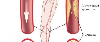
Atherosclerosis is a multifactorial disease. Accompanied by pathologies of the arteries of our organs. Lipid and protein substances accumulate on the vessel wall. Then they grow with connective tissue and even become calcified. As a result, the vessel narrows and blood flow is disrupted. Due to poor circulation, organs suffer.
A lot of attention is paid to bad cholesterol. It is believed that this is what causes the plaques to appear. Is this true and should we be afraid of cholesterol? Cholesterol is used as a building material for every cell in our body. And the brain generally consists of cholesterol for the most part. Regardless of whether we eat foods high in cholesterol or not, cholesterol is always present in the lumen of blood vessels.
Atherosclerosis is believed to be an age-related disease. But the trouble is that it is a silent disease. At the initial stages it is asymptomatic. This can go on for many years. As it progresses, it can cause a number of completely different symptoms and ultimately leads to serious consequences: myocardial infarction or stroke. It is clear that when it comes to the blood vessels of the brain or heart, atherosclerosis manifests itself more clearly.
It is very important to understand that atherosclerosis is not characterized by the appearance of one plaque in one place. This is a systemic disease.
How is an atherosclerotic plaque formed?
The first picture shows the structure of the artery in cross section. The inner lining of the vessel (endothelium) consists of endothelial cells that fit tightly together and line the entire inner surface. Next is the middle shell, which consists of either elastic fibers or muscle fibers. And finally, the loose outer shell (adventitia).
Figure 1. Cross-sectional structure of the vessel. Normal state of the interstitium.
What happens during the development of atherosclerosis? The internal junction of endothelial cells is disrupted, and gaps appear in the inner wall of the vessel. We remember that cholesterol is always present in the lumen of the vessel. You already guessed what happens next: cholesterol rushes into the gaps in the endothelium as a building material. Calcium salts can join cholesterol, it begins to become overgrown with connective tissue, and can calcify. And his growth doesn't stop there. Plaques grow, blocking the lumen of the vessel more and more.
Figure 2. Disruption of interstitial structure. The appearance of an atherosclerotic plaque.
For the normal functioning of a large vessel, each level of its wall must be both supplied with blood through small blood vessels and drained through lymphatic vessels. It is these small lymphatic vessels that drain the entire thickness of the artery wall, and they remove everything pathological to larger lymphatic collectors and further to the lymph nodes.
Figure 3. Blood supply to the vessel wall and drainage through the lymphatic system.
Once the lymphatic system stops draining the artery wall, plaque begins to grow. Thus, atherosclerosis is not a cholesterol problem, but a problem of disruption of the lymphatic system, which stops draining the vessel and the plaque grows.
Where can atherosclerosis develop?
Everyone knows the pathologies of the organs of the head and neck, which are supplied with blood in the carotid artery basin. Most often, we observe the occurrence of plaques in this region: in the common, internal and external carotid arteries. And through this blood line, the brain, visual organs, thyroid gland and others are supplied with blood. A catastrophe can develop in the vessels of the heart, in any artery of this pool. Finally, very often we observe the development of atherosclerosis in various places in the blood vessels of the lower extremities.
At first we see mild symptoms. If we are talking about the head and neck, it could be noise in the head, dizziness, headaches. Atherosclerosis of the coronary arteries can manifest as coronary heart disease. Sharp pain occurs in the lower extremities when walking or exercising. There is a need to stop and wait.
Lymphatec approach to the treatment of atherosclerosis
We have a solution to treat this disease even in the early stages, without waiting for the plaque to grow. We can influence the lymphatic system so that it drains the vessel walls and removes atherosclerotic plaques.
Let us give an example of two similar clinical cases in our clinic. Both patients with obliterative, i.e. advanced atherosclerosis of the lower extremities. The surgeon's view: both patients require urgent amputation. One patient was 60 years old, the second was 80.
The second patient agreed to our treatment. He passed away at the age of 92, and he lived with his leg for 12 years thanks to our treatment technology.
Another patient agreed to amputation. Today we continue to accept patients for treatment after a preliminary analysis of the situation.
You can learn about treatment cases from our articles.
- Atherosclerosis of leg vessels. Case of Yuri's treatment
- Atherosclerosis of the vessels of the neck and heart. Analysis of Valery's treatment.
Video performances
And finally, remember that atherosclerosis is a silent, asymptomatic disease. Please, if you are over 40, take care of your health. Carry out an ultrasound examination of the blood vessels, primarily the neck and legs. If necessary, conduct a study of the heart vessels. Today all research methods are available. And if you or your loved ones already have a health problem such as atherosclerosis, do not ignore it.
LEARN ABOUT TREATMENT POSSIBILITIES