Local therapist
Ablyazov
Irshat Ravilevich
21 years of experience
Local therapist of the highest category. Member of the Russian Scientific Medical Society of Therapists.
Make an appointment
Thrombocytopenia is a separate disease or a symptom of another disease that is characterized by a low platelet count (less than 150,000 units per microliter). The condition is accompanied by high bleeding, as well as slow stopping of bleeding from small vessels.
Symptoms
Symptoms of thrombocytopenia include:
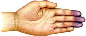
- Purpura. This is the name for hemorrhages in mucous tissues or skin. In fact, it looks like red spots - most often they can be seen in places of strong contact with clothing. The spots do not hurt, they can be pinpoint or quite extensive - including bruises of different colors (depending on the time of formation).
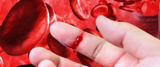
- Regular nosebleeds. Since small vessels are weakened, bleeding can begin due to a cold, sneezing, or minor injury. Bright red blood may flow for ten minutes or more, putting the patient at risk of losing a lot of blood.
- Bleeding gums, prolonged bleeding after tooth extraction. The mechanism is the same: due to weak blood vessels, the blood cannot stop quickly in case of small or large injuries.
- Bleeding of internal organs. For example, blood is found in the gastrointestinal tract, when the stool becomes colored, or in the genitourinary system - then the urine becomes colored. Both symptoms are very alarming and require immediate contact with specialists.
- Too heavy and long menstruation. Moreover, bleeding can be observed on other days of the cycle.
All of these symptoms may indicate other diseases - both concomitant with thrombocytopenia and independent. What complicates the situation is that there are a lot of reasons and mechanisms for the development of this condition. In medicine, there are several types of this disease.
How to recognize low platelet levels
Sometimes it is discovered by chance, for example, during a general blood test, for which the therapist sends the patient as part of a preventive examination.
Such incidentally discovered thrombocytopenia may not have symptoms. But more often, a lack of platelets makes itself felt with characteristic signs.
Thus, the first symptom is usually Thrombocytopenia: Causes, Symptoms & Treatment / Cleveland Clinic bleeding from the nose or from a cut that cannot be stopped. Other common signs of low platelet levels include:
- bleeding gums;
- traces of blood in the stool (in this case it looks black, tarry), urine, vomit;
- too long and heavy menstruation in women;
- petechiae, small bleeding that most often appears on the legs and looks like a red or purple rash;
- purpura, bruises of a crimson (purple) color that appear easily and as if by themselves;
- rectal bleeding.
Classification of thrombocytopenia
Let's talk about the causes of thrombocytopenia that underlie the different types of this disease.
Hereditary thrombocytopenias
These conditions are based on diseases of genetic origin. These may be May-Hegglin anomaly, Bernard-Soulier syndrome, Wiskott-Aldrich syndrome, TAR syndrome, as well as congenital amegakaryocytic thrombocytopenia.
Each of these diseases has many characteristics. For example, Wiskott-Aldrich syndrome occurs exclusively in boys and occurs in four to ten cases per million. Bernard-Soulier syndrome can only develop in a baby who has received the altered gene from both parents, and May-Hegglin anomaly can develop in 50% of cases, provided that only one parent has the prerequisites for this. As a rule, all of these diseases have among their symptoms not only a low level of platelets, but also a number of other pathological conditions, and therefore require special treatment.
Productive thrombocytopenia
They are also caused by a huge list of reasons and factors of various origins. All of them are associated with the fact that platelet formation processes in the red bone marrow are disrupted.
For example, this includes some types of anemia, cancer metastases, acute leukemia, and taking cytostatic medications. There are even factors such as radiation or alcohol abuse.
For example, in acute leukemia, a disease of the circulatory system causes a mutation in the bone marrow stem cell. Because of this, it divides and begins to displace hematopoietic cells. As a result, the number of not only platelets, but also leukocytes, erythrocytes, and lymphocytes decreases. This condition has its own name - pancytopenia. That is, immune thrombocytopenia in this case is part of it.
If we are talking about the effect of medications, then most often a decrease in platelet levels is caused by drugs from the following groups:
- antidiabetic,
- anti-inflammatory,
- antipsychotics,
- antithyroid,
- antibiotics,
- diuretics,
- anticonvulsants.
Of course, not all medications and not in all cases are capable of causing such a reaction, but this is another reason to think about the fact that you should not prescribe any medications yourself or take them without the supervision of your doctor.
Moreover, there is also such a factor as increased sensitivity to certain medications. Usually it is initially determined by a peculiar genetic predisposition, but this is quite rare.
When we talk about radiation, we are talking not only about a person’s presence in dangerous areas, but also about treatment with radiation therapy. When tumors are destroyed, the blood-forming cells of the red brain can be affected, so a condition where platelet levels drop can become a side effect of cancer treatment.
The development of symptoms of thrombocytopenia due to alcohol abuse most often occurs against the background of binge drinking and is associated with a high level of ethyl alcohol in the blood. It has negative and long-lasting effects on the bone marrow and usually causes a temporary decrease in platelets. If binge drinking becomes frequent and too long, there is a risk that the changes will be irreversible.
Causes
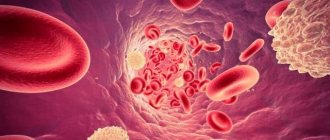
Platelets are formed elements of blood, nuclear-free flat plates ranging in size from 1 to 2 microns. They are formed from megakaryocytes (precursor cells) in the red bone marrow.
Megakaryocytes are relatively large cells. They have long processes and are almost completely filled with cytoplasm. During maturation, tiny fragments of their processes break off and enter the bloodstream. These are platelets. According to scientific data, from 2000 to 8000 platelets can be formed from one donor cell.
Thrombopoietin, a protein hormone produced in the kidneys, liver and muscles, is responsible for the development and growth of megakaryocytes. It is transported through the bloodstream to the red bone marrow, where it ensures the formation of megakaryocytes and platelets. The more platelets there are, the more the process of thrombopoietin formation is inhibited. This allows you to maintain the number of blood cells at the same level.
If any of these steps fail, the number of platelets in the peripheral blood decreases. Thrombocytopenia develops.
Taking into account the causes and mechanism of development, the disease comes in different forms:
- hereditary;
- productive;
- destruction;
- consumption;
- redistribution;
- breeding.
Hereditary thrombocytopenias
Hereditary thrombocytopenia is observed with various congenital anomalies. Its causes are genetic mutations:
- Wiskott-Aldrich syndrome. Caused by mutations that cause the red bone marrow to produce very small platelets (less than 1 µm in diameter). Due to their abnormal structure, they are quickly (within a few hours) destroyed in the spleen.
- May-Hegglin anomaly. A very rare genetic pathology, due to which the process of separating platelets from megakaryocytes is disrupted. As a result, the number of blood cells decreases, but their sizes become gigantic (6-7 microns). In parallel, there is a disturbance in the formation of leukocytes (leukopenia).
- Congenital amegakaryocytic thrombocytopenia. Typically, this form of thrombocytopenia is diagnosed in infants. It is associated with impaired platelet production by the bone marrow.
- Bernard-Soulier syndrome. Thrombocytopenia in a child appears only if he has inherited a defective gene from both his mother and father. The disease manifests itself in early childhood. It is characterized by the formation of functionally incompetent giant platelets (6-8 µm), unable to attach to damaged vascular walls and communicate with each other.
- TAR syndrome. A very rare cause of congenital thrombocytopenia, combined with the absence of both radius bones.
Productive thrombocytopenia
The group of productive thrombocytopenias includes pathologies of the hematopoietic system associated with disruption of the process of platelet formation in the red bone marrow. Causes of diseases:
- Acute leukemia. Stem cells mutate, and a large number of clones appear that are unable to perform specific functions. Gradually, clones displace hematopoietic ones from the red bone marrow. Not only the number of platelets decreases, but also the number of leukocytes, lymphocytes and erythrocytes.
- Myelodysplastic syndrome. The bone marrow cannot produce enough healthy cells. Hematopoietic cells multiply rapidly, but their maturation processes are disrupted. As a result, many functionally immature cells are formed (among them platelets) that undergo apoptosis (self-destruction).
- Aplastic anemia (the body produces very few new blood cells).
- Cancer metastases. Tumor cells in the final stages of cancer begin to leave the primary focus and spread throughout the body. Settling on tissues and organs, they begin to actively reproduce. This leads to the displacement of hematopoietic cells from the red bone marrow.
- Myelofibrosis. The stem cells mutate and the bone marrow is replaced by scar tissue. Foci of hematopoiesis develop in the liver and spleen (the size of the organs increases greatly due to this).
- Radiation. Ionizing radiation has a destructive effect on the hematopoietic cells of the red bone marrow. They begin to mutate.
- Alcoholism. Alcohol inhibits the processes of hematopoiesis in the red bone marrow, which reduces the content of platelets, leukocytes and red blood cells in the blood.
- Cytostatic drugs. Used to treat tumors. They can inhibit hematopoiesis in the bone marrow, which reduces the number of platelets.
- Allergic reactions to certain medications (diuretics, antibiotics, antipsychotics, anticonvulsants and anti-inflammatory drugs, antidiabetic drugs).
- Megaloblastic anemia. They develop with a deficiency of folic acid and vitamin B12.
Thrombocytopenia destruction
The cause of thrombocytopenia in this case is the increased destruction of platelets in the spleen (can also be in the lymph nodes, vascular bed or liver). Pathology is observed when:
- thrombocytopenia of newborns;
- Evans-Fisher syndrome;
- some viral diseases (viral thrombocytopenia);
- post-transfusion thrombocytopenia;
- taking medications (drug-induced thrombocytopenia);
- idiopathic thrombocytopenic purpura.
Thrombocytopenia of newborns
Occurs if there are antigens on the surface of children's platelets that are not on maternal platelets. Antibodies produced by the mother's body enter the newborn's bloodstream and destroy his platelets. The described process can last up to 20 weeks of intrauterine development. As a result, the child may be born with thrombocytopenia.
Fisher syndrome
It is a consequence of systemic diseases - autoimmune hepatitis, rheumatoid arthritis, systemic lupus erythematosus. Also, Evans-Fisher syndrome can develop against a background of relative well-being. Then they talk about idiopathic thrombocypenia.
Viral thrombocypenia
Once entering the body, viruses actively multiply. Antibodies are formed on the surface of the affected cells, and the cell's own antigens also change. The affected cells are destroyed in the spleen. Among the viruses that can cause thrombocypenia are measles, rubella, influenza, and chickenpox.
Post-transfusion thrombocytopenia
Post-transfusion thrombocytopenia is a consequence of platelet or blood transfusion. They are characterized by pronounced destruction of platelets in the spleen. The cause of the disease is that foreign platelets enter the body during a transfusion, to which antibodies begin to be produced.
Drug-induced thrombocytopenia
Some medications can bind to antigens on the surface of blood cells. Antibodies are produced to the formed complexes, which causes platelets to begin to be destroyed in the spleen. Among the “provoking” drugs are Quinidine, Meprobamate, Chloroquine, Ranitidine, Heparin, Cimetidine, Gentamicin, Ampicillin, etc.
Idiopathic thrombocytopenic purpura
Idiopathic thrombocytopenic purpura (also known as autoimmune thrombocytopenia or essential thrombocytopenia) is characterized by a sharp decrease in the level of platelets in the peripheral blood. At the same time, the composition of other blood elements is not disturbed.
Thrombocytopenia destruction
Here we are talking about the active destruction of platelets, which is usually observed in the spleen. Such processes are also caused by a large number of factors. These include taking certain medications, some viruses, Fisher-Evans syndrome, etc.
Idiopathic thrombocytopenia deserves special consideration. This is the case when it is not possible to establish the cause even with a huge number of possible options. At the same time, doctors note that even in such a situation, the action of provoking factors is observed. For example, the problem appears after bacterial and viral infections, preventive vaccinations, use of a number of medications, hypothermia or prolonged exposure to the sun.
This condition also occurs in newborns. It begins to form in the womb, when antibodies in her body cause the destruction of platelets in the child.
Fisher-Evans syndrome is also not an independent cause, since it itself is a consequence of various systemic diseases. This syndrome occurs in systemic lupus erythematosus and rheumatoid arthritis, but in some cases it can be an independent deviation. As for viruses, rubella, chicken pox, measles, and influenza can lead to a decrease in platelet levels. Sometimes this is a reaction to vaccination, although such a mechanism is described quite rarely and almost never occurs.
Thrombocytopenia consumption
Here the mechanism is as follows: platelets, for some reason, begin to be activated in the vascular bed. In response to this, the red bone marrow begins to actively produce them - and as a result, its capabilities are slowly depleted. After this, the level of red cells begins to fall.
One of the causes of consumption thrombocytopenia is disseminated intravascular coagulation syndrome. It stands for disseminated intravascular coagulation syndrome. And it can develop against the background of severe tissue destruction during operations, burns, injuries, due to severe infections, chemotherapy, organ transplants, etc. That is, there are many underlying reasons that can cause a decrease in the level of platelets in the blood.
Clinical cases of thrombocytopenia
Home Useful articles Clinical cases of thrombocytopenia
S.V. Kuleshova, A.A. Altaeva, E.A. Kuznetsova
Clinical cases of false and true thrombocytopenia in outpatient practice.
Federal State Budgetary Institution "Polyclinic No. 2" of the Administration of the President of the Russian Federation, 119146, Moscow, Russia
The article is devoted to the laboratory nuances of diagnosing thrombocytopenia in outpatient practice. Using the example of two different patients, the importance of knowledge of basic research in the differential diagnosis of false and true thrombocytopenia is shown.
Key words: thrombocytopenia, EDTA-dependent thrombocytopenia, platelet count according to Fonio.
In the activities of an outpatient clinical diagnostic laboratory, a general blood test accounts for up to 30% of all tests performed.
Typically, a general clinical blood test consists of data from a hematological analyzer, and if there is a deviation in the leukocyte formula, a differentiated count of leukocytes in a blood smear using light microscopy.
The current level of hematology analyzers allows you to provide up to 70% of test results without smear microscopy.
However, the doctor should not blindly trust laboratory analyzers, but should be the final authority, after whose review and approval the results will be validated.
There are no small details in the work of a clinical laboratory diagnostics doctor. In the flow of work, choosing from the data of a hematology analyzer those that require additional research is not easy. An example of the need for a wide range of knowledge and skills of a CDL doctor is thrombocytopenia - a decrease in the number of platelets in the absence of other abnormalities in the count of formed elements and in the blood smear. Thrombocytopenia includes conditions in which the number of peripheral blood platelets is less than 150 x 109/L. A platelet count of 50x109/l still allows abdominal surgery while maintaining hemostasis. Platelet count ≤ 20 x109/l. refers to critical values from the point of view of clinical laboratory diagnostics [1]. For such patients, according to the national standard, transfusion of platelet concentrate is indicated [2]. To calculate the number of platelets, the following are recognized as unified:
- blood count using a hematology analyzer;
- counting in smears using the Fonio method.
A decrease in platelet count in a blood test may be true and will then require immediate action based on the severity of the thrombocytopenia. A decrease in platelet count in a blood test may not be true. Then it will reflect the individual characteristics of the patient or will be the result of an error at the preanalytical stage. In addition to true thrombocytopenia, a decrease in the number of platelets is possible, which usually occurs due to untimely passage of blood through the analyzer, or due to spontaneous aggregation of platelets. False decreases in platelet counts can occur in a wide range of disorders, such as autoimmune diseases, viral and bacterial infections, and chronic inflammatory diseases. Spontaneous platelet aggregation can be a manifestation of factors such as immunological (antiplatelet antibodies), chemical (anticoagulants) and physical (temperature). Venous blood is preferable for performing a complete blood count. Today, potassium salts of ethylene-diamine-tetra-acetic acid (EDTA) are used as an anticoagulant. EDTA-dependent thrombocytopenia is a consequence of the interaction of antiplatelet antibodies with platelet antigens in the presence of EDTA and when exposed to low temperatures. According to foreign authors, EDTA-dependent pseudothrombocytopenia accounts for 0.07-0.11% of all blood tests [3]. EDTA-dependent pseudothrombocytopenia is manifested by a decrease in the number of platelets, and these phenomena progress as the time elapses after blood collection increases. To avoid aggregation, it is recommended to pass blood through the analyzer in the interval of 0-5 minutes. or 1 hour or more after blood collection [4]. In the interval of 5 min. — 1 hour, temporary platelet aggregation occurs, which can lead to a false decrease in platelet levels in the blood sample. Immediately after blood collection, the possibility of spontaneous platelet aggregation is excluded.
We want to demonstrate the importance of such a routine technique as platelet counting using the Fonio method. And also two clinical cases emphasize the need for a clinical laboratory diagnostics doctor to be not only an analyst, but also to work with a microscope.
The first clinical case is common in outpatient practice.
Patient M., born in 1962. I applied for a certificate from the pool as part of the annual medical examination. At the time of examination, he is not actively complaining. General condition is satisfactory. Body temperature 36.6°C. Consciousness: clear. Skin: pink. Visible mucous membranes are pink. The food is ok. Lymph nodes: not enlarged. The thyroid gland is not enlarged. There is no swelling. The osteoarticular system is without pathology. The tones are sonorous, the rhythm is correct. Pathological noises are not heard. Heart rate 68/min. SBP 120/80 Hg. DBP 120/80 mm Hg Abdomen: not enlarged, involved in breathing, soft and painless on palpation. The liver is not enlarged. Physiological functions are normal. Urinary system: Urination is not impaired. The effleurage symptom is negative on both sides.
When performing a general blood test, a sharp decrease in the number of platelets was revealed - up to 15x109/l.
| GENERAL BLOOD ANALYSIS No._________ Date of referral February 5, 2014 12:59 | |
| F.,I.,O.M, born in 1962 Institution of the Federal State Budgetary Institution "Polyclinic No. 2" Diagnosis upon referral: K29.30 | ist. bol. No. 287 On doctor's orders |
| Parameter name | Result | Norm | Unit |
| ESR according to Panchenkov | 5 | 2 — 10 | mm/h |
| Hemoglobin (HGB) | 145.00 | 130.0 — 160.0 | g/l |
| Red blood cells (RBC) | 4.75 | 4.00 — 5.00 | 1012/l |
| Hematocrit (HCT) | 40.6 | 40 — 48 | % |
| (MCV) Average volume of erythr. | 85.5 | 80 — 103 | fl |
| (MCH) Average heme content in one erythrocyte | 30.5 | 26 — 34 | pg |
| (MSHC) Average heme concentration in one erythrocyte | 35.7 | 30 — 38 | g/dl |
| Platelets (PLT) | 15 | 150 — 400 | 109/l |
| Lymphocytes (LYM) | 35.7 | 5 — 55 | % |
| Bas., eos., monocytes (MXD) | 5.1 | 1 — 20 | % |
| Neutrophils (NEUT) | 59.2 | 5 — 95 | % |
| LYM# - absolute number of lymphocytes | 2.1 | 0.8 — 2.7 | |
| MXD# - absolute number of basof., eosin., monocytes | 0.3 | 0.1 — 1.5 | |
| NEUT# - absolute number of neutrophils | 3.5 | 1.2 — 5.3 | |
| RDW-SD- distributor erythrocyte size | 40.7 | 33.4 — 49.2 | |
| Relative volume of large thrombi. (P-LCR) | 47.7 | 13 — 43 | % |
| Mean platelet volume (MPV) | 13.4 | 9 — 13 | fl |
| RDW-CV-red blood cell distribution by particle size | 18.5 | 10.8 — 14.9 | |
| PDW(platelet distribution weight) | 24 | 9.8 — 18.0 | % |
| White blood cells (WBC) | 5.90 | 4.00 — 9.00 | 109/l |
| Rod-nuclear (neutrophils) | 3 | 1.00 — 6.00 | % |
| Segmented nuclear (neutrophils) | 59 | 47.00 — 72.00 | % |
| Eosinophils | 1 | 0.50 — 5.00 | % |
| Lymphocytes | 34 | 19.00 — 37.00 | % |
| Monocytes | 3 | 3.00 — 11.00 | % |
The analysis was performed on a Sysmex KX-21N hematology analyzer manufactured by Roche Diagnostics (Switzerland). The microscopy specimen was prepared and stained using a HemaTek automated blood smear stainer, manufactured by Bayer Diagnostics. When calculating the leukemia formula, frequent and large accumulations of platelets were detected, which suggested pseudothrombocytopenia. The patient was called for a repeat blood test. Blood was drawn and slides were prepared for platelet counting according to Fonio. The unified method of counting in blood smears (according to Fonio) is based on calculating the number of platelets in stained blood smears per 1000 red blood cells, followed by calculation per 1 μl (or 1 l) of blood, based on the known content of red blood cells in this volume [5]. Prepared, fixed and stained preparations according to Romanovsky-Giemsa were microscoped with an immersion lens, counting the number of platelets in thin areas of the preparation where red blood cells are located in isolation. In each field of view, the number of red blood cells and platelets was counted by moving the smear until 1000 red blood cells were counted. Then we recalculated the number of red blood cells obtained from the analyzer.
The blood was passed through the analyzer at 4 minutes:
| FULL NAME. M, born 1962 Institution of the Federal State Budgetary Institution "Polyclinic No. 2" Diagnosis upon referral: K29.30 | ist. bol. No. 287 On doctor's orders |
| Parameter name | Result | Norm | Unit |
| ESR according to Panchenkov | 5 | 2 — 10 | mm/h |
| Hemoglobin (HGB) | 145.00 | 130.0 — 160.0 | g/l |
| Red blood cells (RBC) | 4.7 | 4.00 — 5.00 | 1012/l |
| Hematocrit (HCT) | 40.6 | 40 — 48 | % |
| (MCV) Average volume of erythr. | 85.5 | 80 — 103 | fl |
| (MCH) Average heme content in one erythrocyte | 30.5 | 26 — 34 | pg |
| (MSHC) Average heme concentration in one erythrocyte | 35.7 | 30 — 38 | g/dl |
| Platelets (PLT) | 225 | 150 — 400 | 109/l |
| Lymphocytes (LYM) | 35.7 | 5 — 55 | % |
| Bas., eos., monocytes (MXD) | 5.1 | 1 — 20 | % |
| Neutrophils (NEUT) | 59.2 | 5 — 95 | % |
| LYM# - absolute number of lymphocytes | 2.1 | 0.8 — 2.7 | |
| MXD# - absolute number of basof., eosin., monocytes | 0.3 | 0.1 — 1.5 | |
| NEUT# - absolute number of neutrophils | 3.5 | 1.2 — 5.3 | |
| RDW-SD- distributor erythrocyte size | 40.7 | 33.4 — 49.2 | |
| Relative volume of large thrombi. (P-LCR) | 21.7 | 13 — 43 | % |
| Mean platelet volume (MPV) | 9.4 | 9 — 13 | fl |
| RDW-CV-red blood cell distribution by particle size | 12.5 | 10.8 — 14.9 | |
| PDW(platelet distribution weight) | 11.8 | 9.8 — 18.0 | % |
| White blood cells (WBC) | 5.90 | 4.00 — 9.00 | 109/l |
| Rod-nuclear (neutrophils) | 1,5 | 1.00 — 6.00 | % |
| Segmented nuclear (neutrophils) | 58 | 47.00 — 72.00 | % |
| Eosinophils | 1 | 0.50 — 5.00 | % |
| Lymphocytes | 35 | 19.00 — 37.00 | % |
| Monocytes | 4,5 | 3.00 — 11.00 | % |
The normal platelet count was confirmed by the Fonio count – 235x109/l.
This pseudothrombocytopenia did not pose a hemorrhagic or thrombotic risk to the patient.
The following example is an illustration of true thrombocytopenia, which, according to the standard of patient management of the American Society of Hematology, is subject to hospitalization for intravenous immunoglobulin and glucocorticosteroids [6].
Patient L, born in 1991. I went to the clinic to see a dermatovenerologist about hemorrhagic rashes and bloody discharge from the nose. I feel satisfactory. From the anamnesis it became known that after a long stay in the cold, for two hours at a temperature of -24 0C, he independently began to take Paracetamol + Phenylephrine + Pheniramine + Ascorbic acid prophylactically. Two days before treatment, the first rashes appeared on the skin of the legs, which over the next days spread throughout the body. Denies chronic diseases. Denies bad habits. Allergy history is not burdened.
Objective condition of the patient: The condition is relatively satisfactory. Body temperature is 36.1 C. The skin is of normal color, hemorrhagic rashes of the type of multiple petechiae measuring 1-3 mm. on the skin of the trunk, limbs, oral mucosa. There is no swelling. Peripheral lymph nodes are not enlarged. The pharynx is moderately hyperemic, there are no plaques. Respiratory system: There is a pulmonary sound over the lungs percussion, vesicular breathing on auscultation, no wheezing. Chd 16 in 1 min. Cardiovascular system: Heart sounds are sonorous and rhythmic. The pulse is equal to heart rate - 82 per 1 min. Blood pressure 110/70 mmHg. Digestive system: The tongue is moist, covered with a white coating. The abdomen is soft, painful in the right and left hypochondrium. Liver + 2 cm, painful on palpation. The spleen is enlarged. The stool is normal. Urinary system: Urination is not impaired. The effleurage symptom is negative on both sides.
Basic laboratory data:
A general blood test performed on a Sysmex KX-21N analyzer (Sysmex KX-21N) manufactured by Roche Diagnostics (Switzerland) revealed leukopenia, relative lymphocytosis and a complete absence of platelets.
| FULL NAME. L, 05.11.1991 Institution of the Federal State Budgetary Institution "Polyclinic No. 2" Diagnosis upon referral: L95.80 | ist. bol. No. 8 On doctor's orders |
| Parameter name | Result | Norm | Unit |
| ESR according to Westergren | 10 | 2 — 15 | mm/h |
| Hemoglobin (HGB) | 138.00 | 130.0 — 160.0 | g/l |
| Red blood cells (RBC) | 4.61 | 4.00 — 5.00 | 1012/l |
| Hematocrit (HCT) | *37.4 | 40 — 48 | % |
| (MCV) Average volume of erythr. | 81.1 | 80 — 103 | fl |
| (MCH) Average heme content in one erythrocyte | 29.9 | 26 — 34 | pg |
| (MSHC) Average heme concentration in one erythrocyte | 36.9 | 30 — 38 | g/dl |
| Platelets (PLT) | *0 | 150 — 400 | 109/l |
| Lymphocytes (LYM) | *73.1 | 5 — 55 | % |
| Bas., eos., monocytes (MXD) | 2.4 | 1 — 20 | % |
| Neutrophils (NEUT) | 24.5 | 5 — 95 | % |
| LYM# - absolute number of lymphocytes | 2.2 | 0.8 — 2.7 | |
| MXD# - absolute number of basof., eosin., monocytes | 0.1 | 0.1 — 1.5 | |
| NEUT# - absolute number of neutrophils | *0.7 | 1.2 — 5.3 | |
| RDW-SD- distributor erythrocyte size | 33.9 | 33.4 — 49.2 | |
| RDW-CV-red blood cell distribution by particle size | 12.2 | 10.8 — 14.9 | |
| White blood cells (WBC) | *3.00 | 4.00 — 9.00 | 109/l |
| Rod-nuclear (neutrophils) | 3 | 1.00 — 6.00 | % |
| Segmented nuclear (neutrophils) | *18 | 47.00 — 72.00 | % |
| Eosinophils | 5 | 0.50 — 5.00 | % |
| Lymphocytes | *69 | 19.00 — 37.00 | % |
| Monocytes | 5 | 3.00 — 11.00 | % |
When counting platelets using the Fonio method, their number was 4x10⁹ l.
Based on severe thrombocytopenia, clinical picture and anamnestic data, the patient was urgently hospitalized at the Central Clinical Hospital of the UD PRF. Blood and bone marrow studies revealed pronounced disturbances in megakaryocytopoiesis and the autoimmune nature of thrombocytopenia. A diagnosis of autoimmune thrombocytopenic purpura was made.
We believe that the above examples were useful for repeating the algorithm for performing a general blood test and interpreting thrombocytopenia.
If the doctor’s internal attitude that all thrombocytopenias are false and do not need additional research wins, then the life of the next patient will be in danger. The platelet critical values must be among the laboratory's other approved critical laboratory values. And responsibility for them must be borne personally by the clinical laboratory diagnostics physician.
The introduction of new technologies into laboratory practice is inevitable, centralization of laboratory services is a global trend, but basic knowledge and skills should not fade into the background in the daily work of a clinical laboratory diagnostics doctor.
- National Standard of the Russian Federation Clinical laboratory technologies. Ensuring the quality of clinical laboratory tests GOST R 53079.3-2008 4.7.3
National'nyj Standart Rossijskoj Federacii Tehnologi laboratornye klinicheskie. Obespechenie kachestva klinicheskih laboratornyh issledovanij GOST R 53079.3-2008 4.7.3
- National Standard of the Russian Federation Donor blood and its components. GOST R 53470-2009.
National'nyj Standart Rossijskoj Federacii Krov' donorskaja i ee komponenty. GOST R 53470-2009
- Alan D. Michelson Platelets. Academic press.2012;989-1011.
- Methodological recommendations Hematological analyzers. Interpretation of blood test. 2007.
Methodology rekomendacii Gematologicheskie analizatory. Interpretacija analisa krovi. 2007.
- Directory. Laboratory research methods in the clinic. Edited by V.V. Menshikov. Medicine. 1987.
Reference. Laboratory metody issledovanija v clinic. Pod red.VVMen'shikova. Medicina. 1987.
- George JN, Woolf SH, Raskob GE, Wasser JS, Aledort LM, Ballem PJ, et al. Idiopathic thrombocytopenic purpura: a practice guideline developed by explicit methods for the American Society of Hematology. Blood 1996;88:3-40.
- Kuleshova Svetlana Vyacheslavovna, head of the clinical laboratory of the Federal State Budgetary Institution "Polyclinic No. 2" of the Administration of the Russian Federation, Moscow, 119146, st. 2-ya Frunzenskaya, 4, (assistant of the Department of Clinical Laboratory Diagnostics, Faculty of Internal Affairs, Russian National Research Medical University named after N.I. Pirogov)
- Altaeva Alexandra Andreevna, Candidate of Medical Sciences, Head of the Dermatology and Venereology Room of the Federal State Budgetary Institution "Polyclinic No. 2" of the Administration of the Russian Federation
- Kuznetsova Elena Alekseevna, doctor of the clinical care department of the Federal State Budgetary Institution "Polyclinic No. 2" of the Department of Public Administration of the Russian Federation
Thrombocytopenia redistribution
If we explain this mechanism very simply, it looks like this: due to some reasons, the patient’s spleen enlarges. And if under normal conditions it stores up to 30% of platelets, then in an enlarged state it retains up to 90% of these cells. At the same time, the system does not see that there are not enough platelets and does not trigger a compensatory mechanism.
The mechanism is also based on various causes: cirrhosis of the liver, some infections such as tuberculosis, autoimmune diseases (systemic lupus erythematosus), tumors of the circulatory system, alcoholism.
Causes of thrombocytopenia
Among the various reasons causing this serious disease, the main ones are:
Thrombocytopenia dilution
This condition is usually observed in people who receive a large number of transfusions of various liquids: for example, plasma, plasma substitutes, etc. As a result, the level of platelets decreases due to the banal dilution of the blood.
Considering the huge number of reasons that can cause immune thrombocytopenia (and we have not named all the options), a patient with its symptoms should immediately consult a doctor who will find the essence of the problem and prescribe effective treatment.
Diagnostics
Since there are so many reasons, it is not possible to name diagnostic measures in detail - everything will depend on the specific situation. But we can highlight the main components of diagnosis:
- General blood analysis.
- Checking bleeding time (Duke test).
- Determining the time it takes for blood to clot.
- Red bone marrow puncture. The method allows you to identify quantitative and qualitative changes in cells.
- Determination of antibodies in blood.
- Research for genetic diseases.
- Ultrasound of internal organs. Most often, the liver and spleen are studied immediately, but this list can be very large.
- MRI. Used to study internal organs and blood vessels.
Diagnosis of the disease begins with a visit to a general practitioner, who, based on a survey, initial examinations and tests, prescribes visits to other specialists.
Treatment and severity
Treatment of thrombocytopenia most often comes down to identifying the primary disease and its treatment, as well as eliminating various unpleasant symptoms. In the process, it is also important to take into account that the severity of thrombocytopenia varies:
Mild degree. It assumes that the platelet concentration is from 50 to 150 thousand units per microliter. The condition of the capillaries is usually normal, bleeding does not develop. Usually, treatment is not necessary at this stage - doctors recommend waiting and monitoring the development of the patient’s condition.
Average degree. In this case, the concentration is 20-50 thousand platelet units per microliter. Under this condition, bleeding gums and nosebleeds are observed. If injuries and bruises occur, serious hemorrhages form in the skin. At this stage, drug therapy may be prescribed.
Severe degree. The platelet concentration is below 20 thousand, hemorrhages are spontaneous and severe. At the same time, the patient himself may feel quite comfortable, does not complain of feeling unwell, but tests show that he needs help.
Since the treatment of thrombocytopenia requires the most thorough and detailed diagnosis, it is important to choose a modern clinic that will help you. JSC "Medicine" in Moscow is just the place where you can get qualified help from the best specialists. The clinic has everything necessary for both examination and treatment of each patient.
Treatment of thrombocytopenia
Thrombocytopenia is treated by a hematologist. First, the patient’s condition is assessed and the severity of the hemorrhagic syndrome is studied.
- With mild severity (50,000-150,000 platelets per μl of blood), the capillary walls are in normal condition, so blood does not leave the vascular bed. Severe bleeding does not develop. Therefore, the problem can be solved without medications. The cause of the disease is being determined. The doctor adheres to a wait-and-see approach. The patient is not hospitalized.
- With moderate severity (25,000-50,000 platelets per μl of blood), hemorrhages in the oral mucosa and nosebleeds are observed. With minor bruises, large bruises form. If there is a risk of bleeding (presence of a stomach ulcer, playing professional sports, etc.), drug therapy is carried out. The patient can undergo treatment at home.
- With severe thrombocypenia (less than 20,000 platelets per μl of blood), spontaneous bleeding occurs in the mucous membranes of the mouth and skin. Hemorrhagic syndrome appears. Prescription of complex drug therapy is mandatory. The patient is hospitalized.
Treatment of thrombocytopenia with drugs
Drug treatment for any form of thrombocytopenia is aimed at:
- stopping bleeding;
- eliminating the cause;
- treatment of the disease that caused the problem.
Patients are usually prescribed medications:
- "Prednisolone";
- "Intraglobulin", "Imbiogam";
- "Revolade";
- "Vincristine";
- "Etamzilat";
- "Depo-Provera";
- vitamin B12.
Non-drug treatments for thrombocytopenia
Treatment of thrombocypenia can be carried out using therapeutic and surgical measures. Most often, hematologists resort to:
- Transfusion therapy. The patient is transfused with donor blood (plasma or platelets).
- Removal of the spleen. The spleen is the main source of antibodies in immune thrombocytopenia and the main site of platelet destruction, so sometimes it is completely or partially removed. In most patients, after removal of the spleen for thrombocytopenia, the condition returns to normal and the clinical symptoms of the disease disappear.
- Bone marrow transplant. First, the patient is prescribed large doses of anticancer drugs that suppress the immune system. This is done in order to prevent the immune system from reacting to the introduction of donor bone marrow, as well as to destroy tumor cells if we are talking about a tumor of the hematopoietic system.
Treatment of thrombocytopenia during pregnancy
Thrombocytopenia in pregnant women requires proper treatment. Typically, expectant mothers are prescribed short courses of glucocorticosteroids (Dexamethasone, Prednisolone). In the third trimester, they accelerate the maturation of the fetal lungs, which makes early delivery possible.
If glucocorticosteroids are ineffective, immunoglobulin is administered intravenously. During the entire pregnancy it is used three to four times, and then again immediately after childbirth. In emergency situations, platelet transfusions are used.
If all the measures taken do not eliminate the symptoms of the disease, the spleen is removed in the second trimester (surgery is especially often resorted to if severe gestational thrombocytopenia is diagnosed in the first trimester). Ideally, surgery should be performed laparoscopically.
The issue of delivery is always decided on an individual basis. Thrombocytopenia during childbirth can lead to severe bleeding, so doctors carefully weigh the pros and cons. A cesarean section is considered less traumatic for the female body.
Treatment of thrombocytopenia with folk remedies
Thrombocytopenia is treated with the leaves of nettle, yarrow, and strawberry. Herbs are brewed with boiling water and drunk chilled, 1/4 cup 3-4 times a day.
Sesame oil has proven itself well for thrombocytopenia. It should be taken one tablespoon 3 times a day.