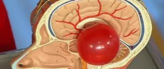
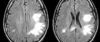
Brain cancer is a process of pathological transformation of brain cells or the surrounding meningeal, glandular and bone tissues. As a result of transformation, abnormal cells begin the process of uncontrolled division, which causes the formation of a tumor that compresses healthy tissue and increases intracranial pressure. In conditions of limited space in the cranium, even a benign neoplasm, if left untreated, can lead to disability or even death.
Kinds
Brain tumors are divided into:
- primary – developing directly from intracranial tissues;
- secondary – developing from metastases spread by a tumor located in another organ.
Tumors are classified according to the location of their formation:
- neuroepithelial, or gliomas – appearing directly in the brain tissue;
- meningiomas - developing in the tissues of the membranes of the brain;
- neuromas – originating in the cells of the nerve fibers of the skull;
- pituitary adenomas – rarely showing malignant signs;
- metastases from other organs;
- dysembryogenetic neoplasms that develop during the embryonic period of a child’s development.
Causes of tumors
At the moment, medical science does not fully understand the reliable causes of the formation of cancer. Doctors name factors that increase the risk of tumor formation. An important role is played by the age and gender of a person. Some types of cancer, such as meningiomas, are more often diagnosed in women over 45-50 years of age. This is due to the characteristics of the female endocrine system and hormonal levels. For example, during pregnancy, some neoplasms progress more intensively. Brain tumors in men, as a rule, have a more severe course and complex prognosis, because in this case the tumors are highly malignant.
Other provoking factors include:
- adverse environmental impacts: radiation, hazardous industries, living in contaminated areas, etc.;
- constant contact with toxic elements;
- infection with viruses that can contribute to the development of cancer;
- genetic characteristics (the presence of certain genes in the human genome that increase the risk of developing a brain tumor);
- head injuries of various nature and origin;
- treatment with drugs, for example, hormones, chemotherapy;
- bad habits;
- consumption of low-quality food saturated with carcinogenic elements.
These factors can influence the formation of diseases both separately (for example, heredity) and in combination.
Symptoms
When brain cancer appears, symptoms in the early stages are local and depend on the location of the tumor. The disease manifests itself:
- impaired sensitivity in certain parts of the body, deterioration of orientation in space;
- memory impairment;
- decreased muscle activity up to complete paralysis;
- convulsive seizures of an epileptic nature;
- hearing damage and decreased speech recognition function;
- partial or complete visual dysfunction;
- partial or complete loss of the ability to write and speak;
- weakness, fatigue;
- pathological changes in the production of hormones from the pituitary gland or hypothalamus;
- changes in character, emotional imbalance;
- decrease in intellectual level;
- the appearance of hallucinations.
The first signs of brain cancer appear as minor changes in one or more of these areas, but the deterioration progresses over time. With a significant increase in pressure inside the skull, general cerebral symptoms appear:
- persistent headaches of high intensity that cannot be relieved with analgesics;
- dizziness caused by compression of the cerebellum or decreased blood supply to brain structures;
- vomiting independent of food intake.
What brain diseases are precancerous?
Precancerous diseases include most benign neoplasms and diseases that precede malignant tumors. Precancers of the brain are characterized by certain changes in the cellular structure. If negative factors continue to influence, then damaged cells with some probability can become cancerous. Science has not fully established a specific list of precancerous diseases that cause the formation of dangerous tumors, so we can only say with confidence that any processes and diseases affecting the brain should be under the control of specialists and not become chronic.
When treating brain tumors in Israel, special attention is paid to the most accurate diagnosis of patients presenting with any symptoms or suspicion of a tumor in the skull.
Causes and risk factors
The exact causes of brain cancer are still unclear, but there are certain risk factors that significantly increase the likelihood of developing tumors:
- predisposition inherited from parents or other close relatives;
- work in hazardous production;
- long-term exposure to radiation;
- decreased immune system function;
- infection with Epstein-Barr virus or cytomegalovirus;
- past traumatic brain injury;
- male gender, old age, white race.
The presence of one or even several of the listed signs does not mean that a person will necessarily develop a tumor in the brain, but the risk of developing the disease for these people is increased compared to others.
Treatment methods
Brain tumors are not only difficult to diagnose, but also difficult to treat. The surgical operation is technically complex, and the risk of complications after such an intervention is quite high. Treatment with chemotherapy and targeted drugs is also not always effective.
Thanks to the advent of modern intelligent equipment, neoplasms, including inoperable ones, are very effectively treated using radiosurgery and other innovative radiation therapy technologies.
Detailed information about modern treatment methods.
Stages
Since signs of brain cancer do not appear immediately after the onset of the disease and remain subtle for a long time, accurately determining the stage of tumor development is a rather difficult task. Oncologists distinguish four stages of the disease.
- Pathological changes affect a small number of cells localized in a limited area of the brain. The patient experiences periodic headaches, weakness, constant drowsiness, and dizziness often occurs.
- The tumor grows, penetrates nearby tissues, and begins to compress individual centers of the brain. This can be manifested by convulsive attacks, periodic vomiting, and digestive disorders.
- Due to the rapid growth of the tumor, surgical removal of malignant tissue becomes almost impossible. Symptoms intensify, cognitive impairment appears, and coordination of movements worsens. A characteristic symptom of the third stage is horizontal nystagmus, or constant horizontal movement of the pupils.
- The tumor grows so much that the patient experiences constant headaches and gradually loses the functions of maintaining vital processes. In the absence of treatment or its low effectiveness, the patient falls into a coma, followed by death.
Types of tumors
Neoplasms that form in the brain can be:
- Benign – tumor tissue does not contain cancer cells. Neoplasia of this type is characterized by clear edges and slow growth. They are much easier to treat surgically, but can become malignant, that is, turn into cancer;
- Malignant tumors contain abnormal cells that can divide uncontrollably and metastasize to other organs. In this case, the disease is difficult to control; in the later stages, surgery is impossible. Treatment in this case is difficult, has a high cost and, as a rule, poor prognosis.
Brain glioma
Glioma
This type of tumor is diagnosed more often, accounting for approximately 60% of all types of cancer that form in the brain. The tumor is formed from glial cells. In most cases, this is a primary neoplasm, most often formed in the area of the chiasmata or ventricles, but it can also affect the brain stem; pathogenesis rarely extends to the bones of the skull, however, in rare cases this is possible. The size of gliomas is usually up to 14 cm. Metastases are rare, growth is usually slow, but due to penetration into adjacent tissues, the boundaries of the lesion are difficult to distinguish.
Three varieties are known
- astrocytoma – diagnosed more often than others (it accounts for about 50% of gliomas);
- oligodendroglioma – up to 10% of cases;
- ependymoma – less than 7%.
According to the World Health Organization classification, there are 4 stages of glioma:
- The first stage is benign. It is characterized by slow growth. Example: giant cell astrocytoma.
- Second stage. Features: borderline malignancy with slow tissue growth, easily progresses to stages 3 and 4. In this case, only one symptom of cancer is identified - cellular atypia.
- Third and fourth stage. In this case, the tumor has all the signs of cancer, for example, necrotic changes in glioblastoma multiforme.
Brain astrocytoma
Removal of a large GRADE II WHO astrocytoma of the right temporal lobe (before surgery)
This malignant neoplasm of varying degrees of aggressiveness is diagnosed quite often. The beginning is given by degenerated glial cells - astrocytes (hence the name - astrocytoma). There are:
- Common astrocytomas: protoplasmic, fibrillary and hemisrocytic.
- Special astrocytomas: cerebellar, piloid, microcystic and subependymal.
Astrocytomas are distinguished by the stage of malignancy:
- The first stage (benign tumor) includes piloid or pilocytic astrocytoma of the brain.
- Second stage of malignancy: fibrillary tumor is also an ordinary neoplasia, second degree of malignancy.
- The third stage is already cancer. It includes anaplastic astrocytoma of the brain.
- The most life-threatening are glioblastomas (giant cell glioblastoma and gliosarcoma), which are classified as stage 4 and are characterized by rapid growth and the ability to metastasize.
In addition to the ones mentioned, there are other types of astrocytomas, for example, ependymoma (develops from ventricular cells), oligodendroglioma (forebrain) - grow quite slowly without the formation of metastases, and glioma of the brain stem.
Meningioma of the brain
Removal of a large middle cranial fossa meningioma before surgery
Pathogenesis begins with pathogenic cells in the superficial membrane that surround the brain tissue. The neoplasm is oval in shape, grows slowly, and is therefore considered benign. Meningiomas are diagnosed in 15% of cases (of all brain tumors). The risk of degeneration into cancer is about 3-4%. The clinical signs that appear are caused by increased intracranial pressure and tissue compression. With malignancy (degeneration of somatic cells into carcinogenic ones), meningiomas become aggressive. Usually more than one lesion forms and may spread to the spinal cord. After surgical treatment, there is a high probability of relapses. As a rule, pathology is diagnosed by chance, because symptoms appear only when the lesion reaches a large size. There are no early signs of the disease.
Note. Meningiomas often form in females.
Other types of brain tumors
There are quite a lot of types of neoplasms. Here are some of those that are diagnosed relatively often (besides those mentioned above):
- medulloblastoma is a brain cancer that originates from the structures of the cerebellum;
- germinoma - formed from germ cells, most often it is detected in the first half of a person’s life;
- Craniopharyngeoma - develops in the pituitary gland at the base of the brain;
- Shawnoma – formed from degenerated Schwann cells;
- tumors in the pineal gland area.
Diagnostics
It is difficult to determine the presence of a brain tumor based on symptoms, especially in the early stages. Taking a biopsy specimen for histological examination is very difficult, since this requires a full-fledged neurosurgical operation. During the examination, the neurologist prescribes the following types of diagnostics:
- electroencephalography to detect brain disorders;
- Echo-EEG to check intracranial pressure;
- ophthalmoscopy to exclude deterioration of visual function due to ocular pathologies;
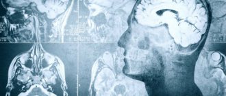
- CT scan of the brain is the main study that allows you to detect a tumor, determine its size, the presence of hemorrhages, calcifications and other pathological changes in tissues;
- MRI of the brain is a complementary study to CT, necessary to clarify changes in the soft tissues of the brain;
- MRI of the vessels of the head - to study changes in the structure of the blood supply to the brain and other tissues;
- PET scan of the brain - to determine the level of malignancy of the tumor.
If a decision is made to perform surgery, a histological examination of the removed tumor tissue is required to determine the histological type of tumor and the level of cell differentiation.
Attention!
You can receive free medical care at JSC “Medicine” (clinic of Academician Roitberg) under the program of State guarantees of compulsory medical insurance (Compulsory health insurance) and high-tech medical care.
To find out more, please call +7, or you can read more details here...
Diagnostic methods
To make a diagnosis, doctors prescribe various types of scans (CT, MRI), laboratory tests, and functional tests.
The use of the latest generation computerized complexes allows specialists to take tumor tissue for examination under a microscope without resorting to a full-scale operation. At the same time, the risk of complications is reduced to a minimum even when the tumor is located in a hard-to-reach place.
Detailed information about modern diagnostic methods and capabilities.
Treatment
Regarding the treatment of brain oncology, you should contact a neurologist or neurosurgeon. Based on the stage of development and location of the tumor, the doctor selects the most appropriate treatment methods in each case.
- Neurosurgery. Removal of a malignant neoplasm is the most effective way to guarantee complete relief from the disease. Intervention is possible at the first and partially at the second stage of the disease, but much depends on the location of the tumor and its proximity to vital centers of the brain. The efficiency of surgeons has increased thanks to the use of laser and ultrasound technology.
- Radiation therapy. Removal of malignant cells using ionized radiation is used as an auxiliary means after surgery, and also as a main means in cases where intervention is not possible. The intensity and duration of irradiation in each case is selected individually, in accordance with the size and location of the pathological area, the type of altered cells and other characteristics.
- Chemotherapy. It is difficult to select drugs for the treatment of brain tumors due to the presence of the blood-brain barrier. As a rule, they are prescribed in combination with radiation therapy, and the course consists of several drugs that complement each other. Courses lasting 1-3 weeks are carried out under constant monitoring of blood changes.
- Cryosurgery. The destruction of tumor tissue by freezing is beneficial in cases where traditional surgery is difficult due to the proximity of vital centers or the deep location of the pathological area. Cryosurgery can be used as a primary method or as an adjunct to conventional surgery, laser and chemical treatments.
- Symptomatic treatment. If the tumor is detected too late, and any of the listed methods do not achieve results, the patient is prescribed therapy aimed at reducing symptoms and improving quality of life. These are glucocorticosteroids, sedatives and antiemetics, NSAIDs for pain relief, as well as narcotic analgesics.
Brain tumors: symptoms, risk factors, diagnosis and treatment
Neurosurgery March 23, 2017
A brain tumor is a growth or abnormal growth of cells in or near the brain. There are several types of brain tumors. They can be noncancerous (benign) or cancerous (malignant). Tumors can also develop from brain tissue (primary) or form in other parts of the body and then spread to the brain (secondary or metastatic).
The rate of tumor growth in the brain can vary greatly. The growth rate, as well as the location of the tumor in relation to other brain structures, determines its effect on the functioning of the nervous system.
Treatments for brain tumors depend on the type of tumor, as well as its size and location.
MAIN SYMPTOMS
Signs and symptoms of a brain tumor can vary greatly depending on its size, location, and growth rate. Main signs and symptoms caused by a brain tumor may include:
- The appearance or change of pain in the head
- Headaches that become more frequent and severe
- Nausea or mouth for no apparent reason
- Vision problems such as blurred vision, double vision, or loss of peripheral vision
- Gradual loss of feeling or ability to move arms or legs
- Balance problems
- Speech problems
- Disorientation in time and space
- Personality and behavior changes
- Seizures, especially if they have not happened before
- Hearing problems
When should you see a doctor?
Make an appointment with your doctor if you have persistent signs and symptoms that bother you.
CAUSES OF BRAIN TUMORS
Tumors that arise directly in the brain
Primary tumors arise directly in the brain or in tissues located close to it—the membranes covering the brain (the meninges), the cranial nerves, the pituitary gland, and the pineal gland. Primary tumors form when errors (mutations) appear in the DNA of normal cells. Mutations allow cells to grow, divide quickly, and survive when healthy cells die. The result is a collection of abnormal cells that form a tumor.
Primary brain tumors are less common than secondary tumors, in which cancer appears in other parts of the body and spreads to the brain.
There are several types of primary tumors and each has its own name, depending on the cells that form it. For example:
- Gliomas. These are tumors that form in the brain or spinal cord and include astrocytomas, ependymomas, glioblastomas, oligodendrogliomas, and oligoastrocytomas.
- Meningiomas. A meningioma is a tumor that develops in the membranes surrounding the brain and spinal cord (the meninges). Most meningiomas are cancerous.
- Acoustic neuromas (schwannomas). These are benign tumors that grow on the nerves that control balance and hearing and carry nerve impulses from the inner ear to the brain.
- Pituitary adenomas. These are mostly benign tumors that form in the pituitary gland at the base of the brain. These tumors can affect pituitary hormones, which affect the entire body.
- Medulloblastoma. It is the most common malignant brain tumor in children. Medulloblastoma forms in the lower part of the brain and spreads through the cerebrospinal fluid. These tumors are less common in adults but do occur occasionally.
- Primitive neuroectodermal tumors are rare malignant tumors that develop in embryonic (fetal) cells in the brain. They can appear in any area of the brain.
- Germ cell tumors. Germ cell tumors can occur in childhood, in the area where the reproductive organs form. But sometimes germ cell tumors move to other parts of the body, such as the brain.
- Craniopharyngiomas. These are rare benign tumors that arise near the pituitary gland, which produces hormones and controls many body functions. While a craniopharyngioma slowly grows, it can affect the pituitary gland and other structures located near the brain.
Cancer that starts in other areas of the body and spreads to the brain
Secondary brain tumors (metastases) are tumors that form against the background of a cancerous tumor that has arisen in another organ and spreads (metastasizes) to the brain.
Secondary brain tumors are most common in people with cancer. But, in rare cases, a metastatic brain tumor may be the first sign of cancer in any organ.
Secondary brain tumors are much more common than primary ones. Although any malignant tumor can metastasize to the brain, the most common metastases are:
- Breast cancer
- Colon cancer
- Kidney cancer
- Lungs' cancer
- Melanoma
RISK FACTORS
In most patients with primary brain tumors, their cause is unclear. But doctors have identified a group of factors that increase the risk of developing a brain tumor. Risk factors include:
Age. As you age, your risk of developing brain tumors increases. They are most common in older people. However, the tumor can appear at any age. There are even some types of brain tumors that occur almost exclusively in children. Exposure to radiation. Individuals exposed to ionizing radiation have an increased risk of developing brain tumors. Examples of ionizing radiation are radiation therapy used to treat cancer and radiation exposure from the explosion of atomic bombs. Other types of radiation, such as electromagnetic fields from high-voltage power lines and radiation from cell phones and microwave ovens, have not been scientifically proven to be associated with brain tumors. Family history. A small proportion of brain tumors occur in people with a family history of tumors or genetic syndromes that increase the risk of developing brain tumors.
DIAGNOSTICS
If a brain tumor is suspected, your doctor may recommend a series of tests and procedures, including:
Neurological examination. A neurological examination may include tests of vision, hearing, balance, coordination, stamina and reflexes, among other things. Abnormalities in one or more functions may indicate parts of the brain are being affected by the tumor.
Medical imaging. Magnetic resonance imaging (MRI) is often used to diagnose brain tumors. In some cases, a contrast agent may be injected through a vein in the arm during the examination.
A number of specialized MRI components —including functional MRI, perfusion MRI, and magnetic resonance spectroscopy—can help the doctor evaluate tumors and determine treatment plans.
Other medical imaging studies may include computed tomography (CT) and positron emission tomography (PET).
Tests done to detect cancer in other organs . If it is suspected that a brain tumor may be caused by cancer in other organs, your doctor may order a series of tests and procedures to determine the origin of the cancer. An example would be a CT scan of the chest to look for signs of lung cancer.
Collection and examination of abnormal tissues (biopsy). A biopsy may be part of surgery to remove a brain tumor or may be performed as a needle puncture.
A stereotactic biopsy can be done to examine tumors in hard-to-reach or sensitive areas of the brain that are easily damaged by surgery. The neurosurgeon makes a small hole in the skull and inserts a thin needle through it. The tissue is then removed using a needle, often guided by CT or MRI (neuronavigation).
The collected material is examined under a microscope to determine whether it contains cancerous or benign cells. This is the most important information for making an accurate diagnosis and prognosis, and, most importantly, for determining further treatment.
TREATMENT
Treatment for a brain tumor depends on its type, size and location, as well as the patient's health and preferences. Surgical intervention.
If the location of the brain tumor allows surgery, the neurosurgeon will remove as much of it as possible.
In some cases, tumors may be small and easily separated from surrounding tissue, allowing for complete surgery. In other cases, the tumors cannot be separated from surrounding tissue or are located near sensitive parts of the brain, making surgery very risky. Then the neurosurgeon removes the maximum part of the tumor located in the safe zone.
Even removing part of the tumor helps relieve signs and symptoms of the disease
Surgery to remove a brain tumor carries certain risks, such as infection and bleeding. Other risks depend on the location of the tumor. For example, removing a tumor near the optic nerve risks vision loss.
Radiation therapy
Radiotherapy uses ionizing radiation—X-rays or protons—that destroy the tumor. The beams may come from a machine outside the body (external beam radiation) or, in very rare cases, the radiation source is placed inside the body near a brain tumor (brachytherapy).
The external beam of radiation can be focused only on the tumor localization area or directed to the entire brain. Whole-brain radiation is most often used to treat cancer that has spread from other organs. The side effects of radiation therapy depend on the type and dose of radiation received. Common side effects during or after radiation include fatigue, headache, nausea, and vomiting.
Chemotherapy
Chemotherapy uses drugs that destroy cancer cells. Medicines may be given orally as tablets or may be given into a vein (intravenously). The most commonly used drug to treat brain tumors is temozolomide (Timodal), available in tablet form. Other types of chemotherapy drugs are also available, prescribed depending on the type of tumor.
The side effects of chemotherapy depend on the type and dose of drugs the patient receives. Chemotherapy can cause nausea, vomiting, and hair loss.
RECOVERY AFTER TREATMENT
Because brain tumors can occur in the parts of the brain that control motor skills, speech, vision, and thinking, rehabilitation may be a necessary part of recovery.
A neurosurgeon can use the following rehabilitation methods:
- Physical therapy to restore lost motor skills or muscle strength
- Occupational therapy to help a patient return to normal daily activities and work after treatment for a brain tumor or other illness
- Speech therapy performed by a speech therapist in case of speech impairment (speech pathology).
Author: Danu Adrian, neurosurgeon, highest category
Forecasts
The success of treatment depends on the timely detection of the disease. If therapy is started at the first or second stage of brain cancer, the five-year survival rate is, according to various estimates, 80-85% of patients. If the disease is detected late, a favorable outcome is possible for approximately 31% of patients. However, it should be remembered that cancer is one of the least studied areas of medicine, so you should never give up your chances of life, even in the most advanced cases of the disease.
Organization of treatment in Germany
At any stage of a brain tumor, you can receive high-quality treatment in Germany. It is organized for foreign patients by Booking Health. We are leaders in the field of medical tourism, and BookingHealth.com, the service for booking treatment abroad, was the first and so far remains the only one to receive an ISO quality certificate. This indicates the high quality of the services provided.
We will help you:
- Choose the best clinic for brain cancer treatment
- Organize the earliest possible start of treatment in Germany before the tumor reaches a large size
- Collect and translate all necessary documents into German
- Get to Germany, as well as from the airport to the clinic that will treat you
- Save up to 70% on the cost of medical services
- Protect yourself from additional expenses (they will be covered by insurance)
To use Booking Health services, leave a request on the website. We will call you back within a day to agree on all the details of your treatment in Germany.
Questions and answers
How do you know if you have brain cancer?
In the initial stages of the disease, symptoms of brain cancer can easily be confused with signs of ordinary fatigue, a mild cold, or a neurological or cardiac disease. To be sure that there is no disease, it is necessary to undergo a CT scan, which allows you to detect a tumor in the initial phase of development.
Is there a cure for brain cancer?
With timely detection of a tumor, timely and high-quality surgical treatment, one hundred percent restoration of health is possible.
What causes brain cancer?
A tumor in the brain tissue develops because one or more cells begin the process of abnormal division. In this case, the pathologically altered cells lose their original function and are only capable of rapid division, which is why the size of the tumor grows rapidly. It has not yet been possible to establish what mechanisms trigger this process.
Attention! You can cure this disease for free and receive medical care at JSC "Medicine" (clinic of Academician Roitberg) under the State Guarantees program of Compulsory Medical Insurance (Compulsory Medical Insurance) and High-Tech Medical Care. To find out more, please call +7(495) 775-73-60, or on the VMP page for compulsory medical insurance
Reviews of doctors providing the service - Back and spine injuries
In 2000, Andrei Arkadyevich performed spinal surgery on me.
Four days in the clinic and I have been living a full life for 20 years without restrictions on movement and I remember with gratitude Dr. A.A. Khodnevich. God bless him. And in 2000 he could walk no more than 10 meters. Read full review Viktor Alexandrovich
20.05.2020
Low bow to Alexander Semenovich Bronstein and Andrei Arkadyevich Khodnevich. I arrived at CELT on July 2, 2021 with extreme pain that I endured for 10 days. Hernia C6-C-7. I was given two blockades in Ivanovo, about 9 complex IVs, I lost 6 kg in a week and was in a panic, I didn’t see a way out and nothing happened to me... Read full review
Elena Nikolaevna L.
20.10.2019
