Hypoxemic crisis (dyspnea-cyanotic attack)
A shortness of breath-cyanotic attack is an attack of hypoxia (paroxysmal shortness of breath with severe cyanosis) in a child with blue-type congenital heart disease, most often with tetralogy of Fallot. It occurs as a result of a sudden decrease in pulmonary blood flow associated with spasm of the outflow tract of the right ventricle of the heart, an increase in blood discharge from right to left and hypoxemia in the systemic circulation.
Attacks of hypoxia develop mainly in young children - from 4–6 months to 3 years. Provoking factors for a dyspnea-cyanotic attack can be psycho-emotional stress, increased physical activity, intercurrent diseases accompanied by dehydration (fever, diarrhea), iron deficiency anemia, neuro-reflex excitability syndrome with perinatal damage to the central nervous system, etc.
A dyspnea-cyanotic attack is characterized by a sudden onset. The child becomes restless, moans, cries, and cyanosis and shortness of breath increase. Takes a forced position - lies on its side with its legs brought to the stomach or squats. On auscultation of the heart there is tachycardia, a decrease in intensity or disappearance of systolic murmur over the pulmonary artery. The duration of a hypoxic attack ranges from several minutes to several hours. In severe cases, hypoxemia, acidosis, convulsions, loss of consciousness, even coma, and death are possible.
ACUTE HEART FAILURE IN CHILDREN IN CRITICAL CONDITION (literature review)
D.N. Surkov, V.I. Snisar Department of Anesthesiology, Intensive and Barotherapy of the Faculty of Professional Education of the Dnepropetrovsk State Medical Academy.
Acute heart failure of varying severity accompanies the course of most critical conditions in children and, therefore, occurs quite often in the practice of intensive care of children [2, 4, 11, 13, 15]. At the same time, there is no coherent system of views on this problem, which would include generally accepted terminology, classification, clear clinical and instrumental criteria for development options and severity, as well as practical recommendations for treatment tactics based on this. In this regard, the purpose of this review was to study the most significant literary data on this issue, as well as an attempt to systematize them from the position of an intensivist. The precise definition of any pathological condition or nosological form is extremely important, since both the diagnosis and the development of the most effective ways to treat this disease depend on the meaning that is put into this definition. Thus, A. Gyton writes that heart failure is a failure of the heart as a pump. However, given that it can last for a long time not only with normal, but even with increased cardiac output (for example, in thyrotoxicosis, beriberi), this definition cannot be considered universal [6]. Heart failure can be considered as a condition in which the heart is unable to pump enough blood to support the metabolism of peripheral tissues [23]. This definition by E. Draunwald et al., in fact, is a variant of the previous one and cannot include forms of heart failure that occur with an increased cardiac output. In addition, it is difficult to say what level of tissue metabolism is required in each particular case. According to F.Z. Meyerson, heart failure is “... this is a condition in which the load on the heart exceeds its ability to do work.” In this case, the excess of blood flow to the heart over the outflow from it is meant, in connection with which, the author himself notes: “This simple definition is not, however, completely accurate, since the discrepancy between the inflow and outflow should relatively quickly lead to lethal disintegration of the function blood circulation and cannot underlie chronic heart failure, which occupies a major place in human pathology” [10]. N.M. presents a unique point of view. Mukharlyamov: “Heart failure should currently be understood as such a pathological condition when the mobilization of compensatory mechanisms is first detected, and then the depletion of the latter with insufficient blood supply to the body” [12]. Critically examining this definition, it should be said that the mobilization of compensatory mechanisms occurs at the beginning of any pathological process that damages the myocardium, and insufficient blood supply to the body can be determined not only by the pathology of the heart muscle, but, for example, by bleeding or vascular spasm, combined or not combined with damage hearts. V.A. Frolov et al. (1994) believe that “heart failure is a condition characterized by a decrease in the reserve capacity of the heart.” Both acute and chronic and even latent forms of heart failure fit this definition. According to the authors, “pathology differs from the norm in that with the development of a pathological process, the biological system, having used up all the possibilities of strictly non-programmed oscillations, begins to function in a narrow direction, maximally mobilizing to achieve the required effect, but this leads to the exhaustion of the body’s reserve capabilities, subsequent pathogenic desynchronization of its functions, overstrain and possible failure of the mechanisms of sanogenesis” [19]. There are several classifications of circulatory failure in general and heart failure in particular. The classification according to N.D. is generally accepted. Strazhesko and V.Kh. Vasilenko (1935), in which acute heart failure is divided into left ventricular (cardiac asthma and pulmonary edema), right ventricular and biventricular. Of particular interest is the clinical classification of acute left ventricular failure by T. Killip and J. Kimball (1967), used, in particular, in acute myocardial infarction [3]:
Class I - there are no clinical signs of heart failure.
Class II - there is moderate shortness of breath, a galloping rhythm and/or congestive wheezing in less than 50% of the area of the lung fields.
Class III - congestive wheezing is detected in more than 50% of the lungs or pulmonary edema develops.
Class IV - cardiogenic shock.
However, such pronounced forms of acute heart failure in children are quite rare, they represent variants of decompensation and require, in fact, resuscitation measures. Chronic heart failure has been studied quite widely and is mainly the prerogative of cardiologists. In the practice of pediatric intensive care, the problem of acute heart failure (AHF) is quite common and complex. According to A.V. Papayan, E.K. Tsybulkina (1984) currently there is no generally accepted classification of acute heart failure in children. The use of the classification of chronic heart failure for this purpose cannot be unconditional, since in acute conditions the disease itself, even at rest, contributes to the development of stage I heart failure [13]. According to E.K. Tsybulkin (1994) AHF is clinically manifested:
- low cardiac output syndrome (LESS) in the form of arterial hypotension and signs of centralization of the blood circulation;
- congestive heart failure (CHF) with overload of the pulmonary or systemic circulation. Signs of congestion in the large circle: peripheral edema, liver enlargement, contouring of the neck veins, ascites, hydrothorax. Signs of stagnation in the small circle: shortness of breath, moist rales in the lower parts of the lungs, clinical signs of pulmonary edema, ineffectiveness of inhalation of high concentrations of oxygen.
The most common causes of SIWS:
- arrhythmic shock - bradyarrhythmias (sinus or due to AV block, ventricular fibrillation, group ventricular extrasystoles) or tachyarrhythmias (excessive tachycardias - Kisch hypermotile toxicosis or acute coronary insufficiency in young children, supraventricular paroxysmal tachycardia, atrial fibrillation and flutter, etc.);
- cardiogenic shock - acute focal (infarction) or total myocardial hypoxia (a condition with hypoxia and acidosis);
- acute pericardial tamponade (wound or rupture of the myocardium, pericarditis, pneumomediastinum and pneumopericardium) or extracardial cardiac tamponade in status asthmaticus III-IV degrees, interstitial emphysema;
- end-stage CHF against the background of decompensated heart defects, myocarditis or myocardia of various origins [20].
In children, the causes of acute heart failure syndrome may be:
- acute bronchopulmonary changes (pneumonia, acute lung injury syndrome [11], atelectasis, hydro- and pneumothorax, etc.), in which the main mechanisms for the formation of heart failure are hypoxia itself and pulmonary hypertension due to intrapulmonary shunting of blood [24, 41];
- any conditions associated (despite normovolemia) with tissue hypoxia: exo- and endogenous toxicoses [15], systemic inflammation syndrome [17, 35], burn disease [2], severe purulent-septic processes, i.e. conditions in which, due to hypercatabolism, the systemic transport of oxygen and glucose does not cover the increased needs of tissues and organs [36]. In such a situation, the minute volume of blood flow increases significantly, which is achieved in children not due to an increase in stroke volume, but rather due to an increase in heart rate. In this case, there is an increase in oxygen consumption by the myocardium, as well as a reduction in diastole time, which leads, on the one hand, to a decrease in ventricular filling in the isovolumic relaxation phase and a reduction in cardiac output, and on the other hand, to a decrease in coronary blood flow, myocardial ischemia and a decrease in its contractility.
According to the severity of A.V. Papayan and E.K. Tsybulkin (1984) distinguishes 3 degrees of acute heart failure:
Acute heart failure of the first degree is characterized by tachycardia and shortness of breath, clearly manifested in the child at rest. The most important symptom is a change in the relationship between heart rate and breathing. In these cases, in children under 1 year of age, the ratio of pulse rate to respiratory rate will be over 3.5; in children over 1 year old - 4.5. There are signs of heart damage: dullness of tones, expansion of the boundaries of relative cardiac dullness.
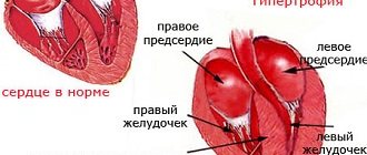
Acute heart failure of the second degree, the most important feature of which should be considered compensatory hypervolemia, depending on the severity, can be divided into 2 conditions: with a predominance of decompensation in only one circle of blood circulation or with total circulatory failure. In case of II A deficiency, if the symptoms of stagnation in the systemic circle predominate, the patient’s liver size increases, and there may be periorbital edema. CVP increases only if decompensation develops quickly, over minutes or several hours. If failure increases gradually over 1-2 days, then central venous pressure may remain normal against the background of progressive liver swelling. The liver in these cases plays the role of a buffer. Muffled heart sounds are required; the boundaries of relative cardiac dullness may expand. If the phenomena of stagnation in the pulmonary circulation predominate, in addition to relative tachycardia, cyanosis increases, the degree of which does not decrease under the influence of oxygen therapy. Scattered fine-bubble rales appear in the lungs, and the accent of the second tone on the pulmonary artery is determined. In case of II B degree deficiency, the listed signs are accompanied by oliguria, peripheral edema, and possible pulmonary edema.
Acute heart failure of the third degree is a hyposystolic form of heart failure with the development of arterial hypotension against the background of clinical overload of the pulmonary circulation [13].
Hemodynamic shock syndrome is represented by acute circulatory failure associated with a drop in the arteriovenous pressure gradient. Three unrelated groups of processes, cardiogenic, vasohypotonic and hypovolemic mechanisms, are the starting point of the pathogenesis of shock syndrome. In addition, clinical practice is often faced with complex processes involving two or three pathogenetic mechanisms simultaneously [30]. There are 4 most important control mechanisms of the human circulatory system for maintaining hemodynamics - the renin-angiotensin system, the autonomic nervous system, local arterial autoregulation and stress vascular relaxation. [36] At the same time, congestive heart failure is characterized by: 1) neurohumoral vasoconstriction, tachycardia; 2) sodium and water retention; 3) an increase in plasma concentration of norepinephrine [40]. In the practice of intensive care of children, acute heart failure syndrome should be distinguished [40], which may be associated with direct damage to the heart muscle (congenital and acquired heart defects, myocarditis [16], poisoning with cardiotoxic poisons [15], etc.), as well as may accompany any critical condition due to developing hypoxia and energy deficiency [41]. In the latter case, changes in the activity of the heart are reversible and are associated, as a rule, with a violation of the systole-diastolic ratios of the cardiac cycle and, as a consequence, with developing transient ischemic and dystrophic changes in the heart muscle. To understand the characteristics of the development of acute heart failure in children, it is necessary to consider such a concept as myocardial oxygen consumption (MVO2). Hemodynamics is a function of cardiac outflow and vascular resistance. In turn, cardiac outflow depends on heart rate and stroke volume. The magnitude of the latter is influenced by preload, afterload and myocardial contractility. The body uses most of MVO2 to support myocardial contractions. In this case, an increase in contractility leads to an increase in oxygen consumption by the myocardium. There is also a direct correlation between heart rate (HR) and MVO2. A 50% increase in heart rate is usually accompanied by a 50% increase in MVO2. Oxygen consumption also depends on the amount of external work done by the heart. The latter can be divided into the generation of aortic pressure (pressure work) and the ejection of stroke volume (volume work). Pressure work (isometric contraction) has a much higher energy cost than volume work (isotonic contraction). It is clear that maintaining blood pressure by increasing stroke volume is more economical. In childhood, an increase in the cardiac index occurs due to an increase in heart rate with little change in stroke volume. Since the myocardial wall tension may not change in this case, the increase in MVO2 for a given increase in heart rate in children is greater than in adults [14]. To date, a huge number of studies, mainly experimental, have been carried out on the essence of biochemical changes in the myocardium, leading to disruption of its contractile activity. The results of these studies indicate that the decisive role in the deterioration of the pumping function of the heart belongs to disturbances in energy utilization caused by changes in the main contractile proteins, manifested by a decrease in the amount of myofibrillar proteins, including proteins of the actinomyosin complex [40], and changes in the physicochemical structure of myosin, leading to to a decrease in its ATPase activity. At the same time, the process of energy accumulation (resynthesis of macroergs) is disrupted due to dystrophic changes in mitochondria. At the same time, the synthesis of ATP, which ensures the functioning of ion channels of cell membranes and the endoplasmic reticulum, is significantly reduced [31]. Under these conditions, the movement of ions through cell membranes, clearly synchronized in the intact myocardium, changes. Mechanistically, the development of heart failure can be considered based on microwave resonance imaging data. The “twisting” motion of the left ventricle is described during systole, which includes clockwise rotation of the base and counterclockwise rotation of the apex. During diastole, a “unwinding” movement occurs. In the normal heart, diastolic "unwinding" continues predominantly during isovolumic relaxation, similar to systolic "twisting" which occurs predominantly during isovolumic contraction. Prolongation of the "unwinding" movement was found in the hypertrophied and hibernated heart. Thus, heart failure is associated with profound disturbances in the mechanical function of the heart, which is manifested by changes in systolic “twisting” and diastolic “unwinding” motion [32]. There are stages in the development of acute heart failure. In this case, in the early stages, distolic relaxation suffers, after which, and often as a result, a violation of systolic contraction occurs [25, 26, 29, 32]. Accordingly, during acute congestive heart failure (CHF) [28], significantly different forms are distinguished, according to the etiology, clinical picture and pathophysiology of left ventricular (LV) dysfunction [27]. Systolic and diastolic dysfunction are characterized by reduced cardiac output with normal (impaired diastolic function) or reduced (systolic dysfunction) LV pumping function. Systolic dysfunction involves a combination of pulmonary congestion with reduced systolic ejection fraction. Impaired diastolic function is accompanied by pulmonary congestion in the presence of a normal or only slightly enlarged ventricle with normal ejection fraction [25, 26]. The relationship between disturbances in left ventricular systolic and diastolic function is well known, but has not yet been fully documented. Moreover, a quantitative classification of diastolic dysfunction is still lacking [29]. Diastole is an energy-consuming process. Calcium ions must be pumped into the sarcoplasmic reticulum against the gradient. In addition, the separation of tropomyosin bridges requires hydrolysis of the adenosine triphosphate molecule. A decrease in the ability of the endoplasmic reticulum to utilize calcium from the cytoplasm of cardiomyocytes leads to an increase in its concentration during diastole and thereby prevents myocardial relaxation [33]. If diastolic function is impaired, the ventricles of the heart cannot fill adequately at normal pressure and require a compensatory increase in it in the atria. The pathogenesis of impaired diastolic function of the myocardium is based on a violation of its relaxation (relaxation) and compliance (compliance) [8]. Impaired diastolic relaxation is one of the earliest changes recorded even before the onset of heart failure. Systolic myocardial dysfunction (decreased contractility) develops at a later date [1]. Therefore, assessment of diastolic function may be a more sensitive measure of myocardial dysfunction than systolic measurements [22]. The study of acute transient disturbance of myocardial diastolic function is promising in terms of identifying predictors of developing acute heart failure and choosing various options for drug therapy aimed at preventing cardiac decompensation, the development of low cardiac output syndrome and cardiogenic shock [22, 27]. An example of an acute disturbance of diastolic function in children is a special variant of toxicosis due to a viral infection, called acute coronary insufficiency or Kishsh toxicosis. The essence of this option is that the dominant factor in its course and outcome is heart failure, which develops as a result of excessive sinus tachycardia in a previously healthy child. Tachycardia is considered excessive due to the fact that due to a significant reduction in diastole time, venous inflow is sharply hampered and stroke volume is critically reduced. These changes primarily affect the coronary blood flow, leading to insufficient blood supply to the myocardium. Heart failure develops in two stages. At stage I, the reduction in diastole time is not accompanied by a decompensated drop in cardiac outflow, although signs of congestive failure in the large circle are already present. At stage II (decompensation), a decrease in diastole leads to hyposystole, accompanied by arterial hypotension, pulmonary edema and coma [20]. Vatolin K.V. et al. it was found that impairment of LV diastolic function is most pronounced in transient left ventricular myocardial dysfunction (LVMD) [4]. In order to control hemodynamics in children in the intensive care unit, invasive methods are traditionally used (direct Fick method, dye dilution method, thermodilution). The severity of the patients' condition and the need for accurate and reliable monitoring of central hemodynamics and its correction justify the risk of invasive intervention. However, given the enormous advantages of non-invasive research methods, they are increasingly used in intensive care and intensive care settings. In the practice of pediatric intensive care, the Kubizek tetrapolar thoracic rheography
(1966) modified.
The method ensures the location of the measuring electrodes in the region of a uniform electric field, and therefore is methodologically more correct. The method involves synchronous recording of ECG, PCG, RG (or carotid SFG) and differential RG, which requires the use of multi-channel recorders. When measuring CO using tetrapolar chest rheography and direct methods (thermodilution, dye dilutions), r, according to the literature, ranges from 0.31 to 0.99. A number of authors, using the method in the intensive care unit, obtained r = 0.94 [7]. However, in patients with heart defects, low compliance with direct methods was found. Moreover, some authors, when simultaneously measuring CO using the method of tetrapolar thoracic rheography and electromagnetic flowmetry during hemodilution, did not obtain not only reliable data on CO in absolute value, but also a reflection of the objective dynamics of the indicator [9]. Echocardiography
is widely used to evaluate children with congestive heart failure. Because of its unique ability to assess left ventricular (LV) size and geometry, LV systolic and diastolic function, right ventricular size and function, heart valves, pericardium, and congenital anomalies, echocardiography is valuable in establishing specific cardiac diagnoses, guiding therapy, and assessment of prognosis in CHF [5, 21]. Echocardiographic measures of ventricular function depend on preload, afterload, and contractility. Therefore, all measures of ventricular function must be meaningful because echocardiographic measurements do not necessarily reflect ventricular contractility. There are no non-invasive methods for determining contractility, but echocardiography allows them to be determined indirectly. The standard is Dopplercardiography, which is not inferior in accuracy to invasive research methods (Zaretsky V.V. et al., 1979; Cross G., Light LH, 1974; Forrester JS et al., 1977; Merrill P., Spenser MD, 1984) [9]. Two-dimensional echocardiography does not reflect myocardial contractility, but rather reliably detects regional wall motion disorders. Analysis of the pattern of regional wall motion abnormalities from the parasternal short-axis position or the apical four-chamber position may provide some information regarding myocardial injury. Methods for studying systolic function include measuring:
- fractions of shortening and septation of valves at point E;
- ejection fractions in M- and B-mode;
- Dopplercardiography to measure the rate of shortening of circular myocardial fibers [21].
Reported use of Dopplercardiography
mitral valve inflow as a means of assessing diastolic function. The three Doppler inflow patterns of the mitral valve were observed to be independent of disease stage. This is because Doppler ultrasound of the mitral valve inflow actually measures the pressure difference between the atrium and the ventricle. The transition between the different patterns is dynamic and can occur over several hours depending on the patient's hemodynamic status and any therapeutic interventions that may have been undertaken. In this case, the first Doppler pattern, pattern I, is formed early in the course of diastolic excursions, when atrial pressure is normal. It is manifested by a decrease in the E wave (passive filling of the ventricle) with a long deceleration time and an increase in the A wave (active filling during atrial contraction). As the pressure in the left atrium increases, the Doppler pattern of the incoming flow takes on a “normal” appearance. The most unfavorable indicator of diastolic excursion is pattern II, in which the E wave is enlarged with a short deceleration time and a very small A wave. The indicated parameters of Doppler patterns of the incoming flow can provide the key to determining diastolic deviations, taking into account that the value of IVRT, Vmax E, E/A reflects the degree of relaxation, and EDV and Vmax A reflect the state of compliance [5, 18, 21] . Calculation of cardiac output can be done non-invasively using continuous two-dimensional and Doppler echocardiography and the use of an equation. Flow for one cardiac cycle, or SV, is calculated as
SV = CSA x V x RR/1000 ml/l,
where CSA (cm2) is the cross-sectional area of the valve through which the flow is measured; V (cm/s) is the average velocity across the valve, and RR is the number of strokes per second. To calculate cardiac output, SV must be multiplied by heart rate [22]. Therefore, real-time cardiac cycle testing is mandatory in pediatric intensive care units [34, 42]. Data from Doppler determination of cardiac output and transmitral patterns will make it possible to differentiate the dependence of certain clinical manifestations on the degree of functional disorders of cardiac activity [5, 21, 38, 39].
BIBLIOGRAPHY
Almazov V.A., Shlyakhto E.V. (1995) Heart failure: current trends in therapy // Kharkov Medical Journal. — 2: 19-22.
Alyes V.F., Andreev A.G., Ulyanova G.I., Granova L.V., Astamirov M.K. (1998) Delivery, consumption and extraction of O2 in the acute period of burn disease in children // Anest. and intensive care unit. - 1: 4-7.
Amosova E.N. (1996) Acute heart failure during myocardial infarction // Journal of a practical physician. - 1: 13-16.
Vatolin K.V., Efimov M.S., Pykov M.I. et al. (2000) Assessment of diastolic cardiac function in intensive care newborns with intracranial hemorrhages // Emergency conditions in children. 6th Congress of Pediatricians of Russia (Moscow). Congress materials. — p.74.
Vorobyov A.S., Butaev T.D. (1999) Clinical echocardiography in children and adolescents: A guide for clinicians. Special literature, St. Petersburg, 423 p.
Guyton A. (1970) Cardiac minute volume. Medicine, M., 64 p.
Zemtsovsky E.V., Guseinov B.A., Izvekova A.V., Polukhina E.L., Chistova I.Ya. (1989) On the accuracy of the rheographic method for determining stroke volume of blood // Cardiology. — 6: 75-79
Korkushko O.V., Moroz G.Z., Gidzinskaya I.N. (1992) Study of diastolic function of the heart in the clinic // Cardiology. - 5: 92-95.
Lebedeva R.N., Abbakumov V.V., Borisova I.V., Svirshchevsky E.B., Bondarenko A.V., Chaus N.I., Karavaev B.I. (1989) Modern methods for assessing hemodynamics in an intensive care unit (comparative aspect) // Anest. and resuscitator. - 4: 3-8.
Meerson F.Z. (1968) Hyperfunction, hypertrophy, heart failure. Medicine, M., 112 p.
Milenin O.B., Efimov M.S. (1990) Hemodynamic disturbances in the acute period of respiratory distress syndrome (according to Doppler echocardiography) // Pediatrics. - 4: 3-9.
Mukharlyamov N.M. (1978) Early stages of circulatory failure and mechanisms of its compensation. Medicine, M., 96 p.
Papayan A.V., Tsybulkin E.K. (1984) Acute toxicosis in early childhood. 2nd ed., revised. and additional Medicine, L., 73 p.
Manual of Pediatrics (Emergency and Intensive Care) (1999) / Edited by M. Rogers, M. Helfair. Peter, St. Petersburg, 1120 p.
Savina A.S. (1992) Acute heart failure and its treatment in case of poisoning with cardiotoxic drugs // Cardiology. - 4: 67-70.
Sidelnikov V.M., Volosovets O.P., Kuzmenko A.Ya., Krivopustov S.P. (1992) Functional state of the myocardium during diastole in children with congested heart disease // Pediatrics, obstetrics and gynecology. - 3: 17-19.
Sipriya A.G., Talvik R.M. (1993) Prognostic value of some indicators of central hemodynamics, oxygen balance and intrapulmonary shunt in patients with sepsis in critical condition // Anest. and intensive care unit. — 5: 43-45.
Trekova N.A., Yavorovsky A.G., Flerov E.V., Yumatov A.E., Kovalevskaya O.A., Asmangulyan E.T., Grishin V.V. (1999) The influence of modern methods of induction of anesthesia on the systolic and diastolic function of the left and right heart in patients with coronary heart disease // Anest. and intensive care unit. - 5: 4-5.
Frolov V.A., Kazanskaya T.A., Drozdova G.A. (1994) On the definition of “heart failure” // Pathological physiology and experimental therapy. - 4: 3-7.
Tsybulkin E.K. (1999) Threatening conditions in children. Emergency medical care: Directory. Special Literature, St. Petersburg, 119 p.
Schiller N., Osipov M.A. (1993) Clinical echocardiography. Mir, M., 347 p.
Bengur AR, Meliones JN (1998) Cardiogenic shock // New Horiz. Sci. and Pract. of Acute Med. - 6(2): 139-149.
Draunwald E., Ross S., Sonnenblick E. (1976) Mechanisms of contraction of the normal and failing heart. 2-d Ed. Boston.
Ducas J., Stitz M., Gu S., Schick U., Prewitt RM (1992) Pulmonary vascular pressure-flow characteristics. Effects of dopamine before and after pulmonary embolism // Am. Rev. Respira. Dis. - 146(2): 307-312.
Federmann M., Hess OM (1994) Differentiation between systolic and diastolic dysfunction // Eur. Heart J. - 12(15): 2-6.
Federmann M., Risti B., Hess OM (1994) Left ventricular insufficiency: systolic versus diastolic dysfunction // Schweiz. Med. Wochenschr. - 124(26): 1196-1202.
Gaasch WH (1994) Diagnosis and treatment of heart failure based on left ventricular systolic or diastolic dysfunction // JAMA. - 271(16): 1276-1280.
Hsueh CW, Lee WL, Chen CK et al. (1998) Dopamine and dobutamine have different effects on heart rate variability in patients with congestive heart failure // Chung Hua I Hsueh Chin, Taipei. - 61(4): 199-209.
Kosmala W., Spring A., Witkowska M. (1997) Relationship between systolic and diastolic function of the left ventricle in patients with impaired relaxation of the left ventricle without symptoms of heart failure. Attempt at quantitative estimation of diastolic function in the impaired relaxation stage // Pol. Arch. Med. Wewn. - 98(11): 414-423.
Kovac Z. (1995) Pathogenic role of intracellular energy insufficiency (hypoenergy) in the development of circulatory collapse (hemodynamic shock) // Lijec. Vjesn. - 117(2): 11-15.
Kozlik-Feldmann R., Kramer HH, Wicht H., Feldmann R., Netz H., Reinhardt D. (1993) Distribution of myocardial beta-adrenoceptor subtypes and coupling to the adenylate cyclase in children with congenital heart disease and implications for treatment // J. Clin. Pharmacol. - 33(7): 588-595.
Matter C., Nagel E., Stuber M., Boesiger P., Hess OM (1996) Assessment of systolic and diastolic LV function by MR myocardial tagging // Basic Res. Cardiol. - 91(2): 23-28
McDonagh T., Robb S., Murdoch D. et al. (1998) Biochemical detection of left - ventricular systolic dysfunction // Lancet. - 351(9095): 9-13.
Murdoch IA, Marsh MJ, Tibby SM, McLuckie A. (1995) Continuous haemodynamic monitoring in children: use of transoesophageal Doppler // Acta Paediatr. - 84(7): 761-764.
Myocardial function in the critically ill: factors influencing left and right ventricular performance in patients with sepsis and trauma (1985) // Surg. Clin. North. Am. - 65(4): 867-893.
Schamberger MS (1996) Cardiac emergencies in children // Pediatr. Ann. - 25(6): 339-344.
Schwarzhaupt A., Grossmann C., Kiencke U. (1998) Simulation of metabolic process in human circulation: evaluation of pH value and CO2 balance // Biomed. Tech., Berl. - 43(10): 275-280.
Shoemaker WC (1996) Oxygen transport and oxygen metabolism in shock and critical illness. Invasive and noninvasive monitoring circulatory dysfunction and shock // Crit. Care Clin. - 12(4): 939-969.
Shoemaker WC (1996) Temporal physiological patterns of shock and circulatory dysfunction based on early descriptions by invasive and noninvasive monitoring // New Horiz. - 4(2): 300-318.
Steifa M., Toman J., Spiranova L. (1997) Acute and chronic heart failure // Vnitr. Lek. - 43(2): 105-110.
Stocker F., Wyler F. (1994) Pediatric cardiological emergencies // Ther. Umsch. - 51(9): 601-606.
Velmahos GC, Shoemaker WC, Wo CC, Demetriades D. (1999) Physiologic monitoring of circulatory dysfunction in patients with severe head injuries // World J. Surg. - 23(1): 54-58.