Among diagnostic cardiac procedures that are informative, it is worth highlighting echocardiography or ultrasound examination of the heart. This is a hardware procedure performed using high-frequency sound waves. It is indispensable for accurately identifying problems that are directly related to the work of the myocardium and the entire cardiovascular system.
The effectiveness of echocardiography is directly related to the evaluation of its results. Competent interpretation of cardiac ultrasound is a direct path to a speedy diagnosis and drawing up an effective treatment plan. Entrust the solution to the problem to an experienced specialist, contact us for qualified medical support!
Why is cardiac echocardiography needed?
There are a myriad of reasons why a doctor might recommend an echocardiogram. A harmless and inexpensive procedure is indispensable for:
- constant pain in the chest, rapid heartbeat, disturbing in a calm state, after minor physical exertion;
- hypertension (stable high blood pressure);
- swelling of the lower extremities, when kidney disease is not diagnosed;
- breathing difficulties that occur after physical activity;
- feeling of a foreign object in the chest.
The attending doctor can prescribe a procedure, and then conduct a full interpretation of the ultrasound of the heart, in case of constant dizziness, arrhythmia, atherosclerosis, pericarditis, heart muscle defects, coronary artery disease. Indications are often multiple pregnancy, hereditary predisposition, medical examination at the enterprise.
Ultrasound cardiography is performed for both adults and children. When a baby is suspected of having abnormalities in myocardial development, shortness of breath appears without symptoms of an acute respiratory infection, loss of consciousness is observed, it is imperative to check the functioning of the main organ. The pediatrician can refer for diagnostics when, during the use of a phonendoscope, extraneous sounds were noticed against the background of normal contractile activity.
The above-mentioned cardiac examination also plays an important role for a teenager. The test is aimed at assessing the normal development of the organ during puberty, because it is at this time that a sharp increase in body size is often observed.
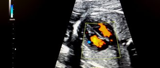
Echocardiography with Doppler analysis
Doppler echocardiography is a variant of ultrasound examination that allows one to evaluate blood flow (velocity and turbulence) in the cavities and large vessels of the heart. The method is based on the Doppler effect: the speed of an object affects the frequency of the supplied signal. This version of echocardiography is highly informative and safe, so it is mandatory for pregnant women and children.
Advantages of the method:
- non-invasive;
- fast processing of information in real time;
- accuracy.
The examination helps to identify:
- pathological blood flows;
- mitral valve prolapse;
- blood clots and cardiac aneurysms;
- defects, congenital and acquired.
Three main modes of echocardiography with Doppler analysis:
- Pulse-wave . Diagnostics of low-speed flows.
- Constant wave . Assessment of blood flow velocity in the aorta (diagnosis of aortic stenosis).
- Color mapping . Visualization of valve and turbulent flows at the site of blockage. Diagnosis of blood clots, aneurysms, atherosclerotic plaques, determination of the nature of the neoplasm.
Interpretation of ultrasound of the heart
In order for the interpretation of cardiac echocardiography to be carried out without errors and completely, the patient’s age, general state of health, and the presence of chronic diseases (pancreatitis, tonsillitis, asthma, vasculitis, etc.) are taken into account. It is impossible to determine the pathology on your own (without knowledge and experience). Only a doctor can correctly assess the answer. There is no place for experiments and guesswork. It is inappropriate to look for answers on the Internet on the websites of companies with a dubious reputation. Trust your health to professionals who provide guarantees and value each patient.
Study parameters
Thanks to ultrasonic waves, you can accurately determine:
- myocardial parameters (sizes of all its parts);
- tissue structure, wall density;
- rhythms, abbreviations, etc.
Imaging will help detect scarring, thrombosis, benign and malignant tumors. The test informs about the condition of the mitral valve, blood volumes and the level of vascular blockage.
By interpreting cardiac echocardiography, the presence/absence of the following diseases can be determined:
- ischemia, when there is a persistent disturbance of blood supply against the background of vascular blockages;
- necrosis, during which tissue death occurs (infarction);
- blood pressure above or below normal (hypo-, hypertension);
- defect, that is, a structural defect of an acquired/congenital type;
- decompensation, when a whole syndrome of failures is noticeable;
- valvular dysfunction;
- rhythmic disruptions;
- rheumatism, when inflammation is observed;
- pericarditis, in which there is inflammation of the membrane;
- stenosis, which indicates a narrowing of the aortic lumen;
- vegetative-vascular dystonia.
Competent image decoding is the basis for success. After all, it is thanks to him that one can establish the fact whether there is a disease or not.
Among the research parameters, several main areas are distinguished. When interpreting cardiac ultrasound in children and persons over 13 years of age, the diameter of the left ventricle and left ventricle, the thickness of the posterior wall of the left ventricle, and more are determined. Each number has a meaning individually. Also, all indicators are taken into account together, because many of them are related to each other and often form a single clinical picture.
How it happens
A painless examination is carried out in a hospital setting or at home, if the situation requires it. The manipulation takes from 20 to 45 minutes. This is a safe way to assess the functioning of the heart muscle and blood vessels, which has no contraindications and is not harmful to health. Step by step it usually looks like this:
- The visitor bares his torso, undresses to the waist and takes a horizontal position, lying on his back with his head towards the diagnostician.
- A special gel is applied to the chest area to help conduct ultrasound waves better.
- A special sensor is used, thanks to which the diagnostician carries out an inspection. The detector is moved slowly across the area being examined to ensure that nothing is missed. The attentiveness of the diagnostician will help with echocardiography decoding.
- The specialist can correct the patient’s position and condition, for example, asking him to hold his breath, roll over, raise his arm, bend his knees, and more.
The response is assessed by a competent specialist, a doctor. It is important that all indicators are taken into account, as well as the individual parameters of the patient’s body. The time of the diagnostic procedure and the state of health, that is, the well-being of the person being diagnosed, are taken into account. Standards and actual figures are reviewed. A comparison is being made. Only after a thorough study can a diagnosis be made and a course of therapy formed. A person who has nothing to do with modern cardiology will not be able to understand the indicators, even if he uses a plate with standard figures for this. Only a properly qualified doctor should be involved in forming a conclusion!
Differences in men, women and children
Normally, data by age and gender is different. EchoCG interpretation helps to analyze the full cycle of myocardial activity (1 systole + 1 diastole). With a heartbeat of 70 beats in 60 seconds, 1 cycle is normally 0.85 seconds.
Adult women and men, as well as children, have different normal values. For example, the heart mass of a representative of the stronger sex aged 25-30 years is about 135 g. In a woman - up to 100 g. The EDV of the left ventricle in men normally reaches 193 ml, in women it does not exceed 136 ml.
The LV wall mass index is an indicator of the ratio of the weight of the organ to the surface area of the human body. The strong half of humanity is characterized by indicators from 71 to 95 g/m2, the fair sex is characterized by a range from 71 to 90 g/m2.
Interpretation of EchoCG results in adults
The diagnostic conclusion is made only by the attending physician. The document in written (sometimes electronic) form is transferred to the patient in the diagnostic room, after which it is recorded in the hospital record. The individual indicators of the person undergoing the examination are entered into a special table, which already contains the normal figures for a specific age and gender.
Trying to decipher an ultrasound of the heart on your own is stupid and unsafe. The combination of several deviations may indicate different pathologies. You can’t do without experience and knowledge here.
| Cardiac parameter | Men's answers | Women's answers |
| LVMM—left ventricular myocardial mass | 135-180 g | 95-142 g |
| LVMI—left ventricular mass index | 71-92 g/m2 | 71-88 g/m2 |
| ESD – LV end-systolic size | 31-42 mm | 31-42 mm |
| ESD – LV end-systolic size | 31-42 mm | 31-42 mm |
| EDV - end diastolic size | 46-58 mm | 45-58 mm |
| Volume of fluid in the pericardial cavity | 10-30 ml | 10-30 ml |
| LV wall thickness in diastole | up to 11 mm | up to 10 mm |
| Blood ejection volume during LV systole | 60-100 ml | 60-100 ml |
| Wall thickness of the RV - right ventricle | 5 mm | 4.8-5 mm |
| LA size - left atrium | 18.5-33 mm | 17.5-33 mm |
| LA end-diastolic volume | 50-82 ml | 38-57 ml |
| End-diastolic volume of the RA – right atrium | 20-100 ml | 20-100 ml |
| Thickness of the IVS - interventricular septum in systole | 5-9.5 mm | 5-9.0 mm |
| IVS thickness in diastole | 7.5-11 mm | 7.5-11 mm |
| Aortic opening area | 20-35 mm2 | 20-35 mm2 |
| Thickness of the outer membrane of the pericardium | 1.2-1.7 mm | 1.2-1.7 mm |
Only a cardiologist can evaluate an ultrasound of the heart; the results are interpreted according to standard values, taking into account the general health of the patient, the medications he is taking and other factors. The size, weight, volume and location of the organ, the condition of the valves and tissues are studied. Parameters for contractions, blood vessels and blood flow, wall thickness and other important nuances are recorded. The speed of blood distribution through the chambers of the heart helps to determine Doppler measurements. It is often performed in conjunction with traditional ultrasound examination.
Decoding EchoCG
After the examination, the doctor draws up a conclusion. First, a visual picture with the presumed diagnosis is described. The second part of the study protocol indicates the patient’s individual indicators and their compliance with standards.
Decoding the data obtained is not a final diagnosis, since the study can be done not by a cardiologist, but by an ultrasound diagnostic specialist.
It is the cardiologist, based on the collected medical history, examination results, interpretation of tests and data from all prescribed studies, who can draw accurate conclusions about your condition and prescribe the necessary treatment!
Norms of cardiac echocardiography in children
In pediatrics, standard indicators are directly related to the patient’s body area. It can be determined using a formula using the child’s height and weight data. During diagnostics, review and note information on:
- cavities of the right/left ventricle, as well as the left atrium;
- wall density;
- aorta, especially its ascending section;
- hemodynamics;
- the septum between the heart ventricles.
Competent interpretation of cardiac echocardiography in children requires care and responsibility in all situations.
| Parameters for diagnosing myocardium in newborns | Male | Female |
| Left ventricular EDR | From 19 to 25 mm | From 18 to 24 mm |
| Left atrium diameter | From 13 to 18 mm | From 12 to 17 mm |
| Left ventricle diameter | From 6 to 14 mm | From 5 to 13 mm |
| Left ventricular ESR | From 12 to 17 mm | |
| MZhP (thickness) | From 3 to 6 mm | |
| Right ventricular wall thickness | From 2 to 3 mm | |
| Blood flow speed | About 1.3 m per second | |
| Ejection fraction varies in infants | From 65 to 75% | |
Please note that sometimes after cardiac echocardiography has been performed, interpretation is carried out directly in the diagnostician’s office. The fact is that often the hardware examination is carried out by the attending physician himself, which significantly saves time for the patient and helps the doctor quickly make the correct diagnosis.
Sometimes the list of parameters in the response is different. This happens if the research was carried out with devices of different manufacture and power, that is, devices with different technical characteristics and capabilities.
Deviations from the norm
There are a huge number of indicators indicating deviations from the norm. Eg:
- Stenosis is said to occur when the valve opening is reduced in diameter, making it difficult to pump blood.
- If the valve leaflets interfere with the reverse blood flow, perform their natural function inadequately, insufficiency is suspected.
- Pericarditis is detected when, with a normal fluid level of 20-30 ml, all 500 ml are traced.
The list of deviations from the norm and possible diagnoses associated with this can be endless. But you shouldn’t take deciphering an ultrasound of the heart so lightly. Here it is inappropriate to guess and assume without the help of a doctor. Mistakes lead to irreparable consequences. Therefore, experiments should be abandoned.
How to do an ultrasound of the heart at the MedicCity clinic?
It is advisable for every person to have an echocardiogram at least once in their life. The fairly low cost of cardiac ultrasound is another reason to prefer this particular diagnostic method.
At MedicCity you can undergo all types of cardiac diagnostics - ECG, EchoCG, bicycle ergometry, HOLTER, ABPM, etc.
Just type in a search engine: “Ultrasound of the heart, Savelovskaya metro station, Dynamo metro station.” Or call us at: +7 (495) 604-12-12.
The doctors of our multidisciplinary clinic will do everything possible to relieve your heart pain!