- Home >
- Directory >
- Interpretation of ultrasound of the heart
Has your doctor prescribed an ultrasound of your heart?
Or is the echocardiography already over, and the results are in your hands? In order not to miss the pathology, do not try to interpret the ultrasound of the heart yourself. Don’t risk your health, take the protocol to a specialist! If you are interested in what parameters and how the survey results are assessed, refer to this article!
How to determine pathology?
The main methods that can be used to determine the condition of the left atrium include: electrocardiography (ECG) and echocardiography (EchoCG).
ECG
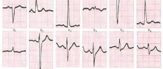
The function of the left atrium on the ECG is assessed by the P wave in leads I, II, aVL, V5, V6.
This method allows you to see:
- Atrial hypertrophy (thickening of the walls). Signs on the cardiogram: increased height and bifurcation of P in I, II, aVL, V5, V6 (the so-called “P - mitrale” - raising the second part of the tooth); negative or biphasic P, P duration is more than 0.1 s.
Hypertrophy is the basis for the occurrence of atrial fibrillation (fibrillation). On the ECG it is expressed by: absence of the P wave, the presence of chaotic f-waves (especially in II, III, aVF, V1, V2), irregular ventricular rhythm. In addition, the proliferation of muscle fibers contributes to the appearance of sinus tachycardia - an increase in the number of impulses arising in the sinoatrial node. On the ECG, the P wave is normal, the RR distance is shortened. - Atrial dilatation (an increase in the size of the cavity against the background of a thinning of the wall) can be suspected using an electrocardiogram only in the presence of arrhythmias.
EchoCG signs
EchoCG or ultrasound (ultrasound) determines the size and performance of the left atrium, which makes it possible to diagnose hypertrophy and dilatation of this section.
The method is used to diagnose coarctation of the aorta, mitral and aortic valve defects, cardiac tumors (myxomas), the presence of which affects the size and function of the left atrium.
Normal ultrasound of the heart
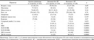
Here are some of the indicators that are taken into account when assessing results:
- Myocardial mass: men - 130-180 g; women - 90-140 g.
- Left ventricle size: At rest - 5.7 cm in men, 4.6 cm in women)
- During contraction – 4.3 cm in men, 3.1 cm in women)
Interpreting the results of a cardiac ultrasound is a serious process that requires the doctor to have fundamental knowledge of human anatomy. Only a specialist can competently assess deviations and understand whether it is worth sounding the alarm. You may need additional examinations.
Take care of your health! Trust the decryption to professionals!
If you have any questions, ask our specialist!
Ask a Question
Preparation for the procedure
No special preparation is required for cardiac echocardiography
It is recommended to follow some important rules:
- On the eve of the procedure, limit the consumption of coffee and black tea.
- Avoid all types of alcoholic beverages several days before the test.
- Do not overwork, do not engage in physical exercise immediately before the ECHO ECG.
Procedure
The procedure is carried out as follows:
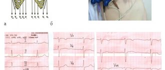
- The patient needs to undress to the waist, freeing the chest;
- Lie down on a medical couch;
- A contact gel is applied to the chest for the procedure;
- Special sensors are located on the chest.
When should it be performed (Echo CG)
The earlier pathologies or diseases of the heart muscle are diagnosed, the greater the chance of a positive prognosis after treatment. An ultrasound should be performed for the following symptoms:
- periodic or frequent pain in the heart;
- rhythm disturbances: arrhythmia, tachycardia;
- dyspnea;
- increased blood pressure;
- signs of heart failure;
- previous myocardial infarction;
- if there is a history of heart disease;
You can undergo this examination not only with the direction of a cardiologist, but also with other doctors: endocrinologist, gynecologist, neurologist, pulmonologist.
Carrying out the procedure
Sensors connected to the echocardiograph transmit ultrasonic vibrations into the chest cavity, where the heart and the largest and most important vessels are located.
Sound vibrations reflected from the heart and blood vessels return to the echocardiograph screen to form an image of the heart, incoming and outgoing vessels. Unlike an electrocardiograph, an echocardiograph shows all the structural features of the heart, and not just a graphical representation of the organ’s activity parameters.
The procedure for taking an echocardiogram can be carried out in two ways. Highlight:
- transthoracic echocardiogram of the heart;
- transesophageal echocardiogram of the heart.
It is very simple to understand how transthoracic cardiac ultrasound is performed. This is a study of the structure of the heart through the muscular wall of the chest cavity. The method is traditional and most commonly used.
If access to the heart by ultrasound waves is difficult (the presence of fatty deposits in the patient, the structural features of the chest and the organs located in it), a transesophageal echocardiogram of the heart, or transesophageal echocardiography, is performed .
The resulting image is of the same high quality and clear. A three-dimensional picture makes it possible to examine all anomalies in the structure, if any.
Contraindications to this type of study can only be diseases of the esophagus (inflammation, bleeding, muscle spasms).
As preparation, avoid eating for 7-8 hours before the procedure.
The duration of Echo - CS is about fifteen minutes.
There is another classification . It is based on the output heart image format. Highlight:
- one-dimensional (M-mode);
- Doppler;
- two-dimensional.
One-dimensional echo ecg (M-mode) – top view of the heart. It makes it possible to characterize the work of the heart and the largest vessel in the human body - the aorta, as well as the structure of the ventricles and atria.
In two-dimensional diagnostics, ultrasonic waves from sensors transmit a two-plane image of the heart to the screen. During the diagnosis, the work of the organ, parameters, and functions of the cardiac structures are analyzed.
ECHO ecg with Doppler analysis determines the dynamics of blood movement in the heart and blood vessels. Doppler echocardioscopy is usually prescribed in conjunction with two-dimensional cardiac diagnostics. There are two types of Doppler analysis:
For a detailed diagnostic study, a contrast agent is used, which more clearly highlights all areas of the heart, their structure, structure.
An analysis with a contrast agent can be prescribed during a repeat study to analyze changes in results and indicators after treatment or to clarify a previously made diagnosis if any doubts arise.
Interpretation of cardiac ultrasound may show different results, which will depend on the presence or absence of physical activity accompanying the study.
Stress echocardioscopy
This type of study allows you to obtain the results of the heart both at rest and when a particular type of physical activity occurs.
This method allows you to recognize coronary heart disease in the first stages, at the very beginning of its development.
Initially, indicators are taken from the heart and blood vessels in normal mode. Having recorded them, the patient is transferred to a state close to stress.
This can be achieved using two options:
- medicinal;
- using dosed physical activity on the patient.
Since a situation dangerous to the patient’s health is artificially created, medical workers who can provide qualified assistance if necessary must be present during the procedure.
Increased heart function can be caused by administering special medications intramuscularly, intravenously or orally. This method is more dangerous due to the possibility of various side effects. To use it, an initial diagnosis of the patient for tolerability of these drugs must be carried out.
Another method is to apply dosed physical activity to the patient. The person being studied needs to perform a series of exercises before the study or perform certain exercises on a simulator with connected echocardiograph sensors.
Why is cardiac echocardiography needed?
There are a myriad of reasons why a doctor might recommend an echocardiogram. A harmless and inexpensive procedure is indispensable for:
- constant pain in the chest, rapid heartbeat, disturbing in a calm state, after minor physical exertion;
- hypertension (stable high blood pressure);
- swelling of the lower extremities, when kidney disease is not diagnosed;
- breathing difficulties that occur after physical activity;
- feeling of a foreign object in the chest.
The attending doctor can prescribe a procedure, and then conduct a full interpretation of the ultrasound of the heart, in case of constant dizziness, arrhythmia, atherosclerosis, pericarditis, heart muscle defects, coronary artery disease. Indications are often multiple pregnancy, hereditary predisposition, medical examination at the enterprise.
Ultrasound cardiography is performed for both adults and children. When a baby is suspected of having abnormalities in myocardial development, shortness of breath appears without symptoms of an acute respiratory infection, loss of consciousness is observed, it is imperative to check the functioning of the main organ. The pediatrician can refer for diagnostics when, during the use of a phonendoscope, extraneous sounds were noticed against the background of normal contractile activity.
The above-mentioned cardiac examination also plays an important role for a teenager. The test is aimed at assessing the normal development of the organ during puberty, because it is at this time that a sharp increase in body size is often observed.
What is this department and where is it located?
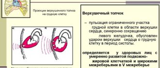
Anatomy
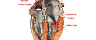
The structure of the left atrium, like the right, is similar to an irregular cube.
Chamber walls:
- The anterior one protrudes and forms the left ear, which is adjacent to the pulmonary trunk on the left.
- Rear.
- Upper.
- Internal - takes part in the formation of the interatrial septum. It has a thinner part that corresponds to the oval fossa.
- The lower one forms the basis of the left ventricle.
- Outdoor.
The LA wall is thinner than the right one. The inner surface of the auricle is lined with pectineus muscles, the rest of the atrium is smooth.
Four pulmonary veins drain into the LA (two from each lung):
- Top right.
- Bottom right.
- Top left.
- Bottom left.
They carry arterial blood from the lungs. The openings of these veins are located on the posterior wall of the left atrium and do not have valves.
Function
Main functions of the left atrium:
- Depositing. The chamber is a container that receives blood from the pulmonary veins.
- According to the pressure gradient, it conducts blood into the left ventricle after the mitral valve opens.
- Helps complete filling of the left ventricle through its contraction.
- When the atrium walls stretch, the pressure rises, which stimulates the formation of natriuretic peptide (NUP). The biologically active substance reduces circulating blood volume and blood pressure. It has been proven that NUP prevents the development of cardiac hypertrophy.
- There are many baro- and mechanoreceptors located in the LA. The former respond to an increase in central venous pressure, which in turn leads to the activation of the latter, which contribute to the development of tachycardia (accelerated heartbeat).
Indications for the procedure
For example, athletes are required to undergo an ECG of the heart.
An echocardiogram is a regular test that every person for whom sports is a profession must undergo.
Particular attention is paid to the following sports:
- all types of weightlifting;
- marathon running;
- extreme sports.
Patients who have previously been diagnosed with:
An echocardiogram is also mandatory for pregnant women. A lag in weight gain is an indicator that may indicate congenital organic or functional heart defects in young children. Echocardioscopy can confirm or refute this diagnosis.
In an adult, especially after sixty to seventy years, an echocardiogram of the heart can show serious age-related changes in the structure and functioning of the organ. Carrying out such a procedure once a year allows you to identify the problem and provide timely assistance.
Most adults and adolescents are recommended to undergo cardiac echocardioscopy every one and a half to two years, even if there are no complaints and the previous ECG was normal.
Echocardiography shows that there are deviations from the norm in the structure of the central circulatory organ and that attention should be paid to them.
What diseases can ultrasound of the heart detect?
Diseases that are usually detected after a cardiac echo:
- heart failure;
- tachycardia (acceleration of the heart muscle);
- bradycardia (slowing of the heart muscle);
- pre-infarction condition;
- previous heart attack (damage to the heart muscle);
- inflammatory diseases of the muscles of the heart and pericardial sac;
- angina pectoris in the initial stages of development;
- congenital organic heart defects;
- aneurysm of the aorta, aortic arch, and vessels of the pulmonary trunk.
It is not worthwhile to engage in self-treatment or diagnose yourself based on information from the Internet. Diagnosis should only be carried out by an experienced cardiologist.