With age, many people begin to worry about varicose veins and other vascular diseases of the lower extremities. This is not surprising - after all, the main burden of movement falls on the legs. The higher the load when moving, the more difficult it is for the veins and arteries of the legs. That is why most often the pathological condition of the vessels of the lower extremities is observed in women who prefer high heels (even young ones), as well as in overweight people and those who work “on their feet” - stand for a long time or walk a lot.
In order to assess the condition of the blood vessels in the legs, doctors recommend giving preference to modern ultrasound techniques. This procedure does not cause discomfort and has a high diagnostic value.
In what cases is ultrasound of the lower extremities performed?
Ultrasound examination of blood vessels can be carried out for preventive purposes or in the presence of disturbing symptoms. Early diagnosis allows you to begin timely treatment and avoid complications, incl. life-threatening.
Direct indications for the diagnostic procedure are the following:
- Rapid fatigue of the legs;
- Discomfort and pain when walking for a long time;
- Pronounced vascular network;
- Periodically occurring feeling of numbness;
- Change in skin tone;
- Constantly cold feet;
- Blue fingers;
- Severe causeless itching;
- Increased sensitivity of the skin;
- Convulsive spasms.
Women who have been using hormonal contraceptives for a long time should be especially careful when diagnosing blood vessels - with long-term use, oral contraceptives can cause an increased risk of blood clots. Vigilance is also necessary for the following factors:
- Recent stroke;
- Diabetes;
- Various bad habits;
- Endocrine diseases;
- Weak blood vessels;
- Unbalanced diet, lack of nutrients.
Indications for the procedure
Ultrasound of the leg arteries is performed for diabetes mellitus, atherosclerosis, arterial thrombosis, and after a myocardial infarction. The following symptoms may indicate a deterioration in their condition:
- numbness of the toes or their acquisition of a bluish tint;
- cramps in the calf muscles;
- pain in the legs when walking;
- the appearance of “spider veins”;
- swelling of the legs;
- high blood pressure and cholesterol levels;
- Constantly cold lower extremities (compared to other parts of the body).
Possible contraindications to the study
There are no direct contraindications to ultrasound examination. However, it is worth understanding that the reliability of the results will decrease significantly if the patient moves his legs during the manipulation. Therefore, people with mental disorders, neurological and other pathologies, in which it is difficult to keep the lower limbs motionless, should consult with a doctor about how best to undergo the study - in some cases, taking sedatives may be recommended.
Advantages of the multidisciplinary clinic "Alfa Health Center"
- Accurate result. Our medical center is equipped with modern ultrasound equipment General Electric LOGIQ 8. It allows you to obtain images without interference. The arteries of the lower extremities can be seen very clearly on ultrasound, which, together with the doctor’s experience, makes it possible to make an accurate assessment of their condition.
- A wide range of services is offered by doctors of different profiles. If ultrasound data needs to be supplemented with another diagnostic method, or the diagnosis requires treatment by a specialist, you will immediately be able to receive the necessary services.
- Affordable price. Our clinic has branches in major cities of Russia. Prices for ultrasound of the veins and arteries of the lower extremities in each region always remain affordable.
Do you need preparation for an ultrasound?
Ultrasound examination of the vessels of the lower extremities does not require any serious preparatory measures. But for the information content of the study, it is advisable to adhere to the following recommendations:
If you take any medications (including herbal ones) on an ongoing basis, you must inform your doctor about this. Some of them can affect the condition of blood vessels and pressure. In this case, before the study, it is necessary to limit their intake (unless, of course, these are vital drugs).
From your usual diet, you should exclude foods that can affect the condition of blood vessels and the speed of blood flow. These include tonic drinks, various energy drinks, chocolate, strong coffee and tea.
The day before the upcoming test, you must stop drinking alcohol.
10 hours before the procedure, refrain from smoking.
Before the study, it is necessary to remove hair from the lower extremities.
How does an ultrasound scan of the leg arteries work?
The patient is placed on the couch on his back to the right of the doctor. The stomach and both legs are completely exposed. The ultrasound sensor is lubricated with gel for better contact with the patient's skin. The study begins with a linear sensor from the groin area. The common femoral arteries are assessed. If the blood flow through them is main, further examination of the superficial and deep femoral arteries, popliteal and leg arteries is carried out, for which the patient must be turned onto his stomach. To assess blood flow to the foot, the dorsalis pedis artery and the posterior tibial artery behind the ankle are examined. In case of critical ischemia, the patient may be asked to lower the leg down to improve blood circulation. During the study, coloring of the arteries is noted in the color Doppler mapping mode; in the usual B-mode, the structure of the artery wall is studied, the presence of atherosclerotic plaques and pathological dilations (aneurysms) is noted. The speed of blood flow through the arteries must be measured. To assess the possibility of using saphenous veins for shunts, their diameter, course and patency are determined. After examining the arteries in the legs, it is necessary to examine the condition of the abdominal aorta and iliac arteries. To do this, another (convex) sensor is taken and the doctor removes large vessels through the abdomen. It is necessary to assess the patency, condition of the wall of the aorta and iliac arteries, the speed and nature of blood flow through them.
In our clinic, ultrasound of the arteries of the lower extremities is constantly used during surgical operations. Ultrasound diagnostics is necessary to identify pathological discharges along the shunt when performing femoral-tibial bypass surgery in situ, to assess the resistance of the shunt and the dependence of blood flow through the arteries in the lower leg and foot on the operation of the shunt. Without these studies, it is impossible to adequately assess the success of vascular reconstruction.
Ultrasound algorithm
To begin with, a person is asked to prepare the field being studied, that is, to remove clothes below the waist. The patient lies down on the couch. The lower limbs should be shoulder-width apart. The ultrasound doctor applies a thin layer of a special gel, along which the sensor slides. To increase the information content of the study, the doctor may ask the person to change their body position, for example, lie on their stomach, turn on their side, or stand on their feet. An ultrasonic sensor reads the condition of the vessels and displays an image on the monitor in front of the specialist. If necessary, the ultrasound doctor adjusts the frequency of sound radiation and analyzes the data obtained. The study evaluates the vessels of the popliteal, lesser subcutaneous, sural and fibular regions, as well as the posterior surface of the legs.
The duration of the study does not exceed 20-30 minutes.
How is an ultrasound of the veins of the lower extremities performed?
An ultrasound examination is important if you suspect the presence of serious diseases, such as thrombosis or varicose veins. A specialist will determine with 100% accuracy the area where pathological processes occur (formation of blood clots, slowing of blood flow, expansion and inflammation, damage to the walls).
To perform an ultrasound, you do not need to prepare and shave hair from the area, limit physical activity, follow a diet, etc. The procedure does not take much time and does not require complex and uncomfortable preparation.
Since ultrasound of the veins of the lower extremities is performed to determine the disease, excellent quality equipment is required. The beam must be accurately reflected from the red blood cells. Thanks to this, the doctor sees what is happening in the veins, vessels and arteries. Therefore, in a few minutes you can assess the condition of valves, deep and superficial veins, blood flow speed, etc.
What is duplex scanning?
The best modern diagnostic method is ultrasound scanning, since the quality of the results obtained is maximum. In the West, preference is given to color mapping. It increases the accuracy of the information if you need to track the speed of blood flow towards and away from the sensor (marking is done in red and blue). The method allows you to perform the following tasks:
- Monitor damage and deformation of the venous walls.
- Find out the speed of blood flow in the deep and superficial veins.
- Check the functionality of the valves.
- Assess the dimensions and density of the thrombus (determine the degree of thrombosis).
Classical and minimally invasive operations are always performed with 100% accuracy, as ultrasound of the veins of the lower extremities is performed in combination with duplex scanning.
Before visiting a specialist, be sure to take a shower. There is no need to shave your leg hair or deep clean your skin. It is advisable not to drink alcohol on the eve of an ultrasound examination. The procedure is performed as follows:
- In the office you undress from the waist down (down to your underwear).
- Ultrasound gel is then applied to the legs.
- The specialist checks the condition of the veins, valves, vessels and arteries using a device with a sensor.
- If necessary, the intensity of pressure and the power of the beam are increased to more clearly see the condition of the deep veins.
- Your body position changes—your doctor may ask you to stand up or lie down to see how the blood flows. For more information, you will need to hold your breath (take a deep breath).
Now you know exactly how an ultrasound scan of the veins of the lower extremities is performed. The session lasts a few minutes and you will not experience any discomfort. There is no pain. During the change in ultrasound intensity, you will not feel anything (many people fear pain or discomfort).
Do not forget that only a competent phlebologist with extensive experience can decipher the result. Therefore, immediately after a duplex scan or ultrasound, go to an appointment with a specialist so that he can make a final conclusion.
The First Phlebology Center will determine the condition of the veins of the lower extremities and prescribe an effective course of treatment for you. You can sign up for an examination right now by phone.
Dopplerography of the vessels of the lower extremities
Often, a comprehensive ultrasound examination with Doppler is performed, which helps to assess the speed of blood flow. As a rule, additional manipulation is indicated in the following cases:
- If the slightest blow causes hematomas and extensive bruises;
- With pallor and cyanosis of the skin;
- If your feet are always cold, they freeze even in warm shoes.
The rules for preparing for the study remain the same as for conventional ultrasound of the vessels of the extremities.
Who is indicated for vein ultrasound?
An ultrasound examination is performed if a person has venous disease or symptoms associated with it:
- Large protruding veins are visible under the skin on the lower extremities;
- A characteristic star-shaped vascular pattern appeared;
- Severe swelling is observed in the mornings or evenings;
- After physical activity, your legs hurt, there is heaviness and discomfort;
- Wounds appear on the skin that do not heal for a long time;
- Feet are constantly cold;
- The skin on the legs changes color.
Experts recommend that you undergo an ultrasound examination of the veins in your legs once a year. It is recommended to undergo diagnostics twice a year if you have diabetes or obesity, if you have bad habits, as well as if you are taking combined oral contraceptives or if there have been cases of vascular diseases in your family.
Typically, a referral for an ultrasound is given for existing diseases:
Vein thrombosis
This is a pathological process that is characterized by an increased risk of blood clots.
This disease is dangerous - it can even be fatal. To avoid this, you need to start treatment immediately and be constantly monitored by a doctor. Postthrombophlebitic disease.
It is a complication of thrombosis.
Varicose veins and venous insufficiency.
These diseases are characterized by impaired circulation in the veins.
Varicothrombophlebitis.
It manifests itself in severe pain and, if left untreated, leads to serious complications.
Angiodysplasia.
Loss of vascular functionality. It can only be treated surgically. Ultrasound in this case is necessary to monitor the effectiveness of treatment.
Triplex ultrasound
Today, this method of ultrasound examination of the vessels of the lower extremities is considered the most modern. It allows you to observe a three-dimensional image of the vessel on the screen with detailed visualization. This diagnostic procedure allows us to identify the following pathological processes:
- thrombophlebitis;
- atherosclerosis;
- destruction and structural anomalies of vascular sections;
- angiopathy;
- vasculitis;
- postthrombophlebitic diseases.
The duration of the study is approximately one hour.
Decoding and results
Ultrasound is an affordable imaging method that does not use harmful radiation. During the diagnosis, the doctor determines the thickness of the vascular membranes, pulsation index, and vascular resistance. The minimum and maximum speed of blood movement is also determined.
After conducting the study, the specialist receives accurate data that indicates the condition of the vascular system. It defines a number of important parameters and reflects them in the protocol. Based on such information, the doctor gives an opinion, according to which the vascular surgeon develops treatment tactics.
Using ultrasound examination of the vessels of the lower extremities, various diseases are detected:
- stenosis is a term that refers to the narrowing of blood vessels in the circulatory system. The development of such a pathology leads to undesirable consequences: muscle atrophy, ulcerative formations, pain. Stenosis appears in people who lead a sedentary lifestyle and are overweight. Diabetes mellitus also leads to changes in the structure of blood vessels
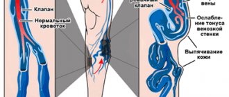
- Varicose veins are a serious disease accompanied by impaired blood flow. The trigger for the development of pathology is considered to be disruption of the venous valves with the subsequent occurrence of blood reflux. The main symptom of the disease is dilated saphenous veins. First, a person is bothered by a feeling of heaviness in the legs and a burning sensation, and then swelling appears. Pregnant women and people who spend a lot of time in one position (office workers, salespeople, drivers, bank tellers) are predisposed to varicose veins.
- Phlebitis is a disease in which the vein wall becomes inflamed. Its main symptoms include soreness, a slight increase in temperature, and redness of the skin. Phlebitis can be chronic or acute. The appearance of the disease is provoked by varicose veins, infection, and mechanical damage to the vessel. When diagnosing phlebitis, the doctor prescribes a set of therapeutic agents
- thrombosis - the formation of blood clots inside blood vessels. This is a pathological process because the free flow of liquid connective tissue through the system is limited. The key signs of the disease are severe swelling, pain and induration. Thrombosis may be asymptomatic. In such cases, the disease is detected by ultrasound. This problem cannot be ignored, because it leads to serious consequences.
Ultrasound of the vessels of the lower extremities is a study with which you can detect the disease in time and prescribe effective treatment. To carry it out, equipment with wide functionality is used. This is a guarantee that the results will be reliable.
ADVANTAGES AND DISADVANTAGES OF THE METHOD
Patients who have been prescribed an ultrasound examination of leg veins may be interested in learning information about the disadvantages and advantages of this method:
- The procedure is informative and allows to identify a wide range of pathological conditions of the vessels of the lower extremities;
- It is completely safe for the body, ultrasound can be performed an unlimited number of times;
- It is not painful and does not even cause discomfort;
- Suitable for people of any age.
- The only disadvantage we can note is the relatively high cost, on average from 1000 rubles.
