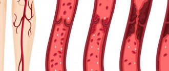
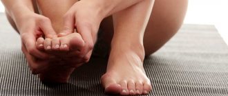
Rheovasography is a method for studying arterial and venous blood flow in the upper and lower extremities using a rheograph apparatus.
The procedure is painless and comfortable and takes 10–15 minutes. Two pairs of electrodes are placed on the limbs - above and below along the blood vessels. The electrical resistance between these areas is measured. The data is processed by a computer program and decrypted by a doctor.
Electrical resistance depends on the degree of blood filling of the vessels, their expansion or narrowing. The stronger the blood flow, the lower this indicator. An increase in resistance indicates difficulty in blood flow. Based on the nature of the diagram, the angiologist judges the characteristics of the arterial supply and venous outflow in the extremities.
Indications for the procedure
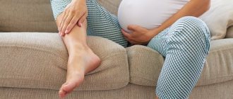
Rheovasography of the lower extremities can be prescribed for the following symptoms:
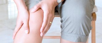
- Numbness of the legs.
- Cramps.
- Change in skin color of the lower extremities.
- Loss of sensation.
- Edema.
This diagnostic procedure is mandatory for diseases such as:
- Atherosclerosis.
- Thrombophlebitis.
- Rheumatic diseases.
- Diabetes.
- Phlebeurysm.
- Blockage of the bloodstream.
- Obliterating endarteritis, etc.
Limitations of the rheovasography method of the lower extremities
Unlike duplex scanning, which does not require any preparation or special conditions, rheovasography of the lower extremities requires compliance with a number of conditions and careful attention to external factors.
The rheovasography schedule of the lower extremities may be inaccurate in the presence of heart failure, vein occlusion (vein obstruction), or local thrombosis. Also, these rheograms are distorted when exposed to external factors: the presence of pressing force (tight bandage or plaster on the leg, tight, tight clothing), low temperature in the office, patient mobility during the examination.
For rheovasography of the legs to be informative, a number of conditions must be met.
- A day or more before, stop taking any vascular medications that can affect blood circulation;
- Two hours before the test, smoking patients should quit nicotine;
- Immediately before rheovasography of the lower extremities, the patient should rest for approximately 15 minutes;
- During vein diagnostics, the patient should remain as still as possible, unless the doctor asks otherwise.
Samples
Interpretation of study results will not be complete unless samples are performed. The purpose of the tests is determined by the need to determine the reserve of the blood flow system.
Two types of samples are used:
- Chemical (other names: nitroglycerin, medicinal). To conduct a drug test, the patient is asked to take a nitroglycerin tablet. After a five-minute wait, rheovasography is performed. The data obtained are compared with the results of a previously performed RVG, when the patient did not take nitroglycerin. The test is necessary to differentiate between functional vascular spasm and organic constriction. If there is an increase in the rheographic index and elasticity index, the test is considered positive, and the violations are classified as functional.
- For diagnostic purposes, the physical (compression) test is considered much more important. To carry it out, a cuff is placed on the patient's thigh, after which air is injected into it. Immediately after removing the cuff, another rheovasography is performed. Attention is paid to the time of complete recovery after compression. Based on the results of a physical test, one can judge the reserves of blood outflow from the veins.
Key Indicators: What They Measure
The RVG results show a curve. Wave interpretation is based on qualitative and quantitative values. These include: the steepness of descents and ascents, intervals, amplitude, peaks.
Based on the values, control indices are derived, which are:
- rheographic - it controls the strength of blood flow in the arteries;
- arterial walls shows IE;
- IPS – peripheral resistance index;
- numerical value of venous outflow.
The most basic indicator is considered to be RI - rheographic index.
Decoding the results
Obtaining research results does not take much time.
Within 30 minutes, a specialist can decipher the received line and display the indices. RVG decoding is output in Ohms. As mentioned above, RI shows the total volume of filling of blood vessels. With readings of 0.05 Ohm and above, we can say that their condition is normal.
With deviations of one unit, mild deficiency is observed. If RI is sharply reduced, the patient is diagnosed with a blood supply disorder.
Standards for vascular tone data without deviations are calculated from 0.4. Values that do not reach the specified units are already considered violations. The lower the indicator, the lower the tone of the vascular wall.
The outflow index shows the speed of blood movement in the venous canal. RVG should range from 0.2 to 0.5. Anything that exceeds this line indicates serious illness.
The IPI responsible for counteracting vascular channels should be in the range of 0.2-0.45. In case of obvious blood flow pathologies, the index may be higher or lower than the norm.
Additional samples
Violations that were recorded during RVG do not always indicate damage.
The resulting index may have a functional nature of deviations. Often such results are obtained when preliminary preparation is not followed. There are two additional samples. They help determine what is really happening, a temporary spasm or pathology:
- Nitroglycerin. Using the drug, the rheovasography procedure must be completed several times. The first occurs before the drug is used, and the second after its action. The doctor receives data from two studies and conducts an analysis. If comparable results do not differ significantly, then we can conclude that there is a pronounced pathology on the face. In the case where the drug had an effect and the indicators improved, one-time disturbances caused are assumed.
- Compression. The essence of the procedure is to use a cuff that compresses the required area of study. As in the case of the RVG drug, the patient undergoes it twice. Before and after applying the cuff to the limbs. This test helps determine how quickly blood supply is restored when the deep veins are blocked.
The latest rheovasography devices have a large number of positive aspects. They greatly simplify the work of specialists. The system itself calculates the results, which is much faster than routine calculations.
At the same time, the patient does not receive any radiation or other negative effects on his body. The method is more than safe and informative. Due to this, this diagnosis is relevant in medical practice.
How is RVG performed?
The procedure is performed at a certain temperature. Cold indoors causes the arteries to narrow, and too high a temperature causes them to widen. The patient is asked to lie on his back. The area to be examined is degreased with alcohol, a pad moistened with a conductive solution of sodium chloride (5-10%) is placed on the skin, 2 metal electrodes are symmetrically attached and secured with rubber bandages.
- When is ultrasound of the vessels of the upper extremities necessary and how is it done?
The mechanism of action of RVG is to pass an electric current through the examined area of the body. The strength of the impulses is minimal and completely safe for humans. The examination of the vessels of the lower extremities is carried out using high-frequency current (10 mA). Passing through the body, electrical impulses encounter various resistances and create voltage fluctuations, which are recorded by sensors and transmitted to a recording device (rheograph). The parameters are reflected on the rheovasogram - a curved line where the rise and fall of the surges show the inflow and outflow of blood.
RVG readings are not the final and only signs of the disease, but they serve as the basis for choosing methods for examining the patient.
A little information about the rheograph
The device is based on a generator that generates current. Using a special attachment, measurements are converted into graphic form.
Using appropriate electrodes attached to certain areas of the body, a rheogram is recorded.
What is rheography? At the beginning of the procedure, an electrophoresis-treated napkin is placed on the patient’s skin, through which the electrode reacts better with the body.
The skin is first wiped with medical alcohol; the presence of a fatty layer worsens the result of the process.
What the procedure will show
Having figured out what the essence of the diagnostic method is, people begin to be interested in how they can independently decipher the result they receive. But without the proper knowledge, this is quite problematic, because for most people, a rheogram is some kind of mathematical graph, and the impedance of biological tissues does not mean anything at all.
The basis of the picture is based on two aspects:
- sinusoids, which are presented as a steep rise, which describes arterial blood flow;
- a smooth descent, which is a reflection of the venous blood flow.
But not only this makes it possible to assess the condition of the vascular network in terms of volumes and other features of the incoming blood. It will be necessary to take into account many curves and use additional calculations, where Kedrov’s formula plays an important role.
The diagnostician will definitely take into account the regularity of the verified curve, which implies similarity between several curves. The shape and the number of secondary curves that are in a descending phase are also taken into account.
For example, it is worth analyzing the indicators characteristic of vegetative-vascular dystonia or arrhythmia. The presented ailments will be indicated by adjacent curves with different shapes.
Next, a special index is calculated, which must fit into the average statistical interval. Falling out of it threatens with suspicion of serious pathologies.
The following values specifically help identify diseases:
- level of venous outflow;
- amplitude-frequency indicator;
- measurements of the time interval for pulse wave propagation.
An experienced doctor will take into account various functional tests in order to get a detailed picture of what is happening.
What is rheovasography? Principle of operation and its types
RVG is one of the newest modern non-invasive diagnostic methods.
It is used to examine the blood vessels of the legs and arms. The technique helps to identify areas of arterial blockage that lead to inflammation. It is also possible to examine the vessels and identify disorders associated with their operation.
Principle of operation
The essence of the method is to detect the resistance of a skin area through which a harmless electric current is passed.
In the sensors attached to the body, a certain force and frequency of charge oscillations are set.
If the blood flow is not intense enough, then the resistance of the skin and tissues becomes higher, and vice versa.
The device’s readings are displayed on the monitor and then on a paper tape. Where all changes are recorded in the form of a curved line.
The process of performing the examination and providing information about the results is similar to an ECG. Based on these data, the doctor makes a diagnosis regarding the area being examined.
Kinds
There are two types of rheovasography:
- Detection of blood flow pathologies in the legs.
- Study of the vascular channels of the hands.
Based on disease statistics, diseases of the lower extremities are the most common, therefore rheovasography of this type is used more often.
Central rheography
Varieties of rheography cover a huge number of proposals. But the central variation is consistently in high demand, as it is aimed at accurately assessing the condition of the heart.
The manipulation is based on the study of blood flow coming from the pulmonary artery and aorta. It is these two large vessels that allow you to familiarize yourself in detail with the health of the cardiac region.
Taking into account the blood flow of the lung and right ventricle, it is possible to evaluate the contractile function of the heart muscle.
Normal values in clinical practice are as follows:
- flat ascending part;
- round top with a small depression;
- round top with wave;
- smooth descent.
Carrying out central rheography involves dividing rheograms into certain types, depending on the data obtained:
- hypervolemic;
- hypovolemic;
- hypertensive.
The first version indicates an increased volume of incoming blood. The main indicators indicating such a deviation cover a high and at the same time peaked curve with a steep downward part.
Hypovolemic conditions are no less dangerous, but not all patients know what it is.
The scenario involves a reduced volume of blood flow, which is visible on visualization due to:
- decreasing the height of the curve;
- serifs on the ascending part;
- flat top;
- gently descending parts.
No less dangerous are the biophysical manifestations of the hypertensive subtype. This will indicate increased pressure recorded in the lungs. In the image, deviations can be recognized due to the steep rise of the curve, as well as the gentle descent and round top.