Arteriography of the lower extremities is a method of visualizing the lumen of the arteries by introducing a radiopaque substance into them through a catheter and simultaneous fluoroscopy (video recording of x-rays), with recording and processing of the resulting image using special equipment. In this case, the specialist receives objective information about the anatomical structure of the vascular bed of the area under study, the speed of blood flow, the presence of stenoses (narrowings) and occlusions (complete blockage), the degree of development of collateral circulation in the limb under study.
Based on this information, a council of doctors sets indications for surgery and determines the stages of treatment; taking into account the entire complex of preoperative examination, the choice of revascularization method is made (open bypass, endovascular or hybrid intervention).
Treatment under the compulsory medical insurance policy is possible!
Submit your application
Follow the news, subscribe to our social networks
Details
Advantages of angiography at the Innovative Vascular Center
Arteriography of the lower extremities is performed on a stationary Philips Allura Xper FD20 angiographic complex, or on a Veradius mobile angiograph if performed intraoperatively. The use of modern angiographic systems makes it possible to conduct research at any required frame rate, which gives an objective picture of the nature of blood circulation in the limb.
Our clinic uses CO2 angiography without the use of iodine contrast. This method is well suited for patients with iodine intolerance and allows any operation to be performed using the endovascular method.
To reduce the risk of complications, we perform angiography most often through a peripheral radial approach on the arm, which allows the patient to begin walking immediately after the procedure.
What types are there
The area of interest in MRI determines the type of MR angiography. Main areas:
- head;
- neck;
- lower limbs;
- abdomen;
- pericardium;
- upper limbs;
- small pelvis
MRI angiography of the vessels of the brain, neck, and lower extremities is most often prescribed.
A study of the blood supply system of the central nervous system can prevent the development of organic damage to the cerebral substance. MRI of the brain with MR angiography involves studying the lumen of the arteries and determining the functionality of the veins.
A neck scan is performed to assess the patency and fullness of the great vessels.
MRI of the lower extremities with MR angiography is prescribed for damage to the venous network, inflammatory processes, and thrombosis. Early diagnosis of diseases contributes to the effectiveness of treatment measures.
Angiography of cerebral vessels
Acute and chronic insufficiency of cerebral blood supply is manifested by decreased performance, inhibition of certain functions, and deterioration of well-being. In the absence of treatment, an increase in symptoms is observed:
- cephalgia;
- loss of memory, intelligence;
- paralysis;
- seizures, etc.
Severe disease can lead to disability and death.
MRI angiography of cerebral vessels with contrast is prescribed for diagnostic and preventive purposes. Patients at risk for cerebrovascular pathologies are recommended to undergo regular tomographic examinations.
MRI of the head with vascular angiography allows you to diagnose:
- arteriovenous malformations - there is no intermediate capillary network in the junction zone, the surrounding tissues are atrophic, signs of gliosis are observed;
- carotid-cavernous anastomosis - due to the intensive discharge of blood from the internal carotid artery into the sinus cavity, the pressure in the sinus increases, dilation of the ophthalmic vein occurs on the affected side, and pulsating exophthalmos develops (displacement of the organ of vision forward);
- aneurysms of cerebral vessels - there is a uniform increase in the diameter of the artery in a limited area or protrusion of the wall in the form of a bag (diverticulum);
- intracranial atherosclerosis - against the background of degenerative processes, a narrowing of the lumen of the blood channel occurs with subsequent expansion;
- vasculitis of cerebral vessels - the formation of multiple foci of cerebral infarction is possible;
- stenosis, deformation of the main arteries - pathological tortuosity, coiling (loop formation), kinking (curvature at an acute angle).
Brain MRI with vascular MR angiography is used in the diagnosis of strokes. Magnetic resonance imaging is informative already in the first 6 hours after an attack.
MRI angiography of head vessels
Acute disturbance of cerebral circulation can be caused by ischemic and hemorrhagic phenomena. In the first case, changes in the hemodynamics of the arteries are observed on MRI images of the brain with MR angiography. Intracranial hemorrhages occur when the integrity of the vascular wall is disrupted. Venography images reveal single or multiple hematomas.
Angiography of the lower extremities
The venous system of the legs experiences high loads, which is due to the structural features of the human body. Blood rises through the vessels, overcoming the force of gravity. The veins of the lower extremities have a system of valves that prevent reverse flow.
Vascular diseases of the legs are accompanied by swelling, a feeling of heaviness, and impaired motor function. When blood clots form, there is a risk of the latter breaking off and being transported through the arteriovenous network.
MRI angiography of leg vessels
A serious complication is blockage of the pulmonary circulation by an embolus (high risk of death).
MRI angiography of the veins and arteries of the lower extremities allows you to diagnose:
- thrombosis;
- atherosclerosis;
- thrombophlebitis;
- traumatic vascular lesions;
- stenosis, occlusion of the arteries of the lower extremities;
- diabetic foot syndrome.
Timely detection and treatment of thrombosis during venography reduces the risk of life-threatening complications.
Angiography of the neck
The study of the great vessels makes it possible to assess the blood supply to cerebral structures, soft tissues, the thyroid gland, etc. In the diagnosis of diseases of the central nervous system, complex MRI angiography of the neck with examination of the arteries and veins of the brain is often used.
Scanning reveals:
- thrombosis of intracranial venous sinuses;
- causes of cerebrovascular accident;
- stenosis, occlusion, thromboembolism of the main and brachiocephalic vessels;
- aneurysms;
- vasculitis of neck vessels;
- malformations;
- disruption of venous outflow from the cranial cavity, etc.
MRI of the neck with MR angiography (series of layered photos)
The scan results in detailed images of the vasculature and surrounding tissues. MRI of the brain and neck with MR angiography allows you to assess the patency, fullness of the vertebral arteries, the quality of cerebral blood supply, and the functionality of the internal jugular veins with the main tributaries.
How is angiography of the lower extremities performed?
Angiography is performed by a highly qualified x-ray endovascular surgeon in a sterile cath lab and with sterile consumables. Anesthesiological monitoring is carried out throughout the study.
Under local anesthesia, a puncture of the artery of the arm or leg is performed, after which a thin catheter is inserted into its lumen, through which a radiopaque substance is injected. During the examination, the patient may feel warmth as the contrast is injected, and very rarely there may be pain. The duration of the angiography itself is 10-15 minutes.
When performing angiography, the specialist receives objective and accurate information about the structure and damage to the arterial bed of the area under study. The resulting images are saved electronically and can subsequently be written to disk and given to the patient.
Angiography of the arteries of the neck and brain
Two related vascular studies are related by the fact that the carotid (localized in the neck) and vertebral arteries are the main ones for the blood supply to the brain, so the indications for their examination are similar:
- Headache that persists for a long time;
- Dizziness of unknown etiology;
- Severe causeless drowsiness;
- Frequent or prolonged fainting;
- Traumatic injuries to the neck and skull;
- Presence of vascular pathology in the past.
Possible complications of angiography
Complications after angiography are very rare. However, due to the fact that this study is invasive (penetrating through the natural external barriers of the body) and is accompanied by the introduction of a radiocontrast agent into the systemic bloodstream, associated complications may still develop: contrast-induced nephropathy,
- bleeding
- hematomas and false aneurysms at arterial access sites
- local infectious complications
- allergic reactions to contrast
Our specialists do everything possible to prevent these complications, and also know the necessary treatment methods in case of development.
MR angiography with contrast
Magnetic resonance imaging of the circulatory system involves various scanning modes:
- time-of-flight;
- phase contrast;
- MRI angiography of vessels with an intensifier.
Time-of-flight tomography provides T1-weighted images. The liquid medium under study is brightly colored against the background of the surrounding soft tissue.
Phase contrast MR angiography involves receiving a signal from blood moving through the vessels. The method allows you to determine the characteristics of fast arterial and slow venous flow with equal accuracy. The simultaneous use of time-of-flight and phase-contrast MR angiography helps to differentiate between hematomas and other static areas.
Magnetic resonance imaging with enhancement visualizes the bloodstream in the area of interest. A solution of gadolinium salts is used as a contrast agent. The drug has paramagnetic properties and provides a hyperintense signal.
Contrast for vascular MRI angiography is administered intravenously. The injection is given using a catheter connected to an automatic device. This system allows the “coloring” solution to be supplied at a constant speed.
Layer-by-layer images during MRI angiography of cerebral vessels
Data recording begins after the lumen of the bloodstream is filled with contrast. 1-2 ml of solution is pre-injected, which helps to identify the initial moments of the arterial and venous phases. In this way, the signal registration time during vascular MRI angiography is determined. For further measurements, the peak arterial concentration of the gadolinium solution is used.
Angiograms make it possible to study the nature of the blood supply to a certain area. A broken line with a hyperintense signal indicates stenosis and occlusion of the vascular bed. The consequence of the pathology is ischemia and necrosis of the underlying tissues.
MRI angiography of vessels with contrast does not show calcifications, blood clots, or cholesterol plaques. In case of atherosclerosis, embolism, the images show changes in the lumen, fullness, blood channel, and parietal foci with a hypointense signal.
Contrast MR angiography for MRI diagnostics of neoplasms visualizes arteries and veins in the area of interest, shows the condition of surrounding tissues, and helps determine the nature of the disease.
The hemodynamics of the tumor circulatory network depends on the malignancy of the process. The vessels of a malignant tumor have a large number of anastomoses and bends, so the tumor tissue accumulates and releases the contrast agent more slowly.
Subarachnoid hemorrhage on MRI with MR angiography
The final diagnosis is made by the attending physician. The results of MRI angiography of veins and arteries for vascular neoplasms are supplemented with data from clinical tests, biopsy and other research methods.
What will need to be done in the ward
- You should drink plenty of fluids. This is necessary to compensate for the loss of water that you had during preparation for the study and to quickly remove the contrast agent from your body. For most people, this means drinking at least 6 glasses of water, juice or tea. Check the required volume with your attending physician or x-ray surgeon after the study.
- You must strictly adhere to bed rest for 6-8 hours after the examination and keep your leg straight on the side of the puncture site (when using the femoral approach). If using the radial carpal artery approach, you should remain in bed for 1.5 to 2.0 hours, but limit use of your right arm for the next 24 hours.
The puncture site may be slightly numb for a few days.
The next day the pressure bandage will be removed and you can return to your normal lifestyle. You will receive a conclusion about the study and will be able to discuss the results with your attending physician or x-ray surgeon.
When is CTA contraindicated?
Like all radiation techniques, CT scanning has its limitations. Almost all contraindications for angiography are due to the use of contrast. This substance contains iodine, which can negatively affect internal organs and systems. In addition, iodine is excreted from the body in the urine, but is able to penetrate, among other things, into breast milk, so for nursing mothers such a procedure is prescribed only in extreme cases.
CT scan of the vessels of the lower extremities is not performed in the following situations:
- the patient is in serious condition;
- with renal failure;
- if you are allergic to iodine (checked before prescribing);
- the level of keratin in the blood is increased (checked before prescribing);
- during pregnancy;
- with diabetes mellitus in the stage of decompensation.
Women during lactation may require CTA, in which case feeding is stopped for two days after the procedure. Children under 14 years of age are prescribed such a study only in severe cases, usually replacing it with other types. In case of renal failure, such examination is also avoided, since iodine has a negative effect on the kidneys. But if the patient’s life is at risk, the doctor may prescribe a CTA under strict supervision of renal function.
People weighing more than 200 kg will need an open-type tomograph. Sometimes the equipment is designed to weigh no more than 150 kg. When making an appointment by phone or online, it is better to clarify this nuance immediately.
How to behave after an angiographic examination
After a successful angiography, the patient should spend 6 to 24 hours in a lying position (if puncture of the vessels on the thigh was performed). During this time, he is under observation in the intensive care ward or in the general surgery department under the supervision of on-duty medical personnel. During the specified time, the patient must avoid movements in the hip (or shoulder joint, if access was made through the brachial or axillary artery) on the side of the puncture.
The pressure bandage is removed by a doctor, usually the morning of the next day after the procedure. After removing the bandage, the patient can return to his usual mode of activity, provided he feels well, has normal hemodynamic parameters (pulse, blood pressure) and there are no signs of hematoma in the puncture area. The patient can be discharged from the hospital on the same day.
Preparation for the procedure
The study of the circulatory system is a very painstaking process; it can be influenced by a number of factors that distort the data. Therefore, doctors recommend preparing in advance. When writing a referral, the doctor must warn his patient about the prohibition on taking drugs that reduce blood clotting. Two days before your appointment, it is advisable to give up alcohol, as ethyl alcohol sometimes leads to blockage of small capillaries. You need to stop eating 6 hours before; the diagnosis is carried out on an empty stomach. You need to take with you to your appointment:
- referral for the procedure from your doctor;
- pictures and results of all previous diagnostics;
- extract from the outpatient card;
- other documents that may be relevant to the disease being studied;
- plain drinking water (you will need it after diagnosis).
Upon arrival at the radiologist's office, you need to inform him about concomitant diseases and medications that are being taken. So, he must be warned if he has diabetes, pregnancy, an allergy to iodine, or the presence of prostheses and implants in the legs.
When is CTA prescribed?
Due to the fact that the technique is based on radiation, albeit minimal, this procedure is prescribed only by a doctor. That is, you cannot undergo it simply at will; for this you must have a referral from a vascular surgeon or another treating doctor. Indications for such a diagnosis are various disorders in the legs, suspicions of vascular pathology, and patient complaints.
A doctor can write a referral if the patient experiences:
- pain and lameness after physical activity, weakness in the limbs, trembling;
- causeless pain in the leg muscles;
- numbness of the fingers, feet or certain parts of the limbs;
- coldness in the legs, even if you are warm.
Also, if the doctor does not feel pulsation when palpating the lower extremities, this is the reason for CTA. Any suspected vascular blockage requires a CT scan, as it often leads to serious complications such as heart attack.
During the procedure, the radiologist can see various vascular pathologies. Considers the diameter of the vessel, the presence of a lumen or thrombus, and finds areas of narrowing. Three-dimensional images of the patient show inflammatory processes in the vessels and adjacent tissues. Thanks to this, most diseases, even those not related to the circulatory system, can be detected with CTA of the lower extremities.
Such detailed diagnostics are needed before operations, to confirm or refute the diagnosis. If a disease is detected, tomography allows you to monitor the healing process and adjust treatment in a timely manner. Angiography of the legs is prescribed for:
- atherosclerosis;
- varicose veins;
- injury to arteries or veins;
- aneurysm;
- inflammation of the aorta (Takayasu disease);
- malignant and benign vascular tumors;
- chronic venous insufficiency;
- swelling of the legs of unknown nature;
- arterial thrombosis;
- phlebothrombosis and thrombophlebitis;
- angiopathy in diabetes mellitus.
During the course of treatment, the doctor may prescribe one or more tomography procedures. After surgical interventions, they are also sent to CTA, this allows you to review the results of the operation after some time, prescribe treatment or end it.
Description of the CTA method
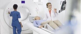
The circulatory system is distributed throughout the body and provides life to every cell. Therefore, failures in its operation quickly affect the human condition. Diagnosing vascular diseases was extremely difficult before the invention of computed tomography. Today, using this method, doctors can examine even small capillaries in detail without making a single incision. The essence of CTA is an X-ray scan of parts of the body. The images created form a 3D model of the circulatory system on a computer monitor.
Content:
- Description of the CTA method
- When is CTA prescribed?
- Where can I get such a diagnosis?
- Preparation for the procedure
- How would this happen
- When is CTA contraindicated?
- Similar diagnostic methods
- Is CTA dangerous?
The innovation of angiography is due to the even greater accuracy of the results than those of a simple X-ray or conventional CT. For clear images, a special amplifier is used - a contrast agent. This is an iodine-based solution that is injected into a vein into the subject. The contrast does not allow X-ray rays to pass through, so in the pictures the circulatory system looks more contrasting, with the smallest nuances.
Unlike other diagnostic techniques, CTA of the vessels of the lower extremities can reduce the proportion of radiation exposure. This technology uses rays collected in a beam, while the exposure time is as short as possible. For examination, only the lower part of the body is placed in the tomograph chamber, which also reduces the level of exposure to X-rays. The procedure consists of several stages: preparation, administration of contrast, obtaining images and deciphering the results.
For the patient, the diagnosis is absolutely painless; the only invasion is the intravenous administration of the drug. The tomography process lasts no more than a minute, then the doctor examines the resulting 3D model in detail and writes a conclusion based on it. The patient receives the results in written and electronic form, from which the attending physician will determine a treatment regimen or further examination.
Where can I get such a diagnosis?
If the patient is treated in a private clinic, a similar service can be provided here. In this case, all that remains is to set a date and complete the session. You can also undergo diagnostics in public medical institutions, despite the fact that a computer tomograph is very expensive equipment. If a doctor from a regular hospital or clinic has prescribed CTA of the vessels of the lower extremities, you need to clarify where exactly the test can be done, perhaps it will be a diagnostic center.
You can do this without leaving your home using the Internet. Such private medical centers always have websites describing their services. Sometimes the cost is indicated here and there is the possibility of online registration. The price for one procedure ranges from 100-200 dollars, depending on the clinic and the tomograph itself.
Indications for angiography of cardiac vessels (coronary angiography):
- angina pectoris - if taking more than three medications does not provide an adequate effect,
- previous myocardial infarction,
- angina pectoris above functional class 2,
- unstable angina,
- new-onset angina,
- progressive angina,
- variant angina (Prinzmetal's angina),
- early post-infarction angina,
- painless myocardial ischemia according to ECG, daily ECG monitoring, stress tests (ischemic depression of the ST segment more than 2 mm),
- atypical chest pain - when ECG or radioisotope research methods give reason to suspect ischemic heart disease,
- ventricular rhythm disturbances that occur during physical activity.
Highly professional and friendly specialists of the Clinic of High Medical Technologies named after. N.I. Pirogov will provide you and your loved ones with the most effective, safe and comfortable methods of diagnosis and treatment of a wide range of diseases.
Conditions in which angiography is contraindicated
Contraindications can be absolute and relative. The doctor makes the final decision based on examination of the patient. If diagnosis is necessary, all risks are assessed, additional preparation is carried out and a decision is made in favor of manipulation. It is important to realize that the benefit of diagnosis can be much greater than the potential risk for a particular condition.
There are no absolute contraindications to cardiac angiography.
The procedure cannot be performed on patients in a decompensated state, when entire organ systems are unable to perform their function. Contraindication is intolerance to the contrast agent. A condition in which angiography is not recommended is pregnancy.


