A clinical blood test is a broad and informative diagnostic method that shows changes in blood plasma and formed elements (erythrocytes, platelets, leukocytes). However, the method is closely related to other diagnostic criteria and is not specific to a particular disease, with a few exceptions. A blood test for cancer can have varying degrees of diagnosis verification - from almost 100% for hematological malignancies to almost zero for solid tumors.
This is due to the fact that oncology can affect absolutely any tissue or organ, and, for example, with a disease of the hematopoietic system, a laboratory technician can see in the analysis the presence of specific altered cells, and with damage to a parenchymal organ (liver, lungs) - only general changes.
In this case, more in-depth tests will be required, which, in addition to the blood count, show changes in biochemical agents. And the newest technological methods also determine the antigenic composition of cells, the presence of specific cancer antibodies and other chemical markers (tumor markers).
Analysis of urine
Cancer of the urinary system manifests itself as blood in the urine.
Urine may also contain ketone bodies, which indicate tissue breakdown. However, these symptoms also accompany diseases not related to oncology, for example, they indicate the presence of stones in the bladder or kidneys, or diabetes mellitus. For diagnosing other types of cancer, urine analysis is not informative. It cannot be used to judge the presence of cancer, but deviations from the norm indicate health problems. If the deviations are serious and confirmed by the results of other basic tests, then this is a reason to conduct special tests to determine cancer.
The exception is multiple myeloma, in which specific Bence Jones protein is determined in the urine.
For the study, morning urine is collected in a sterile container, which can be purchased at a pharmacy. You need to take a shower first.
What does ESR show?
ESR is not a specific indicator for any particular disease, that is, it is impossible to establish a specific diagnosis based on its increase.
This test is considered useful for identifying hidden forms of various diseases and determining the activity of chronic inflammatory conditions. ESR can also serve as an indicator of the effectiveness of therapy.
However, measuring ESR is in no way used to diagnose cancer.
Stool analysis
Blood may also be present in the stool, and it is almost impossible to notice it visually. Laboratory analysis will help determine its presence.
The presence of blood in the stool is a sign of intestinal cancer (most often colon), but it is also a symptom of many benign gastrointestinal diseases. Polyps in the intestines can bleed. Moreover, it should be remembered that polyps tend to degenerate into a malignant tumor. In any case, the presence of blood in the stool is a reason to undergo a more in-depth diagnosis and take tests to detect cancer.
Feces are also collected in a sterile container in the morning.
What blood test shows cancer?
Many patients are convinced that it is possible to detect cancer using a blood test. In fact, there are several types of this diagnostic procedure, starting with a general analysis and ending with an analysis for tumor markers. The following types of cancer diagnosis using blood tests with varying degrees of information content are distinguished:
- general analysis;
- biochemical analysis;
- blood clotting test;
- immunological blood test (for tumor markers).
Even if cancer has not yet manifested itself with painful symptoms, negative changes are already occurring in the body, which can be recorded by a blood test. When a malignant tumor grows, it destroys healthy cells that help the body grow and releases toxic substances. These changes are noticeable even with a general blood test, but they can also be a sign of dozens of diseases not related to cancer.
The most informative is considered to be an analysis of tumor markers - specific substances that are released into the blood as a result of the vital activity of tumor cells. However, given that tumor markers are contained in the body of any person, and their number increases during inflammation, this analysis does not 100% prove the presence of cancer. It only becomes a reason to undergo more reliable tests to determine oncology.
ESR indicators in cancer diseases
How much does the blood sedimentation rate increase in tumor diseases? When neoplasms form, a sharp (up to 70-80 mm/hour) increase in value is observed.
But such a reaction of the body can also occur in the presence of inflammatory processes and other diseases. Thus, exceeding normal values is not a direct sign of a cancer diagnosis.
If the sedimentation index value changes, the patient is sent for a full examination to determine the true reason for the increase or decrease in the value.
Causes of excessive erythrocyte sedimentation - neoplasms:
- breast;
- Cervix;
- ovaries;
- Bone marrow;
- lymph node.
In rare cases, other forms of cancer may be detected and require careful investigation. The sedimentation index in lung cancer may be characterized by an increase in normal values, but if the morphological appearance of leukocytes changes, the disease may not manifest itself for a long time.
Erythrocyte sedimentation time is the main indicator of the presence of various changes occurring in the body. However, each time the normal values change, additional diagnostics must be performed.
In some cases, cancer settling time can be shortened. This may be due to an increase in white blood cells or bile salts during breastfeeding. When the development of cancer cells provokes an increase in the number of white blood cells, the pathology produces two completely opposite effects that compensate for each other. Therefore, the increase in CRP in cancer occurs more slowly.
Will a general blood test show cancer?
This analysis does not provide complete information about the presence of a tumor in the body. However, this is one of the basic tests that helps detect cancer at an early stage, when it does not yet manifest symptoms. Therefore, if you decide which tests to take to check for cancer, then you need to start with it.
The following changes in the structure of the blood may indicate malignant processes in the body:
- decrease in the number of lymphocytes;
- increase or decrease in the number of leukocytes;
- decrease in hemoglobin;
- low platelets;
- increased erythrocyte sedimentation rate (ESR);
- increase in the number of neutrophils;
- presence of immature blood cells.
If a patient, in the presence of one or several of the listed signs at the same time, experiences weakness, quickly gets tired, loses appetite and weight, it is necessary to undergo a more detailed examination.
Blood is donated on an empty stomach or at least 4 hours after eating. The sampling is carried out using a finger.
How to calculate the individual ESR norm in elderly patients?
The easiest way is to use Miller's formula:
For example, the permissible ESR limit for a 60-year-old woman is: (60 years + 10): 2 = 35 mm/hour
When changes are detected in a clinical blood test, the first thing the patient does is go to a general practitioner. A useful point is that ESR is included in the Clinical Blood Test, which means that the doctor simultaneously sees the level of leukocytes, platelets, and hemoglobin. When making a diagnosis, the doctor first chooses between three groups: infections, immune diseases and conditions, and malignant diseases. The doctor interviews and examines the patient, after which, based on symptoms, examination and diagnostic data, he determines further tactics.
If the reason for the increase in ESR has not been identified, the analysis should be repeated after 1-3 months. Normalization of the indicator is observed in almost 80% of cases.
Blood chemistry
The method identifies abnormalities that may be a sign of cancer. It should be taken into account that the same changes are characteristic of many non-oncological diseases, so the results cannot be interpreted unambiguously.
The doctor analyzes the following indicators:
- Total protein. Cancer cells feed on protein, and if the patient has no appetite, then its volume is significantly reduced. In some cancers, the volume of protein, on the contrary, increases.
- Urea, creatinine. Their increase is a sign of poor kidney function or intoxication, in which protein in the body is actively breaking down.
- Sugar. Many malignant tumors (sarcoma, cancer of the lung, liver, uterus, breast) are accompanied by signs of diabetes mellitus with changes in blood sugar levels, since the body does not produce insulin well.
- Bilirubin. An increase in its volume may be a symptom of malignant liver damage.
- Enzymes ALT, AST. Increased volume is evidence of a possible liver tumor.
- Alkaline phosphatase. Another enzyme, an increase in which may be a sign of malignant changes in bones and bone tissue, gall bladder, liver, ovaries, and uterus.
- Cholesterol. With a significant decrease in volume, liver cancer or metastases to this organ may be suspected.
Blood is drawn from a vein. It must be taken on an empty stomach.
Immunological blood test: tumor markers
If we talk about what tests show oncology, then this examination is quite informative and allows you to determine the presence of cancer. It is also used to detect relapses after treatment.
Tumor markers are special types of protein, enzymes, or protein breakdown products. They are released either by malignant tissue or by healthy tissue in response to cancer cells. Now the existence of more than 200 species has been scientifically proven.
Tumor markers are also present in small quantities in the body of a healthy person; their volume increases moderately, for example, with a cold, as well as in women during pregnancy, and in men with prostate adenoma. However, the appearance of certain specific types in large quantities is characteristic of certain tumors. For example, tumor markers CEA and CA-15-3 can signal breast cancer, and CA 125 and HE-4 can signal ovarian cancer. To obtain the most objective result, it is recommended to be tested for several tumor markers.
By increasing the level of a particular tumor marker, it is possible to determine which organ or system is affected by the tumor. Also, this analysis can show that a person is at risk of developing cancer. For example, in men, an increase in the PSA tumor marker becomes a precursor to prostate cancer.
An immunological test is taken on an empty stomach, blood is taken from a vein. Tumor markers are also determined by urine analysis.
Cytological examination
This is the most informative type of laboratory examination, which accurately determines the presence or absence of malignant cells.
The analysis consists of taking a tiny section of tissue in which the presence of a cancerous tumor is suspected, with further examination under a microscope. Modern endoscopic technologies make it possible to collect biomaterial from any organ - skin, liver, lungs, bone marrow, lymph nodes.
Cytology is the study of cellular structure and function. The cells of a cancerous tumor differ significantly from the cells of healthy tissues, so laboratory testing can accurately determine the malignancy of the neoplasm.
The following biomaterials are used for cytological examination:
- imprints from the skin, mucous membranes;
- liquids in the form of urine, sputum;
- swabs from internal organs obtained during endoscopy;
- tissue samples obtained by puncture with a thin needle.
This diagnostic method is used for preventive examinations, clarifying the diagnosis, planning and monitoring treatment, and identifying relapses. It is simple, safe for the patient, and results can be obtained within 24 hours.
Instrumental diagnostics
If a cancer is suspected or a malignant neoplasm is detected, the patient must undergo more detailed examinations to determine the location of the tumor, its volume, the extent of damage to other organs and systems (the presence of metastases), and also to develop an effective treatment program. For this purpose, a complex of instrumental examinations is used. It includes various types of diagnostics, depending on the suspicion of a particular disease.
Modern clinics offer the following types of instrumental examinations:
- magnetic resonance imaging (with or without contrast agent);
- computed tomography (with and without the use of X-ray contrast agent);
- plain radiography in frontal and lateral projection;
- contrast radiography (irrigography, hysterosalpingography);
- ultrasound examination with Dopplerography;
- endoscopic examination (fibrogastroscopy, colonoscopy, bronchoscopy);
- radionuclide diagnostics (scintigraphy and positron emission tomography combined with computed tomography).
These types of examinations make it possible to detect cancer with high accuracy.
Erythrocyte sedimentation rate (ESR)
A test that evaluates the rate of separation of blood into plasma and red blood cells. The rate of separation is mainly determined by the degree of their aggregation, i.e., the ability to stick together.
Synonyms Russian
Erythrocyte sedimentation reaction, ROE, ESR.
English synonyms
Erythrocyte sedimentation rate, Sed rate, Sedimentation rate, Westergren sedimentation rate.
Research method
Capillary photometry method.
Units
Mm/h (millimeters per hour).
What biomaterial can be used for research?
Venous, capillary blood.
How to properly prepare for research?
- Eliminate alcohol from your diet for 24 hours before the test.
- Do not eat for 2-3 hours before the test (you can drink clean still water).
- Stop taking medications 24 hours before the test (in consultation with your doctor).
- Avoid physical and emotional stress for 30 minutes before the test.
- Do not smoke for 30 minutes before the test.
General information about the study
Determination of erythrocyte sedimentation rate (ESR) is an indirect method for identifying inflammatory, autoimmune or oncological diseases. It is performed on a sample of venous or capillary blood, to which a substance has been added that allows it not to clot (anticoagulant). When analyzing ESR using the Panchenkov method, blood is placed in a thin glass or plastic tube and monitored for an hour. At this time, erythrocytes (red blood cells), as having a large specific gravity, settle, leaving a column of transparent plasma above them. The ESR is calculated based on the distance from the upper boundary of the plasma to the red blood cells. Normally, red blood cells settle slowly, leaving very little pure plasma. For this method, a Panchenkov apparatus is used, consisting of a tripod and capillary pipettes with a 100 mm scale.
For capillary photometry (automatic analyzers ROLLER, TEST1), the kinetic “stopped jet” method is used. At the beginning of the ESR analysis, programmed mixing of the sample occurs in order to disaggregate the red blood cells. Ineffective disaggregation or the presence of microclots can affect the final result, since the analyzer actually measures the kinetics of red blood cell aggregation. In this case, the measurement occurs in the range from 2 to 120 mm/h. The results of measuring ESR by this method have a high correlation with the Westergren method, which is the reference method for determining ESR in the blood, and have identical reference values.
The results obtained using the capillary photometry method in the region of normal values coincide with the results obtained when determining ESR using the Panchenkov method. However, the capillary photometry method is more sensitive to an increase in ESR, and the results in the zone of increased values are higher than the results obtained by the Panchenkov method.
An increase in the level of pathological proteins found in the liquid part of the blood, as well as some other proteins (the so-called acute-phase proteins that appear during inflammation) contributes to the “gluing” of red blood cells. Because of this, they settle faster and the ESR increases. It turns out that any acute or chronic inflammation can lead to an increase in ESR in the blood.
The fewer red blood cells, the faster they settle, which is why women have a higher ESR than men. The ESR rate varies depending on gender and age.
What is the research used for?
- For the diagnosis of diseases associated with acute or chronic inflammation, including infections, cancer and autoimmune diseases. Determining ESR is sensitive, but one of the least specific laboratory tests, since an increase in ESR in the blood itself does not allow determining the source of inflammation, in addition, it can occur not only due to inflammation. That is why analysis of ESR is usually used in combination with other studies.
When is the study scheduled?
- When conducting diagnostics and monitoring: inflammatory diseases,
- infectious diseases,
- oncological diseases,
- autoimmune diseases.
What do the results mean?
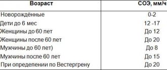
Reference values (ESR norm - table)
| Floor | Age | Reference values |
| up to 15 years | 2 - 20 mm/h | |
| Male | from 15 to 50 years | 2 – 15 mm/h |
| over 50 years old | 2 - 20 mm/h | |
| Female | up to 50 years | 2 - 20 mm/h |
| over 50 years old | 2 – 30 mm/h |
The results of this test must be interpreted in light of clinical data, medical history, and other tests.
Reasons for increased ESR in the blood
- Infectious diseases (usually bacterial causes). ESR can increase in both acute and chronic infectious diseases.
- Inflammatory diseases.
- Connective tissue diseases (rheumatoid arthritis, systemic lupus erythematosus, systemic scleroderma, vasculitis).
- Inflammatory bowel diseases (Crohn's disease, ulcerative colitis).
- Oncological diseases: Myeloma. As a rule, it is accompanied by a very high level of ESR in the blood, because with it pathological proteins are synthesized in large quantities, which cause the formation of erythrocyte “coin columns”.
- Hodgkin's disease is a malignant disease of the lymph nodes. The ESR indicator is usually used not to make a diagnosis, but to monitor the course and effectiveness of treatment of an already diagnosed disease.
- Cancer of various localizations, especially hemoblastosis. It is believed that an extremely high level of ESR in the blood indicates the spread of the tumor beyond the primary site (i.e., metastases).
- Myocardial infarction. When it occurs, damage to the heart muscle occurs, which causes a systemic inflammatory response and, accordingly, an increase in ESR. After a heart attack, the ESR peaks about a week later.
- Anemia. A decrease in the number of red blood cells can lead to an increase in their sedimentation rate.
- Burns, injuries.
- Amyloidosis is a disease associated with the accumulation of pathological protein in tissues.
Reasons for decreased ESR in the blood
- Diseases accompanied by changes in the shape of red blood cells, such as sickle cell anemia or hereditary spherocytosis (they make it difficult for red blood cells to settle).
- Polycythemia (increased number of red blood cells) and conditions that lead to it, such as, for example, chronic heart failure or lung diseases.
What can influence the result?
- ESR in women usually increases during pregnancy and menstruation.
- Oral contraceptives, theophylline, and vitamin A may increase ESR.
- Taking corticosteroids and administering albumin can reduce ESR in the blood.
Important Notes
- ESR analysis is one of the least specific laboratory tests - an increase in ESR in the blood can be observed in many diseases, and a diagnosis cannot be made based on this indicator alone.
- Sometimes high ESR can be observed in healthy people.
- A normal ESR level does not exclude cancer or connective tissue diseases.
Also recommended
- C-reactive protein (quantitative)
- Rheumatoid factor
- Complete blood count (without leukocyte formula and ESR)
- Leukocyte formula
Who orders the study?
Therapist, oncologist, hematologist, infectious disease specialist.