Published: 01/16/2018 Updated: 04/27/2021
An abundance of fatty foods and alcohol does not have the best effect on the functioning of the pancreas. But our well-being depends on how functionally stable it is.
In order not to provoke a pancreatic attack, it is important to remember the factors that negatively affect the condition of the pancreas. And if pain develops, immediately undergo an examination to diagnose pancreatitis.
Diagnosis of pancreatitis
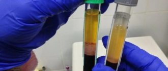
Diagnosis of pancreatitis should begin with determining the main parameters of inflammation in the pancreas. To do this, you need to donate blood, urine and feces to CITILAB:
- 21-20-007 - Alpha amylase. The pancreatic enzyme increases in the blood during acute pancreatitis and necrosis of the pancreas.
- 21-20-009 - Lipase. The amount of enzyme that breaks down fats also increases sharply during the inflammatory process.
- 84-84-005 - Pancreatic elastase in feces. A decrease in the level of this enzyme indicates gland dysfunction.
Urine tests are necessary because the level of enzymes during inflammation increases in the urine an order of magnitude higher than in the blood:
- 21-85-007 - Alpha amylase in urine.
- 21-85-008 — Pancreatic alpha-amylase in urine.
In case of chronic pancreatitis, in order to reduce the likelihood of exacerbation, it is necessary to follow a special diet: give up alcohol, fatty foods, sweets and fast food, and also quit smoking.
Be healthy!
Author:
Baktyshev Alexey Ilyich, General Practitioner (family doctor), Ultrasound Doctor, Chief Physician
Conducting a survey
Diagnosis of pancreatic diseases is one of the difficult tasks of modern medicine. The diagnosis can be established on the basis of anamnesis, clinical pathology, and data from instrumental examination methods.
- Collection of complaints and medical history. Particular attention should be paid to complaints of abdominal pain, nausea, belching, thirst, sudden weight loss, a history of cholelithiasis, a family history of diabetes, etc.
- Inspection. It is necessary to pay attention to the patient’s constitution and skin color.
- Palpation. Normally, the organ is not palpable. When palpated, cysts and swellings can be detected if they reach large sizes.
- Laboratory research. Among the laboratory tests that are important in the diagnosis of pancreatic diseases, the following should be noted:
- examination of duodenal contents (allows you to determine the amount of enzymes);
- examination of stool (color, presence of undigested muscle fibers, etc. matter);
- blood and urine tests.
- Ultrasonography. This is the most accessible and painless method that allows you to conduct an informative examination. Using ultrasound, it is possible to determine the size of an organ, evaluate its structure, identify areas of increased or decreased echogenicity, and determine the diameter of the Wirsung duct.
- CT scan. It is a modern high-tech diagnostic method that is highly informative and allows one to identify pathology in cases where other studies do not produce results.
- The method also allows you to determine the shape and size of the organ, evaluate its structure, identify the presence of even small formations, and also determine the condition of the ducts.
- Endoscopic cholangiopancreatography. This method of radiological diagnosis of pancreatic diseases combines endoscopy and x-ray examination, allows you to study the condition of the ducts, identify the presence of stones and strictures.
- Biopsy. More often, fine-needle aspiration biopsy is used under ultrasound or CT control; it involves taking a piece of organ tissue and then performing a histological examination. Allows you to establish an accurate morphological diagnosis.
The listed examination methods make it possible to determine the anatomical and histological features of tissues and identify dysfunctions, therefore they are used for the differential diagnosis of pancreatic pathologies, allowing the detection of pathology in the early stages.
You can get advice and make an appointment (24 hours a day) by calling:
+7
What pathologies does ultrasound detect?
Using this procedure, you can evaluate changes in the size and contours of the organ, the condition of the ducts, and also diagnose a number of dangerous pathologies:
- acute, chronic pancreatitis;
- congenital pancreatic anomalies;
- cyst, malignant (cancer) and benign neoplasms;
- various inflammations, abscess (purulent inflammation);
- diabetes mellitus, tissue changes caused by diabetes mellitus, pancreatic lipomatosis.
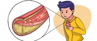
What to do during an attack of pancreatitis
If there is an attack of pain, you must call an ambulance. Symptoms of pancreatitis are similar to appendicitis and heart attack, so it is important not to self-medicate. When waiting for an ambulance, you need to follow simple rules:
- Do not take painkillers. They can hide the symptoms and it will be difficult for the doctor to determine the real picture.
- Lean forward or straighten your back. With these positions, the pain should decrease slightly.
- Do not eat or drink anything until the ambulance arrives.
- No smoking. Nicotine can trigger an attack of pain.
- You need to put cold on the upper abdomen. This will help reduce inflammation a little.
- It is necessary to provide the patient with complete rest: without sudden movements that can cause pain and a new attack of vomiting.
Number for calling an ambulance in Russia: 103. Single number for emergency services (including the Ministry of Emergency Situations, police, traffic police): 112 (works when the phone account is blocked, in the absence of a SIM card).
Pancreatic necrosis.
Pancreatic necrosis is a severe form of pancreatitis, which results in changes in the parenchyma of the pancreas that are degenerative and destructive in nature.
Pancreatic necrosis is more common at a young age, women are most susceptible to it.
Causes of pancreatic necrosis.
Among the causes of pancreatic necrosis, gastroenterologists most often name cholelithiasis, cholecystitis, unhealthy diet, complications after surgery or injury, abuse of alcohol, nicotine, drugs, infections, viruses.
Symptoms of pancreatic necrosis.
Symptoms of pancreatic necrosis are pain in the abdominal area.
Repeated vomiting, attacks of nausea, constant dry mouth, skin pigmentation, attacks of shortness of breath, even at rest, may also indicate pancreatic necrosis; feeling of weakness, mental restlessness and tension.
Methods for diagnosing pancreatic necrosis.
To diagnose pancreatic necrosis, the same studies and tests are used as for pancreatitis.
General rules for preparing for analysis
Before you take pancreatic tests, you need to know how to do it correctly. Doctors usually instruct patients because errors in collecting biological material can lead to significant deviations in the results obtained.
General recommendations come down to several points:
- Studies are carried out on an empty stomach, in the first half of the day. A few days before the tests, you should avoid junk food (fried, spicy, fatty, salty, canned food, coffee, alcohol, carbonated drinks). It is also not recommended to consume legumes that can cause increased gas formation;
- Before taking blood, you must refrain from smoking for at least two hours;
- For problems such as constipation, care should be taken to ensure that toxins retained in the intestines do not affect the test result;
- All containers must be sterile and hands must be thoroughly washed with soap;
- When collecting urine, women must perform genital hygiene, after which it is better to use a tampon to guarantee the purity of the collected material;
- To study a general urine test, you need to take an average portion.
These simple recommendations will help you get tested correctly and avoid possible false results. However, it is worth remembering that sometimes laboratories make mistakes, so if you have the slightest doubt, you should undergo the examination again.
How the research is carried out
Ultrasound of the pancreas is performed using two methods - classical and endoscopic. The first technique is the standard and is prescribed first. Endoscopic ultrasound is required to confirm a questionable diagnosis and in difficult cases.
Classic examination of the pancreas
The method of performing an ultrasound of the pancreas usually takes place through the abdominal wall using a special external sensor. The patient lies down in clothes (without shoes) on the couch with his back, exposing his stomach. The doctor applies a hypoallergenic ultrasound gel, which ensures maximum contact with the device, and then, slowly moving the probe from the central part of the abdomen to the left hypochondrium, examines the pancreas. During imaging, the patient must take a deep breath. The doctor will suggest holding your breath (inflating your stomach) so that the intestines move and nothing interferes with the examination of the pancreas.
To clarify questionable results, the doctor may ask the patient to change his body position (lie on his side or stomach, stand up) and conduct a repeat study. It is possible to obtain erroneous results due to the accumulation of gases in the intestines. To eliminate this problem, the patient needs to drink 2-3 glasses of water. The liquid will act as a “window” and allow you to examine the organs.
The procedure is absolutely painless, the patient does not experience any discomfort or discomfort. The duration is no more than 10-15 minutes.
Endoscopic ultrasound
In some cases, endoscopic ultrasound examination of the pancreas is used to examine inaccessible areas and reduce errors. This option is invasive and does not feel very pleasant. Visualization is performed using a thin flexible endoscope (device) with a video camera and an ultrasound sensor.
The probe is carefully inserted through the esophagus into the stomach and through it into the duodenum. To alleviate the patient’s nervous condition, 30-60 minutes before the procedure, he is given an intramuscular injection of a sedative. EndoUS is performed under anesthesia (local).
Sources
- Acute pancreatitis (Diagnosis and treatment protocols). Authors: S. F. Bagnenko, A. D. Tolstoy, V. B. Krasnorogov, A. A. Kurygin, M. V. Grinev, V. N. Lapshin, V. R. Goltsov. St. Petersburg Research Institute of Emergency Medicine named after. I. I. Dzhanelidze (director - Professor S. F. Bagnenko).
- Clinic of High Medical Technologies named after. N. I. Pirogova St. Petersburg State University. Chronic pancreatitis.
- MSD Handbook. Acute pancreatitis.
- St. Petersburg State Medical University named after Academician I. P. Pavlov. Department of Faculty Therapy, author: Honored Doctor of the Russian Federation, Associate Professor E. V. Kraevsky. Chronic pancreatitis.
Pathological indicators: deviations from the norm visible on ultrasound
| Pathology, temporary changes, disease | Signs on ultrasound |
| Acute pancreatitis | The pancreas is larger than normal (or its individual parts are enlarged); blurry, uneven outline; heterogeneous structure (mainly hypoechoic); The duct of Wirsung is dilated; accumulation of fluid around an organ. |
| Chronic pancreatitis | uneven, blurred contour of the gland; heterogeneous, enhanced structure (hyperechoic); The Wirsung duct is dilated (more than 2 mm); stones are possible - round hyperechoic formations with an echogenic path behind. |
| Cyst or abscess | Echo-negative (black on images) formation with clear, smooth hyperechoic edges |
| Tumor | the part in which the tumor is located is enlarged; the structure is heterogeneous (hypoechoic, hyperechoic or mixed); dilated pancreatic and bile duct. |
| Diabetes mellitus or pancreatic lipomatosis | enhanced echogenic structure; fuzzy, blurry, uneven contour of the organ. |
| Duplication of the pancreas | 2 pancreatic ducts; the isoechoic structure appears uneven. |
| Annular pancreas | The segment surrounding the duodenum is enlarged |
| Metastases | one or more round, hypoechoic (not responsive to ultrasound waves) formations |
Contraindications
In general, ultrasound examination has no contraindications, but there are factors that make the procedure difficult or impractical.
Ultrasound of the pancreas is not performed if:
- allergic reaction to the gel;
- general serious condition of the patient;
- high degree of obesity - the organ is difficult to examine due to the thickness of the fat;
- damage to the skin of the abdominal cavity (wounds, infectious and inflammatory pathologies, fistulas, skin lesions due to systemic diseases).
Contraindications to endoscopic ultrasound:
- bleeding disorders;
- poor patency of hollow organs;
- some diseases of the respiratory and cardiovascular systems (acute myocardial infarction, stroke, bronchial asthma, etc.);
- patient's state of shock;
- burns of the esophagus;
- acute circulatory disorders;
- acute perforated ulcer;
- nodular goiter in the 4th stage;
- upper cervical spine injury.
In each case, for certain pathologies, the doctor determines the possibility of performing an ultrasound on an individual basis.
DIAGNOSTICS OF PANCREAS FUNCTION
LIST OF STUDIES:
- Insulin (IRI)
- Autoantibodies to insulin A-IAA
- C-peptide
- Leptin (eating behavior hormone)
- Glycosylated hemoglobin (HbA1C)
- Cholesterol
- Cholesterol-HDL
- LDL cholesterol
Leptin
Hormone that regulates energy metabolism and body weight
Leptin is a peptide hormone that is secreted by fat cells and is thought to be involved in the regulation of body energy metabolism and body weight. It reduces appetite, increases energy expenditure, alters fat and glucose metabolism, and neuroendocrine function either by direct influence or by activation of specific structures in the central nervous system.
The level of leptin in the blood increases with increasing obesity and decreases with a decrease in the amount of adipose tissue. Normally, an increase in leptin levels suppresses the secretion of neuropeptide Y in the hypothalamus, which is involved in the formation of hunger, and stimulates the activity of the sympathetic nervous system. A decrease in leptin levels after significant weight loss causes an increase in appetite and subsequent weight (body weight) restoration.
Changes in leptin levels are associated with the mechanisms of development of amenorrhea caused by anorexia nervosa, bulimia nervosa, as well as excessive physical activity in female athletes. In these situations, leptin levels are reduced.
It is assumed that leptin concentration plays the role of a physiological signal about the sufficiency of the body's energy resources to perform the reproductive function and affects steroidogenesis in the ovaries. During puberty, the concentration of leptin in the blood increases.
Genetic deficiency of leptin (the synthesis of which is associated with the ob-gene - the obesity gene) in rare cases of hereditary leptin deficiency in humans causes morbid obesity, which can be treated with the use of exogenous leptin.
In other cases, obese people are characterized, on the contrary, by an increase in leptin concentration, which is not accompanied by a corresponding change in eating behavior and energy metabolism. Presumably, this is due to “leptin resistance,” which is associated with impaired transport of the hormone by transport proteins or soluble leptin receptors. Currently, it is considered as one of the factors in the pathogenesis of non-insulin-dependent diabetes mellitus. Excess leptin leads to suppression of insulin secretion, causes resistance of skeletal muscles and adipose tissue to its effects, suppresses the effect of insulin on liver cells, which leads to an even greater increase in glucose levels in type II diabetes.
However, obesity itself does not lead to diabetes with normal pancreatic function.
In addition, high leptin levels create a high likelihood of thrombosis. Research shows that a blood clot begins to form as a result of a special interaction between leptin and its receptors located on platelets, the cells responsible for blood clotting.
It has been established that the connection between the amount of leptin and diseases of the cardiovascular system exists regardless of other risk factors such as smoking, high cholesterol and high blood pressure.
Indications for the purpose of analysis:
- Suspicion of genetic leptin deficiency (previous occurrence of severe obesity);
- In a complex of studies on the problems of weight gain or loss;
- Reproductive function disorders due to reduced nutrition and excessive physical activity;
- In a complex of studies related to the identification of risk factors for cardiovascular diseases;
- Differential diagnosis of type II diabetes mellitus and obesity;
- Recurrent thrombosis.
Preparing for analysis:
At least 8 hours pass between the last meal and blood collection (preferably at least 12 hours). Juice, tea, coffee (especially with sugar) are not allowed. You can drink water.
Material: serum or blood plasma (without hemolysis and lipemia).
Units of measurement: Units of measurement in BioTest: ng/ml.
Reference values: Adults: Women – 1.1 – 27.6 ng/ml; Men – 0.5 – 13.8 ng/ml.
Increased leptin values:
- Obesity, non-insulin-dependent diabetes mellitus;
- Enhanced nutrition.
Decreased leptin values:
- Starvation;
- Weight loss; (body weight);
- Obesity associated with genetic leptin deficiency.
C-Peptide
Biologically inactive marker of carbohydrate metabolism, an indicator of endogenous insulin secretion.
C-peptide is a stable fragment of endogenously produced proinsulin, “cut off” from it during the formation of insulin. The level of C-peptide corresponds to the level of insulin produced in the body.
In the proinsulin molecule between the alpha and beta chains there is a fragment consisting of 31 amino acid residues. This is the so-called connecting peptide or C-peptide. During the synthesis of the insulin molecule in the beta cells of the pancreas, this protein is excised by peptidases and, together with insulin, enters the bloodstream. Until the C-peptide is removed, insulin is not active. This allows the pancreas to store insulin in the form of a pro-hormone. Unlike insulin, C-peptide is biologically inactive. C-peptide and insulin are secreted in equimolar quantities, so determining the level of C-peptide allows assessing insulin secretion.
It should be noted that although the amount of C-peptide and insulin molecules formed during secretion into the blood is the same, the molar concentration of C-peptide in the blood is approximately 5 times higher than the molar concentration of insulin, which is apparently due to different rates of elimination of these substances from the bloodstream . Measuring C-peptide has several advantages over measuring insulin: The half-life of C-peptide in the circulation is longer than that of insulin, so C-peptide levels are a more stable indicator than insulin concentrations. In immunological analysis, C-peptide does not cross-talk with insulin, so measurement of C-peptide makes it possible to assess insulin secretion even while taking exogenous insulin, as well as in the presence of autoantibodies to insulin, which is important when examining patients with insulin-dependent diabetes mellitus.
The level of C-peptide changes in accordance with fluctuations in the level of insulin produced endogenously. The ratio of these indicators may change against the background of liver and kidney diseases, since insulin is metabolized primarily by the liver, and the metabolism and excretion of C-peptide is carried out by the kidneys. In this regard, the determination of this indicator may be useful for the correct interpretation of changes in insulin levels in the blood in cases of liver dysfunction.
Indications for the purpose of analysis:
- Differential diagnosis of diabetes types 1 and 2;
- Prediction of the course of diabetes mellitus;
- Infertility, polycystic ovary syndrome;
- Differential diagnosis of hypoglycemic conditions;
- Suspicion of artificial hypoglycemia;
- Assessment of residual beta cell function in diabetics during insulin therapy;
- Detection and control of remission (juvenile diabetes);
- Diagnosis of insulinoma;
- Assessment of possible fetal pathology in pregnant women with diabetes;
- Assessment of insulin secretion in liver diseases;
- Control after removal of the pancreas.
Preparation for the study: on an empty stomach.
- Material for research: serum.
- Determination method: solid-phase chemiluminescent immunoassay.
- Units of measurement and conversion factors: Units of measurement in the BioTest laboratory - pmol/l
- Alternative units of measurement are ng/ml; Unit conversion: ng/ml x 331 ==> pmol/l
Reference values: 298-1324 pmol/l
Increased C-peptide levels:
- Beta cell hypertrophy;
- Insulinoma;
- Antibodies to insulin;
- Non-insulin-dependent diabetes mellitus (IDDM type II);
- Hypoglycemia when taking oral hypoglycemic drugs (sulfonylurea derivatives);
- Somatotropinoma;
- APUDhome;
- Kidney failure; 9. Eating; 10. Taking medications containing estrogens, progesterone, glucocorticoids, chloroquine, danazol, ethinyl estradiol, oral contraceptives.
Decrease in C-peptide levels:
- Insulin-dependent diabetes mellitus (IDDM type I);
- Insulin therapy (normal reaction of the pancreas in response to the administration of exogenous insulin);
- Alcoholic hypoglycemia;
- State of stress;
Antibodies to insulin receptors (for insulin-resistant type II diabetes mellitus).
Glycated hemoglobin (HbA1c)
A combination of hemoglobin with glucose, which allows one to assess the level of glycemia 1-3 months prior to the study.
It is formed as a result of the slow non-enzymatic addition of glucose to hemoglobin A contained in red blood cells. Glycated (the term “glycosylated” is also used) hemoglobin is present in the blood of healthy people.
The rate of this reaction and the amount of glycated hemoglobin formed depend on the average level of glucose in the blood over the life of red blood cells. As a result of the reaction, several variants of glycated hemoglobins are formed: HbA1a, HbA1b, HbA1c. The latter form predominates quantitatively and gives a closer correlation with the severity of diabetes mellitus.
Glycated hemoglobin reflects hyperglycemia that occurred during the life of red blood cells (up to 120 days). Red blood cells circulating in the blood have different ages. Usually they focus on an average period of 60 days. The level of glycated hemoglobin is an indicator of compensation of carbohydrate metabolism during this period. Normalization of the level of glycated hemoglobin in the blood occurs 4-6 weeks after reaching normal glucose levels. In patients with diabetes mellitus, the level of this compound can be increased by 2-3 times.
In accordance with WHO recommendations, this test is considered optimal and necessary for the control of diabetes mellitus. Patients with diabetes are recommended to test their glycated hemoglobin levels at least once a quarter.
Values may vary between laboratories depending on the analytical method used, so monitoring over time is best done in the same laboratory or at least using the same method. When monitoring diabetes treatment, it is recommended to maintain glycated hemoglobin levels below 7% and review therapy when glycated hemoglobin levels exceed 8% (these values apply only to certified methods for determining glycated hemoglobin with reference limits of 4-6%).
Clinical studies using certified methods show that a 1% increase in glycated hemoglobin is associated with an average increase in plasma glucose levels of approximately 2 mmol/L. Glycated hemoglobin is used as an indicator of the risk of developing diabetes complications. It has been proven that a decrease in glycated hemoglobin values by 1/10 is associated with an approximately 45% reduction in the risk of progression of diabetic retinopathy.
Test results may be falsely affected by any condition that affects the average lifespan of red blood cells. Bleeding or hemolysis causes a false decrease in the result; blood transfusions naturally distort the result; in iron deficiency anemia, a false increase in the result of determining glycated hemoglobin is observed.
Indications for the purpose of analysis
Long-term monitoring of the course and control over the treatment of patients with diabetes mellitus to determine the degree of compensation of the disease.
Preparing for the study
It is advisable to take blood samples on an empty stomach. The study is not advisable to conduct after bleeding or blood transfusions.
- Material for research: whole blood with anticoagulant (EDTA).
- Determination method: borate method.
- Execution time: 1 working day.
- Units of measurement and conversion factors: units of measurement in the BioTest laboratory are % of the total amount of hemoglobin.
- Reference values: 4.5-6.5% of the total hemoglobin content.
Increased HBA1c values:
- Diabetes mellitus and other conditions with impaired glucose tolerance.
- Iron deficiency.
- Splenectomy. A false increase may be due to a high concentration of fetal hemoglobin (HbF).
Decrease in HBA1c values:
- Hypoglycemia.
- Hemolytic anemia.
- Bleeding.
- Blood transfusion.
Which doctor checks the pancreas?
To identify gland pathologies, consultation with several doctors is necessary:
- Therapist. Conducts initial diagnostics, collects anamnesis, finds out the cause of the disease, prescribes additional examinations (ultrasound of the abdominal organs, endoscopy of the stomach, ECG, MRI, blood test, urine test, coprogram). At the same time, he examines the liver and gallbladder for cholelithiasis, cholecystitis and cancer.
- Gastroenterologist. Primary pancreas doctor. It will help you choose the right diet and enzyme preparations when pancreatitis becomes chronic.
- Surgeon. If conservative treatment methods are ineffective, urgent surgical intervention is necessary. Indications for surgery are extensive pancreatic necrosis, cysts, ulcers, perforation of the stomach or intestinal wall, peritonitis.
- Endocrinologist. The help of a specialist is necessary when the inflammatory process spreads to the islets of Langerhans (they synthesize hormones), leading to disruption of the synthesis of insulin, glucagon, and somatostatin. The endocrinologist prescribes hormonal medications and develops a diet.
- Oncologist. Diagnoses pancreatic cancer at an early stage and prescribes surgical and conservative treatment.
- Hematologist. You should contact this specialist if, during a pancreatic examination, damage to the spleen was detected.
To check your child's gland, you need to contact your pediatrician.