Metabolism of iron in the body
Iron is one of the most important trace elements in the human body due to its participation in a wide range of metabolic processes, including oxygen transport, DNA synthesis and electron transport across the cell membrane. Iron homeostasis in the body is maintained by carefully regulating the absorption of iron in the proximal small intestine to compensate for its losses during metabolic processes. A long-term imbalance of iron in the body leads to the development of either hemosiderosis (increased iron concentration) or iron deficiency anemia.
Iron deficiency anemia can be caused by both a decrease in iron absorption in the intestine and an increased rate of loss of the microelement. A decrease in adsorption often occurs due to a decrease in the amount of absorbable iron consumed. Blood loss (hemorrhage) is the most common cause of increased iron loss from the body, but hemoglobinuria with intravascular hemolysis can also cause iron deficiency anemia. Iron malabsorption is a relatively rare cause of anemia associated with severe damage to the small intestine (celiac disease, inflammatory bowel disease, regional enteritis) or with postoperative complications of gastrointestinal surgery.
There are 3 different mechanisms for iron uptake in the proximal intestine. The first mechanism is intended for iron, which is part of the heme, the second - for ferric iron, the third - for divalent iron. In the Russian Federation and Europe, a third of the total iron consumed by the body is heme iron, but the remaining two-thirds enters the body through consumed hemoglobin. Iron in heme is in an unchelated state and is precipitated by many other compounds (phosphates, tannates, oxalates) contained in foods, which interferes with its absorption in the intestine. The solubility and availability of heme for absorption is maintained by the degradation products of globin, which is destroyed by pancreatic enzymes. Heme and non-heme iron are absorbed by enterocytes non-competitively.
Heme enters the cell in the form of intact metalloporphyrin, most likely by a vesicular transport mechanism. Heme is destroyed inside the enterocyte by heme oxygenase, followed by the release of iron. The resulting free heme iron penetrates competitively with non-heme iron through the basolateral wall of the enterocyte to bind to transferrin in the blood plasma.
Trivalent iron uses a different mechanism for penetration into cells than divalent iron. This was demonstrated through competitive inhibition studies using anti-divalet metal transporter-1 (DMT-1) and anti-beta3 integrin blocking antibodies, as well as in transfection model experiments. These studies showed that ferric iron uses beta3-integrin and mobilferrin to enter the cell, and ferrous iron uses the DMT-1 protein.
The route by which larger amounts of non-heme iron are transported into the body is unknown. Most non-heme iron in the human diet is ferric iron.
There are also a number of auxiliary proteins that are also involved in iron adsorption. The iron transport stimulator, hepcidin, increases the absorption of ferric and ferrous iron and plays an important role in the transfer of iron from the enterocyte to the blood plasma. However, the relationship and exact mechanism of action of these proteins is still not completely clear.
The concentration of iron inside enterocytes is directly dependent on the body's need for iron. When specifically staining the epithelium of the small intestine of patients with iron deficiency, iron was not visualized in the enterocytes. However, in people with sufficient iron levels, the coloring of enterocytes was very pronounced. A change in the concentration of iron ions inside enterocytes can cause an increase in the synthesis of receptors or saturation of iron-binding proteins.
Unlike iron deficiency, with increased erythropoiesis (for example, during hypoxia), iron is quickly adsorbed without changing its concentration in the enterocyte. Endotoxins and cytokines change adsorption by a different mechanism, which may be explained by the balance of hepcidin and erythropoietin activity.
Most of the iron that is delivered to the body's cells is bound to transferrin. Transferrin uses two mechanisms to penetrate membranes: the classical transferrin receptor pathway (high affinity, low efficiency) and the receptor-independent pathway (low affinity, high efficiency).
In the classical transferrin pathway, the iron-transferrin complex enters cells along with the endosome. Subsequent acidification of the endosome leads to the release of iron from the protein complex and its entry into the cell cytoplasm. Apotransferrin is then transported from the endosome to the plasma for recycling. The exact mechanism by which iron is delivered into cells via a receptor-independent pathway is not fully understood. Body cells can also deliver iron through mobilferrin and the DMT-1 protein. Their functions in the absence of iron-saturated transferrin are not entirely clear. However, their presence in these cells suggests that they may be involved in the intracellular regulation of iron homeostasis.
Etiology of iron deficiency anemia
Diet factors
Meat is a major source of heme iron, which is less susceptible to the inhibitory effects of other dietary compounds than non-heme iron. The prevalence of iron deficiency anemia is significantly lower in areas where meat is a major component of the diet compared to areas where meat is not a major component of the diet.
Without artificial iron supplements, vegetarians are more likely to develop iron deficiency anemia than people who eat meat products. National programs to increase dietary iron are being actively implemented in many countries where meat consumption is very low and iron deficiency anemia is widespread. However, the fundamental idea is that in most cases, after 1 year of life, a decrease in dietary iron intake alone is not enough to cause the development of iron deficiency anemia, so it is imperative to look for the causes of chronic blood loss. Newborns and infants are the main risk group for the development of iron deficiency anemia due to decreased iron intake.
Hemorrhage
Chronic blood loss, regardless of the cause, causes severe iron deficiency, which in turn leads to iron deficiency anemia. Acute blood loss leads to posthemorrhagic anemia, which is normocytic, as opposed to iron deficiency. In chronic hemorrhage, the bone marrow is hyperstimulated to produce increased hemoglobin, which causes a decrease in the concentration of iron in the body. When iron reserves decrease, hemoglobin synthesis is disrupted and microcytic hypochromic anemia develops.
The maximum change in cellular indices of red blood cells occurs after approximately 120 days due to the complete replacement of previously synthesized normal red blood cells with microcytes. Before this, a dimorphic population of erythrocytes is detected in a peripheral blood smear: normocytes synthesized before hemorrhage and microcytes. This situation is reflected by an increase in the erythrocyte distribution by size (RDW), a change in which is the earliest indicator of iron deficiency anemia.
Hemoglobinuria
Iron deficiency anemia can develop due to loss of iron in the urine. Hemoglobinuria should be suspected if freshly obtained urine is red or red-brown in color, but no red blood cells are detected by microscopy. The differential diagnosis of hemoglobinuria and myoglobinuria is made using 80% ammonium sulfate, which precipitates only hemoglobin, but not myoglobin. Hemoglobinuria can occur with any intravascular hemolytic anemia, or with a rather rare condition such as paroxysmal nocturnal hemoglobinuria. In the early stages of the development of cardiovascular surgery, artificial heart valves were one of the main causes of hemolytic anemia and hemoglobinuria, but recently, with the development of valve production technologies, this complication is becoming less common.
Iron malabsorption
Long-term achlorhydria can cause iron deficiency, since the low pH level in the stomach is required for the elimination of iron ions, which are chelated with mucin and other substances (amino acids, sugars) to maintain solubility and are absorbed in the alkaline conditions of the duodenum. It should also be noted that high consumption of starch can also lead to iron malabsorption and iron deficiency anemia.
Surgeries to remove the proximal small intestine or chronic diseases of this part of the intestine (celiac disease, inflammatory bowel disease) can reduce iron absorption. In patients who have undergone bariatric surgery, postoperative gastric hypochlorhydria impairs iron absorption. At the same time, in patients who have undergone gastric bypass, there is a violation of the reduction of iron to the divalent state, in the form of which ions can be absorbed in the intestinal tract. The situation is worsened by a general reduction in food intake in these patients and often a reduction in meat consumption.
Iron deficiency refractory to treatment
Treatment-refractory iron deficiency is an inherited disease characterized by iron deficiency anemia that is resistant to oral iron supplementation but has a partial response to parenteral treatment. This condition develops as a result of mutations in the TMPRSS6 gene, which lead to overproduction of hepcidin. The disease is characterized by microcytic hypochromic anemia and elevated serum hepcidin levels.
Patients with iron deficiency refractory to treatment are in most cases female. There is a high level of variation in age of onset, disease severity, and response to therapy, even within the same family. The rarest form of iron deficiency, refractory to treatment, is observed in postmenopausal women with androgen deficiency. In this case, a clinical response to treatment is observed only with additional hormone replacement therapy.
How to protect your immune system in the spring and recover from COVID-19?
Kamila Tuychieva
Head of the reception department of the K+31 clinic, general practitioner
– In the spring, many people notice a loss of strength and fatigue. As a rule, this is due to sun deficiency after the winter months and a lack of vitamins. As a rule, proper nutrition, vitamin complexes according to indications, breathing practices, moderate physical activity and good sleep help strengthen the immune system in this case.
It is more difficult for those who have recently suffered a coronavirus infection, which affects many vital organs and systems. Unfortunately, these are not only the lungs, but also the brain, cardiovascular, central nervous system, etc. Therefore, doctors often identify post-Covid symptoms such as asthenia, anxiety, muscle pain, muscle weakness, hair loss and others .
In each individual case, these symptoms vary in duration and severity. It all depends on how the disease progressed, how the person eats, what kind of immunity he has, and whether he leads an active or passive lifestyle. Each specific patient who has recovered from COVID-19 and has post-Covid symptoms is given certain recommendations from doctors. The doctor will also recommend vitamins after Covid. Recovery from coronavirus is individual.
Recommendations for recovery from COVID-19
Asthenia
COVID-19, affecting the central nervous system, also causes asthenia, a condition accompanied by weakness, lethargy, and general malaise. Those who have recovered from coronavirus infection in severe and moderate form require mandatory comprehensive rehabilitation under the supervision of experienced specialists. One of the components of this rehabilitation is therapeutic exercises, which help with muscle pain, muscle weakness, and also stimulate respiratory function.
Anxiety and irritability
After treatment, feelings of anxiety, irritability, aggression or depression may persist. In this case, you should seek the help of a qualified psychologist, especially if the changes affect the quality of life and interaction with others.
Hair loss
The transferred coronavirus can also provoke the occurrence of so-called diffuse alopecia - this is when uniform hair loss is observed. In severe cases, rapid loss occurs, and in milder cases, with greater frequency than usual. The exact data still varies, but, according to some experts, hair follicles do not die, but only fall asleep, so it is possible to restore hair thickness if you consult a qualified doctor who will prescribe the correct treatment.
How to support immunity?
COVID-19 is far from the only viral disease that requires long-term recovery. For example, rehabilitation after infectious mononucleosis, herpes viral infections takes a long time, even severe forms of habitual sore throat, influenza or ARVI sometimes leave unpleasant consequences. Moreover, long-term recovery is usually associated not only with the virus itself, but also with the individual immunological characteristics of the body.
To strengthen the immune system in the spring, we can recommend measures that are aimed at the general rehabilitation of the body. But before doing anything, it is advisable to consult with your doctor.
For those who have recently suffered from coronavirus, such a consultation is necessary, because a specialist will select an individual rehabilitation program and tell you what vitamins to take after Covid. If the coronavirus infection is severe, comprehensive medical care may be required with the involvement of highly specialized specialists - a cardiologist, pulmonologist and others.
Balanced diet
- It is advisable to exclude sweets, confectionery, and yeast products. Replace baked goods made from premium flour with bread made from durum wheat and whole grains.
- It is useful to eat sprouted grains - they are a storehouse of nutrients.
- It is recommended to replace dairy products (milk and cottage cheese) with lactose-free products during the recovery period; you can drink plant-based milk. This is explained by the fact that coronavirus infection usually affects older people. As you age, your body has a harder time digesting dairy products because the older you are, the less enzymes you can produce that are needed to digest lactose. In addition, after an illness, the human body is weakened, so it does not need extra stress on the digestive system.
7 myths about milk and which of them are true? Read HERE
.
- Meat, poultry, fish. Remember that white meats (rabbit, turkey breast) are better digestible than red ones. If you are not allergic, eat fish. Use green vegetables as a side dish.
- Vegetables. Limit vegetables from the nightshade family (potatoes, eggplants, tomatoes).
- Include foods rich in vitamins C and D in your menu. Vitamin D is a powerful immunoregulator, and C can strengthen the barrier function of the respiratory system. Oranges, black currants, and cranberries are rich in vitamin C. Vitamin D can be obtained from appropriate dietary supplements - up to 50 micrograms of vitamin D per day is required. Vitamins are a good way to restore the body.
How to take vitamin D correctly - HERE
.
- Coffee can cause an allergic reaction; it is recommended to replace it with chicory, fireweed, green or black tea.
The main principle of nutrition during the recovery period after Covid is to leave the table with a slight feeling of hunger. In addition, try to eat often (5-6 times a day) and in small portions. Don't eat at night! The fact is that the intestinal immunity “switches on” in the evening and at night, and digestive enzymes are most active in the morning.
Take care of restoring intestinal microflora. Antibiotics can also be used in the treatment of coronavirus infection; the intestinal microflora suffers from this. Therefore, it is important to correct intestinal dysbiosis with probiotics and prebiotics (plant fibers).
Physical activity
The simplest and most accessible type of physical activity for most is daily walks. Start with half-hour leisurely walks. Then gradually increase the time and pace of walking. It also wouldn’t hurt to ask your doctor to choose a set of exercises for you that you can do at home if the weather outside is not very good.
Epidemiology
According to the Ministry of Health of Italy, Belgium, Germany and Spain, the annual incidence of iron deficiency anemia is 7.2-13.9 per 1000 inhabitants per year. Increased incidence is observed in women, young and elderly people, patients with gastrointestinal disorders, pregnant women and women with menometrorrhagia, and people taking aspirin or antacids. In countries where meat is not a major part of the diet, iron deficiency anemia is observed 6-8 times more often than in European countries. This occurs even despite equivalent total iron in the diet, since heme iron from meat products is better absorbed than non-heme iron. In a study of children and adolescents from Sudan and Nepal, iron deficiency anemia was found in 2/3 of those included in the study. Moreover, in some regions, intestinal parasites significantly worsen the course of iron deficiency anemia due to constant blood loss in the gastrointestinal tract.
Clinical manifestations and symptoms
The main symptoms and complaints of iron deficiency anemia are:
- excessive fatigue and decreased ability to perform strenuous physical activity;
- headache;
- muscle cramps of the lower extremities;
- desire to chew ice or chalk;
- decreased learning ability;
- reduced resistance to infectious diseases;
- shortness of breath, chest pain;
- dysphagia;
- pallor of mucous membranes;
- koilonychia;
- atrophy of the tongue papillae;
- angular stomatitis.
Although the diagnosis of iron deficiency anemia is based on the results of laboratory examination, a carefully collected clinical history can greatly facilitate the early detection of this condition, help confirm the etiology of anemia, and also assess the duration of its existence. Iron deficiency anemia develops gradually, and patients most often remain asymptomatic until the body's iron stores are completely depleted, which will lead to disruption of red blood cell synthesis, the functioning of other cells, and, as a result, fatigue and other symptoms.
Half of patients with moderate iron deficiency anemia have pagophagia. Usually the main manifestation of this condition is the desire of patients to live or suck ice, as well as frozen vegetables. Muscle cramps that occur when climbing stairs are also a common early manifestation of iron deficiency anemia. Most often, patients can accurately name the date when these symptoms appeared, which makes it possible to estimate how long the patient has been in a state of iron deficiency.
Fatigue and decreased tolerance to heavy physical activity in iron deficiency anemia are associated with a decrease in the amount of hemoglobin in red blood cells. However, the severity of these symptoms often does not correlate with the level of anemia, which may be due to disruption of other proteins in which iron is a major coenzyme or structural component. A large amount of new evidence supports the idea that iron deficiency leads to deficiency and dysfunction not only of hemoglobin, but also of other enzymes and structural proteins, which leads to the development of muscle dysfunction, pagophagia, dysphagia, decreased mental abilities, weakened immune response to infectious agents and changes in behavior .
Two-thirds of the iron in the body is contained in the hemoglobin of red blood cells. Each gram of hemoglobin contains 3.47 mg of iron. Thus, the loss of a milliliter of blood results in the loss of 0.5 mg of iron. Chronic blood loss associated with parasitic infestations, cancer and other conditions is the most common cause of iron deficiency. Bleeding from the gastrointestinal tract is difficult to notice, and often patients do not understand the significance of tarry stools (melena). Also, excessive menstrual blood loss is often not voiced by patients as a complaint, only in case of cycle disruption. In this case, the presence of clots, pain in the lower abdomen, and the use of several tampons or pads may lead the doctor to think about possible menometrorrhagia.
With anemia, nonspecific pallor of the mucous membranes is observed. Iron deficiency anemia is also characterized by changes in epithelial structures: damage to the esophageal mucosa, koilonychia, glossitis, angular stomatitis and atrophic gastritis. The exact relationship between these conditions and iron deficiency is still unclear. It is assumed that these conditions may be caused by other associated factors. Thus, it was shown that 15% of patients with iron deficiency anemia from European countries experience the previously listed symptoms. At the same time, in patients from the USA, these manifestations of iron deficiency anemia were extremely rare.
Splenomegaly can develop in cases of severe persistent iron deficiency anemia without treatment.
Iron deficiency anemia (IDA). Causes, clinical and laboratory manifestations of IDA.
Iron deficiency anemia is hypochromic anemia, in which the body finds itself in conditions of iron (Fe) deficiency. The iron content decreases in the bone marrow, tissues, blood serum and depot. As a result, the formation of hemoglobin is disrupted, hypochromic anemia and trophic disorders in tissues occur. The development of anemia is preceded by a hidden latent period of Fe deficiency in the body.
On average, the human body contains 4.5 g of iron. Iron is combined with proteins:
1) 60% is contained in hemoglobin and is called heme or heminic Fe - this is functional iron. The function of hemoglobin is to transport oxygen from the lungs to the tissues. Heminic Fe is part of myoglobin, cytochromes, catalase, and lactoperoxidase.
2) Proteins containing iron reserves are ferritin and hemosiderin. Ferritin is a water-soluble protein containing 20% 3-valent iron. There is a lot of it in the liver, muscles, bone marrow, spleen; a little - in plasma.
Hemosiderin is a water-insoluble protein, a derivative of ferritin, which contains even more ferrous iron (about 30%).
3) A protein containing transport Fe - transferrin, belongs to b-globulins, is synthesized in the liver and transports Fe to the right place. Transferin is 1/3 bound to iron and 2/3 free. How much these 2/3 can bind is determined as the total iron-binding capacity (IBC).
% distribution of Fe in the human body:
— in the composition of erythrocytes and erythrokaryocytes of the bone marrow – 65%
– tissue iron – 15%
— Fe reserves – 20%
– transport Fe – 0.1-0.2%
Daily requirement: men – 1 mg per day
women – 2-3 mg per day
12-15 mg of Fe is supplied per day, and 5-10% (0.75-1.5 mg) is absorbed. 2.5 mg of Fe can be naturally absorbed through the gastrointestinal tract. Fe is best absorbed - heme, which is rich in meat products: veal, liver.
Causes of iron deficiency anemia (general):
- iron loss is greater than normal
- insufficient iron intake
- increased iron consumption
1) Fe losses - blood loss, small in volume, but constant and long-lasting. Women are most often affected (menstruation, childbirth, abortion, lactation). Loss of 2 ml of blood » 1 mg of Fe. A woman should not lose more than 60 ml of blood during menstruation, according to hematologists. Gynecologists believe that a woman can lose 100-200 mg. With large blood losses in women, the iron requirement is up to 3 mg per day. 30-40% of women of reproductive age have IDA, and every 2nd woman has a latent period of iron deficiency. During pregnancy, the daily iron requirement is up to 3.5 mg.
In men, blood loss is mainly from the gastrointestinal tract (erosions, diverticula, hernias, ulcers, hemorrhoids).
Iron deficiency anemia occurs with chronic nosebleeds, gingival bleeding, and hematuria.
Iatrogenic iron loss:
1) donation (male donors need Fe - 3-3.8 mg/day, women - 3.7-5.3 mg/day). After each blood donation, it is recommended to take iron supplements for 2 weeks.
2) extracorporeal blood purification (hemodialysis, etc.)
Losses in a closed cavity:
1) endometriosis not associated with the uterine cavity (cavities form in the thickness of the wall of the uterus and other organs; blood is released into these cavities during menstruation, the blood is absorbed, and Fe is converted into hemosiderin (insoluble in water), which is not absorbed.
2) in cysts (same mechanism)
3) isolated pulmonary siderosis (formation of cavities; occurs in the same way as with endometriosis).
In children of the first year of life, young adults, and adolescents, iron deficiency is observed (associated with insufficient initial Fe levels).
If the source of Fe loss cannot be found, iron deficiency anemia is called essential or idiopathic (but until the source of blood loss is discovered).
Clinical manifestations of IDA.
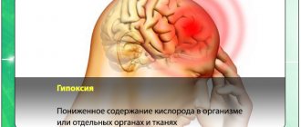
* symptoms of hypoxia (pallor without signs of ectery, weakness, dizziness, palpitations, shortness of breath, fainting)
*symptoms of sideropenia:
- hair splits and falls out
- nails become brittle, changes in the shape of the nails (convexity can be replaced by flattening and even concavity)
- cracks appear in the corners of the lips (jams)
- glossitis, atrophy of the tongue papillae
- distortion of taste and smell (patients like chalk, tooth powder, the smell of gasoline, exhaust gases, acetone, etc.)
- muscle weakness (involuntary urination when coughing, sneezing; in girls, bedwetting).
Laboratory manifestations of IDA.
- decrease in hemoglobin
- low color index
- red blood cells are normal or close to normal
- decrease in hemoglobin content in one red blood cell MCH
- decrease in hemoglobin concentration in the MCHC erythrocyte
- hypochromia, anisocytosis (towards microcytosis), in severe cases - poikilocytosis
- may be reticulocytosis
- decreased serum iron
— OZHS increased
- Ferritin levels are reduced.
Lomanova L.V.
Forecast
In most cases of iron deficiency anemia, the therapy provided has excellent results. However, the situation is significantly complicated by the frequent presence of a severe primary disease that causes anemia (most often oncological pathology). In addition, the prognosis of patients with iron deficiency anemia is significantly worsened by diseases of the cardiovascular system.
Iron deficiency anemia is rarely fatal, but severe and moderate cases can cause sufficient hypoxia to aggravate chronic lung and heart disease.
In children, iron deficiency can cause reduced growth rate, reduced IQ and reduced learning ability.
Stages of iron deficiency anemia
Laboratory examination allows you to determine the stage of iron deficiency anemia.
Stage 1 is characterized by decreased iron in the bone marrow; hemoglobin and serum iron remain within normal limits, but ferritin levels fall below 20 ng/ml. A compensatory increase in iron absorption leads to an increase in the iron-binding capacity of the serum.
During stage 2 , erythropoiesis is impaired and serum iron concentrations fall below 9 µmol/L. There is an increase in the concentration of the transferrin receptor.
At stage 3 , anemia is observed with normal erythrocyte indices and no changes on the smear.
At stage 4, microcytosis and hypochromia develop.
At stage 5 , iron deficiency leads to disruption of tissue function, and symptoms and complaints develop.
Aplastic anemia
Aplastic anemia is a severe blood disease in which suppression of all bone marrow sprouts develops. The causes of the disease are various - from genetic predisposition to the harmful effects of ionizing radiation and various chemical compounds. Clinically, the disease manifests itself as anemic, thrombocytopenic syndrome, as well as severe infectious complications. The diagnosis is made based on the clinical picture, blood tests and bone marrow puncture.
What is aplastic anemia?
Aplastic anemia is a pathological condition of the body in which the number of all three types of blood cells (erythrocytes, leukocytes and platelets) decreases due to a slowdown or complete cessation of their formation in the bone marrow. By origin, aplastic anemia is divided into:
- congenital;
- acquired.
In most cases, inhibition of all three germs of hematopoiesis is observed, however, it has been clinically observed that in different phases of the disease a more pronounced inhibition of one of the germs may be observed
Based on the predominant damage to bone marrow sprouts, the following are distinguished:
- inhibition of one hematopoietic lineage (erythrocyte, leukocyte or platelet);
- inhibition of two hematopoietic germs;
- inhibition of three hematopoietic germs.
Causes of aplastic anemia
The causes of aplastic anemia differ between congenital and acquired anemia.
The following causes of acquired aplastic anemia are distinguished:
- ionizing radiation;
- medications (decaris, analgin, chloramphenicol, tetracycline, butadione, etc.);
- chemical compounds (pesticides, benzene);
- diseases (viral hepatitis A, B and C, Epstein-Barr virus, cytomegalovirus, herpes virus, HIV, parvovirus B19, etc.).
- hormonal disorders of the ovaries, thyroid gland and thymus gland.
Some harmful agents directly affect the bone marrow (ionizing radiation, chemicals and drugs). Others act indirectly through autoimmune mechanisms (viral hepatitis B).
Diagnosis of aplastic anemia
The clinical picture of the disease can largely guide the doctor in the direction of anemia, but the diagnosis must be confirmed or refuted using laboratory tests and paraclinical studies. The most valuable additional studies are:
- complete blood count (CBC);
- biochemical blood test (BAC);
- sternal puncture;
- trepanobiopsy.
General blood analysis
Data from a general blood test for aplastic anemia indicate pancytopenia (a decrease in the number of all three types of bone marrow cells). A decrease in the number of leukocytes is observed mainly due to a decrease in granulocytes (neutrophils, eosinophils and basophils). Thus, the percentage of lymphocytes and monocytes in the leukocyte formula increases relatively. At various stages of the disease, inflammatory signs to one degree or another can be detected. Indicative indicators of UAC for aplastic anemia are:
Hemoglobin (Hb) – less than 110 g/l (normal 120 – 160 g/l). Reduction due to a decrease in the number of red blood cells.
Red blood cells – 0.7 – 2.5 x 10¹²\l (normal 3.7 x 10¹²\l). Decrease in the number of mature red blood cells.
Reticulocytes - less than 0.2% (normal 0.3 - 2.0%). Decrease in the number of young forms of red blood cells.
The color index is 0.85 – 1.05 (the norm is 0.85 – 1.05) indicates the normochromic nature of anemia (the hemoglobin content in the erythrocyte is within the normal range).
Hematocrit (Ht) – less than 30 (normal 35 – 42 in women and 40 – 46 in men). The ratio of the cellular composition of blood to its liquid part. There is a clear decrease in the proportion of cells in the peripheral blood.
Platelets – less than 35 ppm or 100 x 109\l. Decreased platelet count.
Leukocytes – 0.5 – 2.5 x 10⁹\l (normal 4 – 9 x 10⁹\l). Severe leukopenia due to a decrease in the number of granulocytes (neutrophils, eosinophils and basophils).
Band neutrophils – 0 – 2% (normal is less than 6%). Decreased production of young forms of leukocytes.
Segmented neutrophils – 0 – 40% (normal 47 – 72%). Decrease in the number of mature forms of neutrophils.
Myelocytes – 0 – 2% (normally absent). In conditions of granulocytopenia and bacterial infection, a more pronounced than usual shift in the leukocyte formula to the left is observed with the appearance of leukopoiesis precursor cells.
Eosinophils – 0 – 1% (normal 1 – 5%). Decrease in the number of eosinophils.
Basophils – 0% (normal 0 – 1%). Single or complete absence of basophils.
Lymphocytes - more than 40% (normal 19 - 37%). The number of lymphocytes remains normal. Due to a decrease in the granulocyte fraction, relative lymphocytosis is observed (an increase in the proportion of lymphocytes in the blood). Extremely pronounced lymphocytosis can be observed with the accumulation of viral infections.
Monocytes – more than 8% (normal 6 – 8%). The number of monocytes is unchanged and within normal limits. Monocytosis (increased proportion of monocytes in the blood) is explained by a decrease in the percentage of granulocytes in the leukocyte formula.
The erythrocyte sedimentation rate is more than 15–20 mm/hour (the norm is up to 10 mm/hour in men and up to 15 mm/hour in women). This indicator reflects the severity of the inflammatory response in the body.
Anisocytosis is the presence of red blood cells of various sizes in the blood.
Poikilocytosis is the presence of red blood cells of various shapes in the blood.
Blood chemistry
Some types of biochemical blood tests can focus the doctor’s attention on abnormalities in the body that indirectly fit into the three anemic syndromes listed above. Approximate indicators of BAC for aplastic anemia are:
Serum iron is more than 30 µmol/l (normal 9 – 30 µmol/l). Increased serum iron levels due to frequent blood transfusions. High risk of developing hemochromatosis.
Erythropoietin more than 30 IU/l (normal 8 – 30 IU/l in women and 9 – 28 IU/l in men). The increase in erythropoietin occurs for two reasons. Firstly, it is not consumed by the cells of the erythrocyte lineage. Secondly, its synthesis increases compensatoryly in response to anemia.
C-reactive protein – more than 10 – 15 mg/l (normal 0 – 5 mg/l). It is detected during an inflammatory reaction against a background of weakened immunity.
Thymol test - more than 4 (norm 0 - 4). Detects signs of inflammation in weakened immune systems.
Sternal puncture
This type of study is used to visualize bone marrow cells and their percentage. With aplastic anemia, the myelogram will be scanty, the number of cellular elements is significantly reduced. Cambial cells of the erythrocyte and leukocyte series are single or absent. Megakaryoblasts are absent. In rare cases, during puncture it happens to encounter grouped foci of increased cell proliferation as a compensatory reaction of healthy bone marrow to anemia. Such a myelogram may be misleading because it will indicate the absence of aplastic anemia and will therefore be false negative.
Trephine biopsy
Trephine biopsy is a method of removing part of the bone marrow from the patient's ilium wing. The advantage of this procedure over sternal puncture is the possibility of collecting a larger amount of material while maintaining its structure. A larger amount of material reduces the likelihood of a false negative result of aplastic anemia, and studying the structure of the bone marrow allows, in addition to a cytological examination (myelogram), to also conduct a histological examination. Using a blood test and trepanobiopsy results, it is possible to determine the severity of aplastic anemia.
- Aplastic anemia of moderate severity is determined by the following indicators:
granulocytes less than 2.0 x 10⁹\l;
platelets less than 100 x 109\l;
reticulocytes less than 2 – 3%;
bone marrow hypoplasia on trephine biopsy.
- Severe aplastic anemia is determined by the following indicators:
granulocytes less than 0.5 x 10⁹\l;
platelets less than 20 x 109\l;
reticulocytes less than 1%;
bone marrow aplasia on trephine biopsy.
- Extremely severe aplastic anemia is determined by the following indicators:
granulocytes less than 0.2 x 10⁹\l;
platelets are single or absent;
reticulocytes are single or absent;
bone marrow aplasia on trephine biopsy.
Prognosis for aplastic anemia
The prognosis for aplastic anemia largely depends on the timing of detection of the disease. With early detection, there is the possibility of more active intervention in the course of the disease. If detected later, the chances of cure decrease. Congenital Fanconi aplastic anemia is in most cases extremely difficult to treat, since the bone marrow has never been healthy and, accordingly, is very difficult to recover. The presence of congenital developmental anomalies greatly limits the indications for bone marrow transplantation in such patients. In most cases, patients die in childhood from developmental abnormalities or infectious complications. Acquired aplastic anemia has a more favorable prognosis, since in some cases it is reversible after the cessation of the action of the damaging factor on the bone marrow.
Laboratory diagnostics doctor
Novopolotsk city hospital
Kostyuk K.S.
Laboratory diagnosis of iron deficiency anemia
Although the patient's symptoms and complaints can help in the early diagnosis of iron deficiency anemia, the disease is confirmed using various laboratory tests.
Tests for diagnosing iron deficiency anemia:
- general blood analysis;
- peripheral blood smear;
- serum iron, total iron-binding capacity, ferritin.
| Laboratory markers | Changes |
| General blood analysis | Decreased MCV, MCHC; increase in RDW |
| Peripheral blood smear | Microcytosis, hypochromia, thrombocytosis |
| Serum iron | Reduced |
| Total iron binding capacity | Increased |
| Ferritin | Reduced |
Note: MCV—mean erythrocyte volume; MCHC—average erythrocyte hemoglobin concentration; RDW—red blood cell distribution index by size.
General blood analysis
A general blood test allows you to suspect iron deficiency anemia and assess the severity of the disease. In chronic iron deficiency anemia, microcytosis and hypochromia of erythrocytes are observed, characterized by a decrease in the mean erythrocyte volume (MCV) and the mean erythrocyte hemoglobin concentration (MCHC). It should be noted that up to 40% of patients with iron deficiency anemia have normocytic red blood cells, so a normal mean red cell volume does not exclude iron deficiency anemia. The erythrocyte distribution index (RDW) shows the level of variation in erythrocyte size and helps confirm the presence of both normocytes and microcytes in the early stages of the development of iron deficiency anemia, as well as confirm the combined etiology of anemia in the presence of microcytes and macrocytes (iron, B12 and folate deficiency anemia ). An increase in RDW is referred to as anisocytosis.
It should be noted that with iron deficiency anemia, erythrocyte indices change only a few months after the onset of the disease.
An increase in platelet count (more than 450,000/mcd) is often observed, which normalizes with therapy. The white blood cell count is usually within normal limits, but leukocytosis may rarely be observed.
Peripheral blood smear
Examination of a peripheral blood smear is an important step in the diagnosis of patients with anemia. Iron deficiency is characterized by microcytic (reduction in the size of the red blood cell) and hypochromic (decreased hemoglobin concentration in the red blood cell, leading to a “paler” color of the red blood cells) anemia. It should be noted that microcytosis is determined earlier than the change in MCV.
In iron deficiency anemia, codocytes (target cells) are not observed, unlike thalassemia, and anisocytosis and poikilocytosis are most often not expressed. In addition, with iron deficiency anemia, intraerythrocyte inclusions are not observed.
In regions with reduced meat consumption, the coexistence of folate deficiency and iron deficiency is common. In this condition, macrocytes with hypochromic microcytes may be observed in the smear. In this case, the MCV value will be within normal limits.
Ferritin, iron, iron binding capacity
To assess the concentration of iron in the body, studies are carried out on serum iron concentration, total iron-binding capacity and ferritin concentration.
Iron deficiency anemia is characterized by a decrease in the concentration of iron and ferritin and an increase in total iron-binding capacity. However, a complete positive “triad” of laboratory markers occurs at later stages of anemia development. Ferritin is the first to respond to a decrease in iron concentration.
The ferritin molecule is capable of binding 4000-5000 iron atoms at a time, which makes it the main protein storage of iron in the body. Ferritin is found mainly in the cytoplasm of cells of the reticuloendothelial system, as well as in free form in blood serum.
Ferritin concentration directly reflects the total iron content in the body. Thus, serum ferritin concentration is a diagnostic tool for the analysis of iron status. Ferritin concentrations vary greatly among patients of different ages and genders.
In iron deficiency anemia, serum ferritin concentration is one tenth of the normal value. A number of studies have shown that ferritin is one of the most sensitive markers of iron deficiency anemia in the early stages of the disease, however, the results of the study should be interpreted with caution, since in some cases it does not reflect the real picture of the total iron content in the body. Most often this is due to the fact that ferritin is an acute-phase protein and can increase significantly in acute and chronic inflammation, as well as in chronic liver diseases and chronic kidney disease. In this regard, normal ferritin values can be observed when iron deficiency anemia coexists with other chronic pathologies.
Ferritin is also a marker of response and monitoring of iron therapy.
Tests to confirm the etiology of iron deficiency anemia:
Differential diagnosis with anemia of chronic disease
Ferritin is an acute phase protein, and its concentration increases significantly during inflammatory processes, making it impossible to use as a marker for the differential diagnosis of anemia of chronic disease and iron deficiency anemia in some clinical cases. In the late 1990s, a sensitive and early marker of iron deficiency, the soluble transferrin receptor, was described, which increases in proportion to tissue iron deficiency. The main advantage of this marker is that its concentration does not change during the inflammatory response, which makes it an ideal marker for the differential diagnosis of anemia of chronic disease and iron deficiency anemia. Additionally, to increase the accuracy of the study, it is recommended to use the soluble transferrin receptor/Logferritin (sTFR-F). A positive rTGF-F test result is highly likely to diagnose iron deficiency. The sensitivity of the method is 89.19%, specificity is 92.86%.
Hemoglobin electrophoresis
Hemoglobin electrophoresis helps confirm beta thalassemia and other hemoglobinopathies as the cause of microcytic anemia.
Study of hemoglobin in stool
A study of hemoglobin in stool can confirm that chronic gastrointestinal bleeding is the cause of the development of iron deficiency anemia. Modern methods make it possible to detect human hemoglobin in stool with high accuracy and specificity. These methods make it possible to conduct research even in patients who have not previously excluded meat products from their diet. To increase the accuracy of the study, simultaneous testing of fecal calprotectin in stool may be recommended.
The main reason for the appearance of hemoglobin in the stool is malignant tumors of the large intestine, therefore confirmation of iron deficiency anemia and detection of hemoglobin in the stool is an indication for colonoscopy.
Determination of osmotic resistance of erythrocytes
The osmotic stability test of red blood cells is a classic test for diagnosing hereditary spherocytosis. In microspherocytosis, microcytosis and decreased MCV may be observed, but normochromia and normal MCHC values help distinguish this condition from iron deficiency anemia. In rare cases, testing the osmotic resistance of red blood cells helps to accurately determine the etiology of the disease.
Anemia
Anemia is a pathological condition caused by a decrease in the number of red blood cells in the blood and/or a decrease in the concentration of hemoglobin in the red blood cells themselves.
Erythrocytes (red blood cells, RBC) are the largest population of blood cells. Red blood cells contain hemoglobin (Hb), which binds oxygen molecules in the lungs and delivers them to the tissues. In the opposite direction - from the tissues to the lungs - hemoglobin transports carbon dioxide molecules. In other words, hemoglobin ensures the respiration process. The red blood cell has the shape of a disk, concave on both sides. Such a device significantly increases the surface area on which gas exchange occurs. Red blood cells are small in size, which allows them to pass through the narrowest blood vessels (capillaries, arterioles, venules). One red blood cell lives on average up to 120 days.
Low hemoglobin is one of the criteria that must be paid attention to not only when diagnosing anemia itself, but also to assess its severity. A decrease in the amount of hemoglobin in red blood cells occurs when there is insufficient supply of iron during the maturation of cells in the red bone marrow.
There are several classifications of anemia depending on the causes, the mechanisms of anemia development and the severity of changes in blood parameters. They are necessary for the doctor to understand what is causing the current condition. This helps to choose the best tactics for further examination, conduct differential diagnosis and prescribe effective treatment.
The diagnosis and treatment of anemia is carried out by general practitioners, general practitioners, and family doctors. In the case of a severe or malignant course of the disease, the intervention of a hematologist may be required, as well as a more detailed and specialized examination.
There are many reasons for the development of anemia. Anemia is often a complication of an underlying chronic disease.
Iron deficiency anemia develops either when there is insufficient intake of iron in the body, or when the body’s increased need for iron.
Often both factors are present to varying degrees.
The causes of insufficient iron intake may be diseases of the gastrointestinal tract, in which the processes of digestion and absorption of food are disrupted. Consumption of foods containing low amounts of iron or iron in a form that is difficult to digest (exclusively plant-based diet). Some foods (alcohol, milk, coffee) or medications (antibacterial, antacid drugs) interfere with the absorption of iron from food.
The body's need for iron (“iron consumption”) increases significantly in children during periods of active growth and in women during pregnancy and breastfeeding.
Anemia often complicates the course of diseases accompanied by prolonged blood loss (especially against the background of impaired absorption of food):
- peptic ulcer of the stomach and duodenum;
- inflammatory bowel diseases;
- uterine fibroids, endometriosis;
- malignant neoplasms;
- bleeding disorders;
- helminthic infestations.
The human body cannot synthesize iron! We get iron only from food. Not only the amount of iron in foods matters, but also its availability for absorption by the body. Animal products (beef, lamb, liver) contain heme iron. It is absorbed by the body most efficiently. Plant foods (vegetables, fruits, cereals) contain non-heme iron. The human body does not absorb non-heme iron well.
The main reason for the development of iron deficiency anemia, according to experts from the World Health Organization (WHO), is an unhealthy (unbalanced) diet. If you do not consume meat products, then you must remember that iron is absorbed much worse from plant foods. Plan your diet wisely to avoid iron deficiency.
Improves iron absorption:
| Impairs iron absorption:
|
Another type of anemia is a disease caused by vitamin B12 deficiency.
B12 deficiency anemia develops due to a lack of cyanocobalamin.
The reasons for the deficiency may be:
- Insufficient intake of vitamin B12 from food (strict vegetarianism), since cyanocobalamin is found only in meat products, milk, eggs, fish and seafood.
- Impaired secretion of “internal Castle factor” by the stomach;
- Impaired absorption of vitamin B12 in the small intestine;
- Competitive consumption of vitamin B12;
- Increased consumption of vitamin B12;
- Decreased vitamin B12 reserves;
- Violation of vitamin B12 transport;
- The absence of transcobalamin or the appearance of antibodies to it.
Symptoms of anemia:
- Pale skin, cracks, “jams” in the corners of the mouth
- Brittle nails, thinning and striated nails
- Hair loss, fragility, structural damage
- Increased fatigue, decreased performance
- Rapid heartbeat, dizziness, shortness of breath accompany even slight physical activity
Iron deficiency anemia is characterized by
- Perversion of taste: addiction to inedible foods - raw meat, frozen semi-finished products; desire to eat inedible substances - chalk, earth, ice, clay.
- Perversion of smell: addiction to unpleasant odors, for example, gasoline, acetone, paint, etc.
What tests can be done for those who suspect they have anemia? The most accessible and simplest is a general blood test
- Complete blood count (without leukocyte formula and without ESR), venous blood.
If the results of the analysis reveal changes in the number of red blood cells, hemoglobin, hematocrit, erythrocyte indices (MCV, MCH, MCHC, RDW), then it is necessary to consult a doctor to prescribe further examination and treatment.
For reference:
Hematocrit (Ht) is an indicator that characterizes the number of cells per unit volume of whole blood. The hematocrit value depends on the number and shape of red blood cells. A decrease in hematocrit level also characterizes a decrease in the content of red blood cells in the blood.
MCV ( mean corpuscular volume) – the average volume of a red blood cell, reflects the size of the cells. In the presence of long-term iron deficiency, maturing red blood cells become small, microcytes appear in the blood, and the MCV index decreases. If there is a deficiency of vitamins B12 and B9, the maturing cells become large, macrocytes appear in the blood, and this indicator increases. Both conditions negatively affect the main function of red blood cells - the transport of oxygen and carbon dioxide. In the case of macrocytic anemia, their ability to pass through small-diameter vessels is reduced, and their lifespan is reduced.
MCH ( mean corpuscular hemoglobin) - the average hemoglobin content in a red blood cell, reflects the same processes as the previous MCV indicator. With prolonged iron deficiency, red blood cells enter the bloodstream, which during the process of growth and maturation lacked this microelement, and they become not only small, but also insufficiently saturated with hemoglobin. MCH begins to decrease, and a state of hypochromia develops. Red blood cells become pale and their functioning deteriorates. Previously, a color index was calculated to assess the state of hemoglobin saturation of erythrocytes; MCH is its modern, more accurate analogue and allows one to differentiate anemia into normochromic, hypochromic and hyperchromic. In case of deficiency of vitamins B12 and B9, the opposite situation occurs. Maturing red blood cells are oversaturated with hemoglobin, this indicator begins to increase, and a state of hyperchromia develops. Taken together, these two indicators will help the doctor to guess the nature of anemia based only on the results of a general blood test.
MCHC ( mean corpuscular hemoglobin concentration) is the average concentration of hemoglobin in a red blood cell. Reflects the concentration of hemoglobin in a red blood cell. Decreased in iron deficiency hypochromic anemia and diseases accompanied by impaired hemoglobin synthesis at any stage. Increases in hyperchromic B12 and B9 deficiency anemia.
RDW ( red blood cell distribution width) is the width of the distribution of red blood cells by volume. This indicator depends on the average volume of the erythrocyte, therefore, with an increase or decrease in MCV, its increase is observed, which indicates that there are cells of different volumes in the blood, for example, macrocytes and normocytes, or microcytes and normocytes.
For a comprehensive examination, when the cause of anemia is unknown, you can use the Biochemical diagnosis of anemia program, which includes parameters reflecting the metabolism and supply of iron in the body, the supply of vitamin B9 (folic acid) and vitamin B12 (cyanocobalamin), as well as erythropoietin - the hormone that triggers erythropoiesis (maturation of new red blood cells).
- Transferrin - carries out extracellular transport of iron to all cells of the human body.
- Soluble transferrin receptors (sTfR). One of the important diagnostic criteria on the basis of which true iron deficiency can be established. The entry of iron into the cell occurs after the interaction of the iron-transferrin complex with specific proteins - receptors on the cell membrane. The concentration of transferrin receptors depends on two factors - the amount of deposited iron in ferritin and the proliferative activity of the cell. The amount of soluble transferrin receptors is a sensitive indicator of both erythropoiesis activity and iron deficiency. With anemia that has developed as a complication of chronic diseases, this indicator does not change (remains normal). This helps determine the underlying cause of anemia.
- Ferritin is a deposited form of iron, reflecting its total reserve in the body. A decrease in ferritin concentration in the blood is a marker of iron deficiency.
Sources:
- Federal clinical guidelines for the diagnosis and treatment of iron deficiency anemia Official publication Moscow 2015
- Strutynsky A.V. Diagnosis and treatment of iron deficiency anemia of breast cancer “Medical Review” No. 11 of May 14, 2014 p. 839
- Anemia: iron deficiency, B12 deficiency and anemia of chronic diseases. Information letter for hematologists, therapists, interns and residents. Rostov-on-Don, 2013.