The structure of the heart contains tendon threads called chords. Normally, they are attached to the valve flaps through which blood flows, and prevent them from sagging under the influence of gravity. However, sometimes an additional chord may be detected during diagnosis. This is a structural feature of the heart, which does not always indicate an anomaly. But in some cases it can be dangerous to the health and even life of the patient.
What is a chord: location and functions
For normal heart function, it is important that during ventricular systole the communication with the atria is completely blocked. This is ensured by two valves:
- right tricuspid;
- left (also called mitral) - bicuspid.
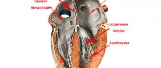
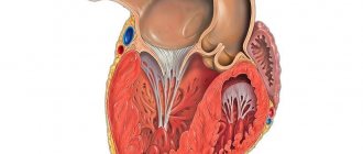
Fleshy trabeculae are attached to the valve flaps, as well as chordae tendineae (cords, heart strings) , formed by dense connective tissue and extending from the papillary muscles. Nature “invented” these structures in order to hold the valve flaps:
- when the cords contract, the valves open;
- at the moment of relaxation of the strings, the valves close tightly, creating a barrier to blood entering the atria.
Sometimes during intrauterine development additional, “extra” fibers are formed. They can be found on both the right and left, but in 90% of cases the left ventricle “suffers.” If such a string goes from the papillary muscles to the walls of the heart or sags freely, cardiologists call it a false chord of the left ventricle (LVF). If it is directed from one wall of the ventricle to another, then such a fiber is called an abnormal cord (ATLV).
The LVDC differs from the mitral valve support string:
- shorter length and thickness;
- structural organization.
A normal, typical fiber is characterized by one central core, in which up to 5 main blood vessels pass. The false chord of the left ventricle is formed by 2–3 rods, separated by connective tissue, inside of which there are microvasculature vessels.
You can also find the following formulation: “additional trabecula of the left ventricle.” Doctors often mean LVDC in this case, although, from an anatomical point of view, the trabecula is a separate structure that continues the cardiac string.
In the English-language medical literature, the term falshe-chordae is used - “false chord”. Some authors recommend replacing the literal translation with “ectopically attached notochord”, as it more accurately reflects the essence of the changes.
Diagnostic examination methods
If systolic murmurs are detected during auscultation (listening), a consultation with a cardiologist and hardware examination are prescribed:
Echocardiography (ultrasound with Doppler ultrasound) is mandatory. Using the Doppler method, you can study the anomaly in more detail: determine the length of the thread and its thickness, the location of attachment, and the speed of blood flow.
An electrocardiogram (ECG) is taken, incl. with exercise, daily heart rate monitoring and bicycle ergometry may be recommended. The doctor prescribes the types of examinations for a patient with an additional chord of the left ventricle individually.
Reasons for the development of the anomaly
The main etiological factor in the occurrence of such an anomaly as an additional chord in the heart of a child is genetic predisposition. The risk increases if the mother has cardiac pathology. But experts do not exclude other reasons for the development of small cardiac anomalies:
- consumption of alcoholic beverages;
- smoking;
- viral and bacterial infections;
- stress;
- excessive physical activity;
- malnutrition;
- environmental pollution;
- radiation.
An abnormality of connective tissue is closely related to its dysplasia. The formation of the fetal cardiovascular system ends by the eighth week of intrauterine development. Obstetricians and gynecologists know that the blood of the unborn child circulates independently already on the 26th day of pregnancy, heart valves are formed at the 6th week, therefore it is in the first trimester that a woman needs to be especially careful about adverse effects.
Symptoms and indications for visiting a doctor
Pediatric cardiologists divide LVDC into those that are hemodynamically significant and those that do not affect health and quality of life. In the vast majority of cases, parents are unaware of the existing anomaly - the child develops absolutely normally.
The clinical picture of LVDC does not have specific signs. Sometimes, even in the neonatal period, a murmur is diagnosed - an acoustic phenomenon in the form of a whistle or creaking, which the doctor hears when listening to the heart. The cause of the noise is the sounds of turbulence as blood moves in the left ventricle or the vibration of an additional heart string.
The manifestation of LVAC is determined by its intracardiac topography and the period of a person’s life. During the so-called growth spurts, when there is an increased increase in body length, children have complaints about:
- excessive fatigue;
- unmotivated fatigue;
- pale skin;
- periodic increased heart rate;
- pain in the area of the projection of the apex of the heart;
- feeling of lack of air.
An electrocardiographic examination can reveal the presence of extrasystoles, conduction disturbances, and signs of left ventricular myocardial hypertrophy. If the occurrence of a minor cardiac anomaly is caused by connective tissue dysplasia syndrome, LVDC is accompanied by the following symptoms:
- asthenic physique;
- low body mass index;
- joint hypermobility;
- history of dislocations;
- deformation of the chest bones;
- poor posture;
- structural changes in internal organs.
Symptoms of LVDC often appear during adolescence. Parents may attribute the teenager’s complaints to the difficulties of the school curriculum, overwork with tutors, overload in the sports section, and prolonged sitting at the computer.
Symptoms
As is clear from the general situation, the clinical picture of an additional chord is absent in most cases.
The patient is unaware of the problem in 90% of situations or even more. That's why he doesn't go to doctors.
Deviations can be detected only by echocardiography results, and that is, by taking a closer look. Only in 10% of situations are there any signs of impairment.
Among them:
- Chest pain. Weak or medium in intensity. They are localized on the left side and can radiate to the shoulder blade, arm, or face. However, not always.
As a rule, they occur after physical activity. They pass at rest. The character is burning, pressing, bursting.
The sign indicates the onset of ischemic processes and becomes more and more distinct as cardiac dysfunction progresses.
- Arrhythmias. Like sudden onset tachycardia. The disturbance is especially noticeable when the load on the body increases. For example, after a brisk walk, climbing stairs, playing sports, even eating.
Suddenly the contraction frequency increases, reaching more than 90 beats per minute.
There may be a feeling of interruptions. Patients describe the condition as “the heart suddenly stops and starts beating again” or “misses contractions.”
These are the phenomena of extrasystole. In some cases, the arrhythmia is represented by paroxysmal (attack-like) tachycardia. There are many options.
- Shortness of breath due to physical activity. For many years, the deviation manifests itself externally as exercise intolerance. The patient cannot walk quickly or play sports. Sometimes the threshold when a symptom arises is so great that a person simply does not reach it. This means that the sign goes unnoticed. Unless the athlete has an extra chord.
- Weakness, drowsiness. Unreasonable lethargy and low performance. As cardiac dysfunction progresses, signs of asthenia become more pronounced. But this takes years.
- Headache, inability to navigate in space. The result of insufficient blood circulation in the brain. Fortunately, the sign does not support critical processes. In addition, other neurological deficits are also detected: problems with intelligence, thinking, memory, decreased visual acuity, and hearing. There are many options.
Clinically, an extra chord corresponds to heart failure and provokes it.
The rate of disease progression is low, extremely insignificant in the majority of cases.
At some point, decompensation may occur. The body will not be able to maintain acceptable functional activity of the heart. The process will begin to develop like an avalanche and lead to disaster.
Fortunately, this is unlikely. Because symptoms will force a person to see a doctor much earlier. In childhood, there are usually no signs.
Manifestation occurs at ages over 15-16 years. It is during this period that subcompensation occurs. The body still supports and stabilizes the functioning of the muscular organ, but it is no longer fully capable.
Diagnostics: how to determine LVHL
When the doctor tells you that your baby has a heart murmur, there is no reason to panic. The condition does not require emergency resuscitation measures or intensive treatment. The main diagnostic method is ultrasound examination. It will help clarify the location and number of additional strands. There are the following types of LVDC:
- according to location in the ventricle - apical, middle, basal;
- in relation to the axis of the heart - diagonal, longitudinal, transverse;
- by number - singular and plural.
If echocardiography has revealed an additional chord in the cavity of the left ventricle in a child of any age, I advise parents to undergo the following examinations:
- A general blood test is not important in terms of diagnosing LVHL, but it will reveal anemia or an existing inflammatory process in the body, which can aggravate the condition of a child with a minor cardiac anomaly.
- Electrocardiography. It is advisable to conduct Holter monitoring, which will exclude arrhythmia with greater diagnostic accuracy.
- ECG tests with physical activity will show how well the child’s heart “copes” with its function.
- Consultations with specialized specialists (orthopedist, endocrinologist, otolaryngologist).
I always explain to parents of infants that sometimes the abnormal chord can lengthen and “grow” into the valve, so during subsequent ultrasounds the doctor does not detect LVDC. In such cases, they say that the baby has “outgrown” this pathology.
University
My son is 36 years old. Recently, an ultrasound revealed that he has multiple accessory chords of the left ventricle. Is it good or bad? The clinic didn’t say anything definite... The Rybakov family, Vitebsk region.
I work in the regional center as a cardiologist. I would like to read in the Medical Bulletin about abnormal chords of the heart: how to identify and then manage such patients? The article will be useful for doctors from the outback... Svetlana N., Vitebsk region.
Evgenia Trisvetova, Doctor of Medicine, answers questions from readers and the MV correspondent Sciences, Professor of the 2nd Department of Internal Diseases of BSMU.
— Evgenia Leonidovna, how do experts explain the appearance of additional chords in the heart?
— I would like to note right away that intravital study of the internal structure of the heart became possible thanks to the invention of imaging devices and the development of ultrasound. In 1981, almost 100 years after the anomalous chordae were mentioned, the first intravital description of anomalous cardiac chordae using two-dimensional echocardiography was published.
Abnormal (false) chords are those that are attached not to the valve leaflets, but to the walls of the ventricles. According to one hypothesis, it is a derivative of the inner muscular layer of the primitive heart, arising in the embryonic period when the papillary muscles are unlaced. According to another assumption, abnormal chordae are muscle trabeculae that are drawn into the cavity of the ventricle during its dilatation or the formation of an aneurysm.
The name “chord” reflects the position of the abnormal cord as a geometric body crossing the chamber of the heart. True chords are fibrous cords consisting of collagen fibers. They receive blood supply and innervation from the heads of the papillary muscles, which are fed and innervated through muscle trabeculae located at the base of the same papillary muscles.
Abnormal chordae arise from inherited defects in collagen metabolism; influence of factors that disrupt embryogenesis with genetic predisposition and cause familial non-hereditary forms.
An abnormally located chord (APX), called a moderator band, was first described during a post-mortem examination in 1893. Subsequently, additional cords in the cavity of the left and right ventricles were designated differently: false tendon, false ligaments, additional chords, muscle cords. Modern sources use the term “abnormally located chord” and false tendon.
- How common is this anomaly?
— During ultrasound examination of the heart in children, the frequency of abnormal chordae ranges from 0.2–21.7%. Andrey Korzhenkov, a researcher at the Novosibirsk Research Institute of Therapy, and his colleagues studied the prevalence of abnormal cardiac chords in adults. In 1991, 859 men aged 25–64 years old underwent ultrasound; signs of abnormal cardiac chords were found in 17.1%. According to the results of our echocardiographic study (2002–2004, 2,401 people), in men 18–25 years old, healthy or hospitalized for diseases of the internal organs, abnormal cardiac chords occurred in 8.3% of cases.
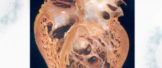
The results of the post-mortem study are interesting. Tatyana Domnitskaya, professor of the Department of Functional Diagnostics at the Faculty of Advanced Training of Medical Workers at the Peoples' Friendship University of Russia, examined the hearts of people who died from various diseases. These were 41 men and 46 women, 57–87 years old. Macroscopic examination revealed abnormal cardiac chords in 14% of cases. Other authors pointed to the occurrence of such chords in 17–39% of cases in women and much more often (61%) in men.
In our work (E.L. Trisvetova, O.A. Yudina, 2006 - Author's note), abnormal chords of the heart were identified in 12.1% of cases (578 deaths 15–91 years old (average age 63.1±1 .08), including 295 (51%) men and 283 (49%) women). In the group of deaths with minor cardiac anomalies (n=98), these chords were diagnosed in 71.4% and 2.08±0.59 cases per 100 examined. The ratio of men to women with abnormal cardiac chords was 1:0.64.
— Which parts of the heart “prefer” abnormal chords?
— A. Korzhenkov and co-authors proposed to determine the relationship of the chord to a specific topographical variant using the division of the walls of the left ventricle into 10 segments. According to the classification of J. Botti, transverse, basal and longitudinal chords are found in 3 sections of the left ventricle: apical, midventricular and basal. The most common ARHs of the left ventricle are in 95% of cases, while in the right they are recognized in only 5% of cases; very rarely abnormal chords of the right atrium are detected. Transverse ones occur in 68%, diagonal - in 30%, longitudinal - in 2% of cases.
ARCHs can be single (81.6%) or multiple. Using ultrasound in young men, we detected ARCH in 8.3% of cases. Moreover, in 99.2% of cases, ARCHs were found in the left ventricle. A post-mortem study of the prevalence of ARH showed that in 17–39% of cases they occur in women and much more often (61%) in men. Age does not affect the incidence of ARH.
— How can a specialist detect abnormal chords?
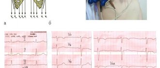
— The most informative method for intravital detection of ARCH is the echocardiographic method. Transthoracic and transesophageal cardiac imaging approaches are used. The sensitivity of transthoracic echocardiography in diagnosing ARCH is 72–87%. The quality of the equipment and the qualifications of the specialist performing the research are important. In practice, both over- and underdiagnosis of ARH are observed. To visualize ARCH, two-dimensional echocardiography is performed from 3 orthogonal projections (sagittal, horizontal, frontal), using the long axis of the left ventricle, the longitudinal axis of chambers 2, 4, 5 and the short axis projection. Parasternal, apical, and subxiphoid approaches are used, allowing one to see all the structures of the heart in real time.
In addition to longitudinal and cross sections, to search for ARH it is necessary to use non-standard approaches and projections. The ARCH criterion is the identification of an echo-dense linear additional formation in the ventricular cavity in 2 mutually perpendicular planes in sectoral scanning mode with confirmation of the results on an M-echocardiogram in the systole and diastole phases. It is necessary to examine the papillary muscles and areas of attachment of the chord to the free walls of the ventricles. The attachment points of the chord are detected in the sectoral scanning mode and the results are confirmed on the M-echocardiogram. An addition to the standard EchoCG protocol has been developed - it is necessary to indicate the main characteristics of the ARCH (localization, topographic variant, echo density in the areas of attachment of the cord, presence/absence of traction of the papillary muscles in systole, thickness and length, changes in the geometry of the left ventricle, the rate of shortening of myocardial fibers in the areas of attachment of the chord ) and other features.
A detailed assessment of the morphometric, topographic and functional signs of ARH is carried out for the differential diagnosis of echocardiographic or clinical manifestations of the abnormality.
The abnormal chordae are located between the following intracardiac structures: the posteromedial papillary muscle and the wall of the left ventricle or the interventricular septum; anterolateral papillary muscle and interventricular septum; papillary muscles; walls of the left ventricle and interventricular septum; walls of the left ventricle. Sometimes abnormal chordae are attached at 3 or more points, forming a membranous structure.
— How should a doctor treat an abnormal finding? Observe the patient, treat?..
— The clinical significance of abnormal chordae has not yet been determined. It is known that people with hereditary and multifactorial connective tissue disorders are characterized by single and multiple abnormal chordae, which are diagnosed along with other minor cardiac anomalies. In this case, clinical manifestations may be caused by multisystem disorders of the structure of connective tissue, including musculoskeletal, skin, ocular, cardiovascular and other visceral signs of dysmorphogenesis, and autonomic disorders.
The actual presence of abnormal cardiac chords may not affect the patient’s subjective sensations and may not bother them. However, if other causes are excluded, they can cause the manifestation of pathological signs. The transverse position of the abnormal chord in the middle third of the left ventricle causes a systolic murmur, which is heard during auscultation of the heart. A long abnormal chord reaching the left ventricular outflow tract is associated with local hypertrophy of the opposite wall of the left ventricle.
Cardiac arrhythmias (ventricular extrasystole, supraventricular/ventricular paroxysmal tachycardia) can occur due to the presence of cells of the conduction system in the abnormal chord or changes in the myocardium at the sites of attachment of the abnormal cord and the appearance of ectopic foci of excitation in the myocardium. The ECG of abnormal left ventricular chordae describes abnormal ventricular repolarization (early repolarization syndrome, nonischemic ST segment depression, flattened or weakly negative T wave), WPW syndrome, and shortened PQ interval.
Short abnormal chords in a transverse position in the cavity of the left ventricle can be injured by the blood flow or torn/ruptured during ventricular diastole. In these places, blood clots are identified, representing a likely source of embolism in the vessels of the systemic circulation. Cases of infective endocarditis that developed as a result of infection of the area of the anomalous chord tear have been described. Changes in the geometry of the left ventricle due to a transverse abnormal chord in the cavity are rare and are compensatory in nature, preventing the expansion of the heart chamber.
Typically, abnormal chordae do not affect the prognosis of life and do not require special treatment. In the case of an echocardiographic “finding,” the patient must be examined to determine whether there are hereditary or multifactorial connective tissue disorders or congenital heart defects.
— Can people with abnormal chordae play sports and have children? Do I need to see a cardiologist?
— If diseases are excluded and there are no clinical symptoms, the abnormal chord is treated as an insignificant feature of the development of the heart. A person leads a normal lifestyle and does not limit himself in physical activity; women plan pregnancy and childbirth. It’s another matter if the doctor diagnosed symptoms of a hereditary or multifactorial connective tissue disorder. Then you need to get specific recommendations from your doctor and follow his advice. If you experience discomfort, shortness of breath, pain in the heart, as well as rhythm disturbances, you need to contact a specialist for additional examination and medical care. Tatyana Skibitskaya Medical Bulletin , February 13, 2015
Doctor's advice: how often to do echocardiography with false chord
For a standard check-up for a minor cardiac anomaly, it is enough to undergo an echocardiography once a year. The examination is scheduled unscheduled if:
- the patient has complaints, including those not directly related to cardiology;
- you notice that the child is growing rapidly;
- there was a sharp weight loss;
- a chronic disease has been diagnosed, such as asthma, gastritis, nephritis;
- the child goes to kindergarten, school, university;
- there is a question about the sports section;
- stress occurred: an exam, the loss of one of the family members, conflict in the children's team and at home;
- pregnancy is planned or determined.
Single LVDC is not considered a pathology. Experts consider them as an anatomical feature of a person. The presence of an additional chord is not a contraindication to attending physical education classes. Young men with abnormal cords can be drafted into the army if they do not have complications or concomitant pathologies. It is only important to clearly tell the child about his condition and warn him that he needs to be attentive to his health.
What complications can occur with a false chord?
The prognosis for single abnormal chords is favorable. Complications are rare, but multiple transverse and diagonal chordae can act as a trigger for the development of diastolic dysfunction of the heart, increasing the risk of thrombus formation, bacterial endocarditis, and can also be combined with other congenital anomalies.
Sometimes LVHL can cause:
- extrasystoles;
- paroxysmal ventricular tachycardia;
- WPW syndrome;
- atrial fibrillation;
- TELA;
- thrombotic lesions of veins;
- ischemic stroke;
- cardiac arrest.
These disorders are observed in adulthood, but a teenager with LVDC should be informed about them. An unfavorable prognosis is characteristic only of non-compact left ventricular myocardium syndrome (LVSM), which is characterized by the presence of numerous additional trabeculae and chordae, combined with changes in the structure of the heart muscle.
Despite these complications, statistics claim: repeated clinical observations have not revealed a relationship between the presence of additional strands in the left ventricle and the risk of cardiovascular mortality.
Possible complications
{banner_banstat8}
These develop extremely rarely. The main cause is heart failure.
A chronic disorder accompanied by a decrease in myocardial contractility, poor nutrition of tissues and structures, and poor blood flow. All other consequences follow from it.
Heart attack, cardiac arrest. Fortunately, these are extremely rare options. In 95% of situations there are no complications for many years or throughout life. But it is not recommended to take risks.
Treatment
As a rule, single LVDC without clinical manifestations do not require treatment. Medicines are prescribed for the development of complications and concomitant heart pathologies. Symptomatic therapy - antiarrhythmic drugs and substances that correct hemodynamics: diuretics, antihypertensives, antioxidants, potassium and magnesium preparations.
Surgical treatment methods (local cryodestruction and excision of left ventricular ventricle) are rarely used: if an abnormal cord causes arrhythmia that threatens the patient’s life.
Lifestyle recommendations:
- give up fast food and sugary carbonated drinks;
- spend at least an hour a day in the fresh air;
- do not sit too long at the computer;
- find time in your personal schedule for physical exercise;
- learn to cope with stress;
- get vaccinated against influenza and other infectious diseases in a timely manner;
It is imperative to discuss the issue of “bad habits” with your teenager. It is important that the young person consciously considers it unacceptable for himself to take a sip of a low-alcohol drink or inhale cigarette smoke.
Pregnancy is not contraindicated for women with LVDC. It does not pose a threat to health, passes without negative consequences for mother and child, you just need to notify the doctor about the existing additional chord.
Forecast
{banner_banstat7}
In most cases - favorable. Even when symptoms develop, many years pass before the process reaches a critical, decompensated phase.
This can only be achieved consciously. Typically, symptoms in the earlier stages are unbearable and significantly reduce the quality of life. There is no choice but to see a doctor.
The survival rate is good, the likelihood of maintaining working capacity is also good. But this does not mean that you need to sit idly by. You cannot do without the consultation and help of a cardiologist.