Clinical significance of hemoglobin determination
In an adult, the normal Hb concentration depends on gender:
- women 11-14g/% or 110-140 g/l;
- men 12-16 g/% or 120-160 g/l.
For men, the norm is higher, since they have greater muscle mass compared to women.
A decrease in hemoglobin concentration is observed in the following conditions and diseases:
- reduced concentration of red blood cells due to iron deficiency, sickle cell anemia;
- insufficient amount of substances that affect the production of red blood cells (folic acid, vitamin B12);
- red bone marrow disease.
Increased Hb concentration is observed in the following conditions and diseases:
- increased production of red blood cells in red bone marrow disease, malignant neoplasms;
- compensatory increase in the number of red blood cells due to cardiovascular and pulmonary diseases;
- a decrease in the amount of blood plasma as a result of which the number of formed elements increases in 1 liter of biological fluid.
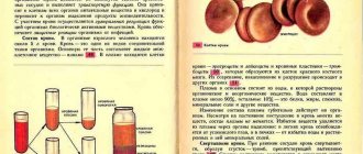
Hemoglobin appears in red blood cells after the loss of the nucleus and the acquisition of cell maturity. The site of red blood cell and Hb disposal is the spleen. The life cycle of 1 cell together is 120 days. After death, iron is released into the blood and supplied to the red bone marrow. With the help of this element, new red blood cells are produced.
Methods for measuring hemoglobin in blood
The following methods exist for analyzing hemoglobin in biological fluid:
- hemiglobincyanide;
- hemichromic;
- Sali;
- ammonia;
After using the appropriate technique, a semi-automatic analyzer is most often used, which measures the wavelength spectrum of the Hb molecule.
The calculation of indicators is carried out not only in the blood, but also in the urine, since minor or significant bleeding is possible in the urinary system.
The semi-automatic hematology analyzer completely eliminates the restoration of medical errors in the report of the required value. But the device will not be able to see and recognize violations of various parameters.
Manual counting is based on the use of a reagent and biological fluid. The method does not count the number of cells, but rather determines their presence or absence. Suitable for urine
Unified methods for determining hemoglobin
Unified methodology - methods of measuring indicators used in most laboratories. They are accurate, fast, practical. For Hb, this is the hemicyanide method with photocolorimetric counting. Allows you to determine the exact amount of the indicator by calculating the wavelength on a specialized semi-automatic device.
The second unified technique is the hymechromic method. Its principle is the same, but higher quality reagents are used that do not harm the health of doctors.
Hemiglobin cyanide method for determining hemoglobin in blood
Hemoglobin has many forms, so calculating its wavelength is very difficult. To simplify the calculation, specialized chemicals are used that convert it into methemoglobin.
To obtain accurate results, it is necessary to use a transforming solution, specialized measuring tubes, and a photoelectrocolorimeter. Hb has a wavelength of 540 m. It is within this limit that the device calculates the given indicator.
The obtained data is substituted into a formula that converts the total amount of indicators into grams per liter (g/l).
Important! At the moment, the method for determining the concentration of hemoglobin in the blood is rarely used, since the reagents used are dangerous to the life and health of laboratory doctors. If the preparation rules are violated, burns of the skin and mucous membranes may occur.
Read in detail about the hemiglobincyanide method in a separate article!
Hemichrome method for determining hemoglobin in blood
Using the technique, Hb is converted into cyanohemoglobin using reagents that are completely safe for humans. For this purpose, cyanide compounds (sodium dodecyl sulfate, lauryl sulfate) are used.
Hemichrome is formed, which is counted at a wavelength of 540 nm. The obtained data is substituted into the formula to convert the indicator to g/l.
Detailed article about the hemichrome method for determining hemoglobin.
Cyanide method for studying hemoglobin in blood
Capillary and venous blood is used for analysis. A transforming solution is added to a human biological fluid. With its help, all forms of Hb are converted into methemoglobin.
5 ml of reagent and 20 µl of biological fluid are poured into a test tube. Wait 10 minutes for the reaction to occur. The resulting liquid is poured into a special cuvette, a photoelectric colorimeter passes rays through it, and counts elements in the wavelength region of 543 nm. The Hb content is calculated using a formula using the obtained indicators.
Sali method for determining hemoglobin in blood
Determining the amount of hemoglobin using the Sali method is rarely used in clinical settings, since more advanced methods exist.
A human biological fluid is obtained, to which hydrochloric acid is added. The solution is thoroughly mixed. This allows Hb to pass into another form - hematin hydrochloride. Add distilled water until the color is identical to the control tube (brown color).
On the lower meniscus of the test tube there will be a number that indicates the content of the indicator in 100 g of blood. To calculate the concentration in 1 liter, the indicators are multiplied by 10. The percentage of Hb is calculated using the proportion:
16,7%=100
- A%=X
- X=A×100/16.7
- X – determined indicator,
- A is the hemoglobin number obtained using a hemometer,
- 16.7 – percentage of hemoglobin in the control sample.
Spectral analysis method for hemoglobin measurement
The most accurate method for determining hemoglobin is to use a spectrophotometer. It detects only 1 of the forms of hemoglobin by capturing its wavelength. There are different forms of Hb in human blood:
- Oxyhemoglobin;
- Deoxyhemoglobin;
- Carboxyhemoglobin;
- Methemoglobin.
To study using a spectrophotometer, it is necessary to convert the indicators into only 1 of the forms. Various reagents are used for this. The indicator is colored in a certain color (red, green). Its wavelength is picked up by the device and counts all the elements in a given trajectory.
Ammonia method for determining hemoglobin
The ammonia method for the quantitative determination of hemoglobin corresponds to the previous methods. The difference is the use of ammonia at a concentration of 0.04%. The sample must sit for a reaction to convert all forms of Hb into one. It is calculated using a semi-automatic analyzer in the form of a spectrophotometer. The solution is placed in a cuvette and installed in the device. The indicator is calculated at a wavelength of 543 nanometers.
Methods for determining hemoglobin in urine
There are 4 types of urine tests. Each of them differs in the reagents used. For research, only a fresh portion of urine is collected. If it infuses, Hb will turn into medhemoglobin, so the reaction will be impossible. Urine is prepared. To do this, add 2 ml of acetic acid and 5 ml of diethyl ether.
- Reaction with guaiac acid. Dilute a pinch of acid, add 3 ml of 96% ethanol. Add 8-10 drops of the resulting liquid to 8-10 drops of diluted urine. If Hb is present in the sample, it turns blue.
- Reaction with amidopyrine. To 2-3 ml of diluted urine add 10 drops of amidopyrine solution and 10 drops of 3% hydrogen peroxide. If Hb is present, the urine turns purple.
- Reaction with benzidine. Add 6 drops of benzidine, 6 drops of 3% hydrogen peroxide, and 4 drops of pure urine to the test tube. If Hb is present, the resulting liquid turns green.
- Use of test strips, tablets. They contain specialized reagents in ready-made form. The strips are dipped into the urine, removed, and the color scale is assessed. The tablets are completely dissolved in human biological fluid. The indicator scale is marked on the packaging.
Determination of hemoglobinpresentation for the lesson
Slide 1
Methods for determining hemoglobin concentration
Slide 2
Assessment of hemoglobin content: clinical significance: ↓ Decrease in hemoglobin concentration: anemia (impaired Hb synthesis, blood loss, hemolysis, lack of red blood cell production, etc.)
Slide 3
↑ Increased hemoglobin concentration: polycythemia, hemoconcentration during dehydration, burns, persistent vomiting; exposure to high altitudes, excessive exercise or agitation;
Slide 4
lung diseases leading to decreased pulmonary perfusion and poor lung aeration; chronic chemical exposure to nitrites, sulfonamides, causing the formation of meth- and sulfohemoglobin. ↑ Increase in hemoglobin concentration:
Slide 5
The term “total hemoglobin” applies to the totality of hemoglobin forms that are present in the blood of healthy people or appear in pathology:
Slide 6
hydroxy hemoglobin (HbO 2); reduced hemoglobin (Hb) (synonyms: hemoglobin (H Hb ++), ferrohemoglobin (Hb 4)); carbo hemoglobin (HbCO 2) (CO 2 - carbon dioxide) Normal forms of hemoglobin
Slide 7
carboxy hemoglobin (HbCO) (syn.: carbonmonoxyhemoglobin (HbCO) 4; met hemoglobin (MetHb), synonyms: hemI globin (Hb +++), ferrihemoglobin (Hb 4 +3); sulf hemoglobin (SHb), synonym - verdoglobin S; hemeIglobincyanide (HiCN) (syn.: cyanmethemoglobin (CNMetHb)); etc. Pathological forms of hemoglobin
Slide 8
ANALYSIS METHODS Most of all in the blood are: oxyhemoglobin, deoxyhemoglobin, carboxyhemoglobin, methemoglobin. Absorption spectra of oxyhemoglobin (HbO2), deoxyhemoglobin (HbH), methemoglobin (HbMet), carboxyhemoglobin (HbCO)
Slide 9
To determine hemoglobin, hemoglobin derivatives are most often analyzed, formed during its oxidation and the addition of various chemical groups to heme, leading to a change in the valence of iron and the color of the solution.
Slide 10
When quantifying hemoglobin by colorimetric methods, the problem arises in choosing a reagent that would convert all hemoglobin derivatives into only one form before photometric analysis.
Slide 11
The best methods for quantitatively converting hemoglobin into its derivatives turned out to be: hemiglobin cyanide (HbCN), hemichrome (HbChr) and hemiglobinazide (HbN3), which, when photometrically, give the smallest determination error among other methods of analysis.
Slide 12
Hemiglobincyanide method (Drabkin method) (1936) Absorption spectrum of cyanmethemoglobin (CNmetHb) Principle of the method: all forms of hemoglobin are converted to hemiglobincyanide (using a transforming reagent (containing potassium iron cyanide, potassium cyanide and sodium bicarbonate) at a wavelength of 540 nm
Slide 13
Hemohyglobincyanide method (Drabkin method) (1932) A 540 HiCN x 16114.5 x 10 -3 x P Hb (g/l) = =367.7 x A 540 HiCN 11.0 x L A 540 HiCN - absorption of hemoglobin solution at a wavelength of 540 nm, 16114.5 - molecular weight of hemoglobin monomer, 11.0 - millimolar extinction coefficient of cyanmethemoglobin, L - optical path length, equal to 10 mm in most photometers, 10 -3 - conversion of the molar mass of hemoglobin to millimolar mass P - blood dilution (1: 251, ratio of 20 μl of blood and 5.0 ml of transforming solution)
Slide 14
The conversion of hemoglobin to hemiglobin cyanide is carried out by its interaction with a transforming solution containing potassium ferricyanide, potassium cyanide, potassium dihydrogen phosphate and nonionic detergent: The detergent enhances the hemolysis of red blood cells and prevents turbidity associated with plasma proteins. Potassium dihydrogen phosphate maintains a pH level at which the reaction takes place in 3-5 minutes.
Slide 15
Potassium ferricyanide oxidizes all forms of hemoglobin into methemoglobin, which forms, with potassium cyanide, hemeIglobincyanide, which has a reddish color, the color intensity of which is directly proportional to the concentration of hemoglobin in the sample. Oxy hemoglobin Deoxy hemoglobin Carboxy hemoglobin Met hemoglobin
Slide 16
Characteristics of the method The hemiglobincyanide method, developed in 1936 by Drabkin, was approved by the International Committee for Standardization in Hematology (ICSH) in 1963. The main advantage of the hemiglobincyanide method is that HbCN (hemiglobincyanide) is a stable derivative of hemoglobin, and all forms present in the blood hemoglobin can be quickly and quantitatively converted into HbCN; The method provides the ability to obtain results with an error NOT exceeding ± 2%.
Slide 17
Safety requirements when working with a solution containing cyanide compounds Despite all the positive parameters of the hemiglobin cyanide method, its big drawback is that it is based on the use of poisonous cyanide compounds;
Slide 18
Instead of potassium cyanide, many use masked cyanide - acetone cyanohydrin, which, during the preparation of the transforming solution, decomposes to form cyanide ion. The nature of the action of acetone cyanohydrin on humans is similar to the action of hydrocyanic acid, but the effect develops more slowly. Acetone cyanohydrin is absorbed through the skin and can cause severe poisoning. Its maximum permissible concentration (MPC) is 0.9 mg/m3, hazard class 2.
Slide 20
Optical design
Slide 21
Dependence of the readings of the MiniGem-540 hemoglobinometer on the time of incubation of blood samples with the transforming solution. The graph shows that only 15 minutes from the start of the lysis reaction, the readings of the device begin to stabilize and then do not change over the next 2 hours. Therefore, hemoglobin measurement should be carried out no earlier than 20 minutes after adding blood to a test tube with a transforming solution, when all the hemoglobin present in the test tube is converted into the final reaction product - cyanmethemoglobin.
Slide 22
Hemichrome method The principle of the hemichrome method is based on the conversion of all forms of hemoglobin into one - hemichrome. The color intensity is directly proportional to the hemoglobin concentration in the sample. Hemoglobin Hemi-chrome Transform. solution: Fatty acids + Ferricyanide K (or Na dodecyl sulfate)
Slide 23
Hemichrome method Absorption spectrum of methemoglobin (HbMet) The maximum of the hemichrome absorption curve is at a wavelength of 533 nm. The typical wavelength closest to 533 nm is 540 nm, at which photometry is carried out taking into account the conversion factor (factor) for 540 nm.
Slide 24
The main advantage of the hemichromic method is that the forms of hemoglobin contained in the blood can be quickly and quantitatively converted into HbChr while the transforming solution is completely harmless
Slide 25
Optical design
Glycated hemoglobin (HbA1c)
Glycated hemoglobin (A1c) is a specific compound of red blood cell hemoglobin with glucose, the concentration of which reflects the average glucose content in the blood over a period of about three months.
Synonyms Russian
Glycohemoglobin, hemoglobin A1c, HbA1c, glycosylated hemoglobin.
English synonyms
Glycated hemoglobin, hemoglobin A1c, HbA1c, glycohemoglobin, glycosylated hemoglobin.
Research method
Ion exchange high performance liquid chromatography (HPLC).
Units
% (percent).
What biomaterial can be used for research?
Venous blood.
How to properly prepare for research?
- Do not eat for 2-3 hours before the test; you can drink clean still water.
- Avoid physical and emotional stress for 30 minutes before the test.
- Do not smoke for 30 minutes before the test.
General information about the study
The glycated hemoglobin (A1c) test helps estimate the average blood glucose level over the past 2-3 months.
Hemoglobin is a protein found inside red blood cells (erythrocytes) that carries oxygen. There are several types of normal hemoglobin, and many abnormal varieties have been identified, although the predominant form is hemoglobin A, accounting for 95-98% of total hemoglobin. Hemoglobin A is divided into several components, one of which is A1c. Part of the glucose circulating in the blood spontaneously binds to hemoglobin, forming so-called glycated hemoglobin. The higher the concentration of glucose in the blood, the more glycated hemoglobin is formed. Having combined with hemoglobin, glucose remains “bound” with it until the very end of the red blood cell’s life, that is, 120 days. The combination of glucose and hemoglobin A is called HbA1c or A1c. Glycated hemoglobin is formed in the blood and disappears from it every day, as old red blood cells die and young (not yet glycated) take their place.
The hemoglobin A1c test is used to monitor the condition of patients diagnosed with diabetes mellitus. It helps assess how effectively glucose levels are regulated during treatment.
For some patients, a hemoglobin A1c test is ordered to diagnose diabetes and prediabetic conditions in addition to a fasting plasma glucose test and a glucose tolerance test.
The resulting indicator is measured as a percentage. Patients suffering from diabetes should strive to keep the level of glycated hemoglobin no higher than 7%.
A1c should be reported in one of three ways:
- as a percentage of the total amount of hemoglobin,
- in mmol/mol, according to the International Federation of Clinical Chemistry and Laboratory Medicine,
- as the average glucose content mg/dl or mmol/l.
What is the research used for?
- To control glucose in patients with diabetes, maintaining its blood level as close to normal as possible is very important. This helps minimize complications to the kidneys, eyes, cardiovascular and nervous systems.
- To determine the patient's average blood glucose levels over the past few months.
- To confirm that the measures taken to treat diabetes are correct and to find out whether they require adjustments.
- To determine uncontrolled rises in blood glucose in patients with newly diagnosed diabetes mellitus. Moreover, the test can be prescribed several times until the desired glucose level is detected, then it must be repeated several times a year to ensure that normal levels are maintained.
- For preventive purposes, to diagnose diabetes at an early stage.
When is the study scheduled?
Depending on the type of diabetes and how well the disease responds to treatment, an A1c test is performed 2 to 4 times a year. On average, patients with diabetes are recommended to have their A1c tested twice a year. If a patient is diagnosed with diabetes for the first time or the control measurement is unsuccessful, the test is ordered again.
In addition, this test is prescribed if the patient is suspected of having diabetes, since there are symptoms of high blood glucose:
- strong thirst
- frequent excessive urination,
- fast fatiguability,
- blurred vision,
- increased susceptibility to infections.
What do the results mean?
Reference values: 4.27 - 6.07%.
The closer the A1c level is to 7% in a diabetic patient, the easier it is to control the disease. Accordingly, with an increase in the level of glycated hemoglobin, the risk of complications also increases.
The results of the A1c analysis are interpreted as follows.
| Glycated hemoglobin indicator | Meaning |
| 4-6,2 % | The patient does not have diabetes |
| 6.5% and more | The patient has diabetes |
| 5,7-6,4 % | Prediabetes (impaired glucose tolerance associated with an increased risk of diabetes) |
According to the clinical recommendations of the Ministry of Health of the Russian Federation of the Russian Association of Endocrinologists NGO “Algorithms for specialized medical care for patients with diabetes mellitus” (2019), an additional diagnostic indicator is the average daily plasma glucose level (ADPL) for the last three months and its correlation with the HbA1c level.
What can influence the result?
Patients with abnormal forms of hemoglobin, such as those with sickle cells, will have low levels of glycated hemoglobin. In addition, if a person suffers from anemia or severe bleeding, his test results may also be underestimated. On the contrary, A1c levels are elevated due to iron deficiency and recent blood transfusion (since liquid blood preservatives contain a high concentration of glucose).
Important Notes
An A1c test does not reflect sudden changes in blood glucose levels. Fluctuations in glucose in patients with labile diabetes will also not be detected by this test.
Also recommended
- Plasma glucose
- Glucose tolerance test
- Fructosamine
Who orders the study?
Therapist, endocrinologist.
Hemoglobin electrophoresis for the diagnosis of hemoglobinopathies
Hemoglobin electrophoresis in alkaline gel allows you to determine the percentage of the main hemoglobin isoforms and screen for hemoglobinopathies. Normally, in the blood of an adult, at least 96.5% of hemoglobin is represented by the HbA isoform, which consists of two pairs of polypeptide chains (globins): 2α and 2β, each of which is associated with heme. Hemoglobinopathies are associated with the presence of an abnormal variant of hemoglobin. Hemoglobin electrophoresis can screen for conditions associated with structural abnormalities of hemoglobin. However, the method does not allow establishing the type and nature of hemoglobinopathy.
English synonyms
Serum Hemoglobin Electrophoresis.
Research method
Electrophoresis and densitometry.
Units
Mg/dL (milligram per deciliter).
What biomaterial can be used for research?
Venous blood.
How to properly prepare for research?
- Do not smoke for 30 minutes before the test.
General information about the study
Hemoglobin electrophoresis in alkaline gel allows you to determine the percentage of the main hemoglobin isoforms and screen for hemoglobinopathies. Normally, in the blood of an adult, at least 96.5% of hemoglobin is represented by the HbA isoform, which consists of two pairs of polypeptide chains (globins) - 2α and 2β - each of which is associated with heme. HbA ensures adequate gas exchange and tissue oxygenation. Fraction HbA2 is a minor form of hemoglobin, has the formula 2α2δ. It is also possible for an adult to have trace amounts of fetal hemoglobin HbF (2α2γ), which is the predominant fraction during the prenatal period. During the first 6 months of life, HbF is replaced by HbA.
Disorders of hemoglobin structure are usually divided into two groups: hemoglobinopathies and thalassemias, although thalassemias are a form of hemoglobinopathies. Thalassemias are disorders of the synthesis of α-, β-, or both hemoglobin chains. There may also be disturbances in the synthesis of δ- and γ-chains. Most variants of α- and β-thalassemia are accompanied by changes in the levels of HbF and HbA2, so the determination of these isoforms can be used to screen for these conditions.
Other hemoglobinopathies are associated with the presence of an abnormal variant of hemoglobin. The most common and clinically significant abnormal hemoglobin variants are: S, D, E, C. The HbS and HbD variants are pathological forms of hemoglobin that exhibit identical migration in an alkaline gel and are the result of point mutations in the gene encoding β-globin. Options E and C with this method migrate as part of the HbA2 fraction; in their presence, the HbA2 fraction reaches more than 25%. The HbS variant, which leads to sickle cell anemia, is of greatest clinical significance.
Alkaline gel electrophoresis of hemoglobin allows screening for conditions associated with structural abnormalities of hemoglobin. However, the method does not allow establishing the type and nature of hemoglobinopathy.
Electrophoresis is a sensitive method that can be used to exclude hemoglobinopathies or determine the tactics of further examination to establish an accurate diagnosis of the disease. The method is characterized by a high intra-staging (SD
The research method is based on electrophoresis of hemoglobin obtained by lysis of washed red blood cells of the subject. After hemoglobin is divided into fractions, the migration zones are stained. The hemoglobin content in the fractions is measured using digital densitometry.
When is the study scheduled?
- Screening for hemoglobinopathies;
- presence of a family history of hemoglobinopathies, family planning;
- microcytic anemia not associated with iron deficiency;
- hemolytic anemia of unknown etiology;
- detection of altered red blood cells (sickle deformation) during microscopic examination of blood.
What do the results mean?
Reference values
| Hemoglobin A | ≥ 96,5% |
| Hemoglobin A2 | |
| Hemoglobin F | |
| Hemoglobin S/D | 0 % |
Important Notes
- The test has high reliability in screening for β-thalassemias, variants leading to sickle cell anemia, and fetal hemoglobin persistence syndromes.
- Identification of α+ thalassemia (single mutation thalassemia) may be difficult when using this method.
Also recommended
02-029 Clinical blood test: general analysis, leukocyte formula, ESR (with microscopy of a blood smear to detect pathological changes)
06-276 Hepcidin-25
06-017 Iron in serum
Who orders the study?
Hematologist, therapist, general practitioner.