The vessels that carry blood from the heart to the periphery of the human body are arteries. Most of these blood tubes contain oxygenated blood. However, there are exceptions: one of the main human arteries, forming the pulmonary trunk, transports blood saturated with carbon dioxide. In addition, there are congenital anomalies in which mixed blood is transported through the network.
A distinctive feature of such vessels is the ability for pulsating contractions that maintain the speed and direction of the flow of biological fluid throughout the body. Their pulsations coincide with the contractions of the heart muscle, due to which the system works as a single mechanism. The diameter of the tubes ranges from 3 cm at the exit from the heart to fractions of a millimeter at the periphery.
Structure
In the general anatomical structure, arteries differ little from other types of vessels. Their walls consist of several layers connected to each other by a membrane:
- The inner layer or intima consists of endothelial cells tightly connected to each other. They contain sensitive cells connected to other layers of the vessel, responding to changes in the internal environment.
- The middle layer or media consists of elastic fibers and smooth muscle cells. It is responsible for changing the diameter of blood vessels. The anatomy of this layer differs in different types of arteries depending on its location in the body. For example, in areas closer to the heart, elastic fibers predominate, while muscles predominate in the vessels of the extremities.
- The outer lining of the artery, or adventitia, consists of several layers of connective cells. It protects the blood tube from external influences.
Vessels of this type are characterized by increased resistance to stretching, since the blood pressure inside them is much higher than in the veins. This causes their anatomical structure to change over time. In large trunks, the inner shell thickens, and in the peripheral ones, the middle and outer layers become denser.
How is the examination carried out?
The doctor examines and interviews the patient and prescribes a comprehensive examination. It includes laboratory tests and vascular diagnostics. Various methods, including instrumental ones, help to assess the condition of blood vessels.
Today, examination of the blood vessels of the legs is carried out using modern equipment. These are angiography, Doppler ultrasound, magnetic resonance angiography and others. Once the diagnosis is made, the patient is prescribed treatment and given recommendations regarding his lifestyle. If the condition is not severe, standard drug treatment and prevention of atherosclerosis are prescribed. If there are vascular diseases of the extremities, the patient is advised to immediately stop smoking, move more, consume less salt and animal fats, and control weight. When the vessels are significantly affected and drug treatment does not bring results, the patient is recommended to undergo surgery. Read more about how obliterating atherosclerosis of the arteries of the lower extremities is treated in the following publication.
Functions
Since arteries carry blood throughout the body, their main function was and remains the transportation of biological fluids. Also, vessels of this type have additional functional properties:
- regulatory - due to the ability to change the diameter of the lumen of the artery, they participate in the regulation of blood pressure;
- metabolic - despite the fact that blood with a relatively stable chemical composition flows through the arteries, active gas exchange occurs in the pulmonary branch: carbon dioxide in the vessels through which blood flows from the heart to the lungs is released, and oxygen molecules are added to the red blood cells;
- protective - the superficial network of vessels prevents critical overheating of the body, expanding and releasing heat to the external environment.
Each of the listed functions is performed under the influence of internal and external factors, chemical and physical changes, to which receptors on the intima react.
Kinds
Anatomical and topographic classification identifies several types of vessels depending on their structure and location. According to the structure of their walls, there are three types:
- Elastic - large tubes (large trunks, aorta), in the middle layer of which elastic fibers predominate. They have the ability to stretch and are most resistant to fluctuations in blood pressure.
- Transitional - medium-diameter tubes (most of the arterial network), in the middle layer of which muscle and elastic cells are equally present. They are distinguished by moderate contractility.
- Muscular - the thinnest branches of the arterial system (arterioles, precapillaries), in the middle layer of which there are almost no elastic moments, but the muscular layer is well developed. They are located at the maximum distance from the heart, therefore, to maintain the direction and speed of blood flow, they contract in waves.
The topographic classification is more branched and is divided into several types depending on their location in the body as a whole, as well as depending on the area of blood supply:
- located on the surface of the body and responsible for the blood supply to the outer membranes and muscles, are called parietal or parietal;
- located inside the body and are responsible for the blood supply to internal organs, called internal or visceral;
- those responsible for transporting blood in areas outside the internal organs are classified as extraorgan;
- penetrating into the parenchyma, lobules and segments, walls of organs, and having branches within this organ, are called intraorganic.
Most of the intraorgan arteries are named after the organ - renal, testicular, coronary, femoral, etc.
In addition, in anatomy there are types of arteries that differ in their branching structure - diffuse and main. The loose type is characterized by frequent bifurcation of the vessel into equal branches, which in turn are divided into 2 even smaller vessels. When examining an artery of this type, it turns out that their shape resembles the crown of a tree. They are found in the membranes of the body and soft tissues, in internal organs. The main vessels look like a straight tube, from which slightly less narrow branches extend at equal intervals. The central trunk gradually narrows, as do its lateral “shoots”. The great vessels represent extraorgan arterial systems.
What is an aortic aneurysm?
The article was prepared by cardiologist Ksenia Nshanovna Borel
The aorta is the largest unpaired arterial vessel in the human body.
In the anatomical structure of the aorta, several sections are distinguished: the ascending aorta and the descending aorta, which, in turn, is divided into the thoracic and abdominal aorta. What are the functions of the aorta?
Figuratively speaking, the aorta is a pipeline that carries out the function of transporting blood.
The aorta allows 200 million (!!!) liters of blood to pass through a person’s entire life! In addition, the aorta regulates blood pressure and heart rate.
A feature of the aorta is its specific structure: the aortic wall contains a large number of elastic and collagen fibers, which determines good extensibility and elasticity. The main arterial trunks depart from the aorta, supplying blood to all organs! It is for this reason that any disease of the aorta can be fatal.
Normally, the diameter of the aorta does not exceed 40 mm and is characterized by a gradual narrowing in the direction from top to bottom. With age, over the course of every 10 years, the aortic root increases by 0.9 mm in men and by 0.7 mm in women, which, in turn, leads to thinning of the aortic wall and increases the risk of its rupture and the formation of an aneurysm.
Why is this happening? This is due to a decrease in the content of elastic and collagen fibers in the aortic wall, which is a manifestation of the “aging” of blood vessels. In other words, older people are at risk of serious damage to the aorta, which can be fatal.
But not all older patients experience aortic damage! This means that there are some additional provoking reasons. Which ones?
The most common of them are factors that change the condition of the vascular wall:
- high blood pressure,
- high levels of blood glucose and cholesterol,
- exposure to tobacco smoke,
- excessive alcohol consumption,
- trauma (as a manifestation of external influence).
Other causes of aortic damage are congenital (hereditary, genetically determined) diseases, which are characterized by thinning of the aortic wall:
- Turner Syndrome,
- Marfan syndrome,
- Ehlers-Danlos syndrome,
- Loeys-Dietz syndrome,
- syndrome of pathological tortuosity of arteries,
- syndrome of a combination of aortic aneurysm and osteoarthritis.
The damage can also be caused by injury (fall from a height, frontal/side collision during an accident).
In addition, patients with a bicuspid aortic valve are at risk for a high incidence of aortic aneurysm.
Attention! Aortic diseases are asymptomatic! It's a ticking time bomb!
If pain appears, this means only one thing - the patient has a very high risk of death, because if it hurts, it means it’s “tearing.” Sorry for this non-medical slang.
The pain varies in intensity. Localized in the chest or abdomen, depending on the location of the aneurysm. The pain may radiate to the spine.
Often the patient loses consciousness because massive blood loss occurs! Provoking factors: a sharp increase in blood pressure, injuries to the chest or abdomen, a sharp increase in intra-abdominal or intrathoracic pressure (coughing, straining, heavy lifting and others).
In this regard, every patient at risk should undergo a screening examination to actively identify problems with the aorta.
If you or your relatives belong to the risk category, then read this text to the end to know how you can prevent sudden death!
Let's talk about aortic aneurysm.
An aneurysm (from the Latin aneurysma - “expansion”) is a saccular or spindle-shaped local deformation of the wall of a vessel or heart.
The most common location of aortic aneurysms is its ascending section. This is a section of the aorta from the left ventricle of the heart and is 5-6 cm long.
The risk of aortic rupture increases when the diameter of the ascending aorta increases to more than 60 mm, and the descending aorta increases to more than 70 mm. Abdominal aortic aneurysms are most often localized in the projection of the origin of the renal arteries and are diagnosed when its diameter increases to more than 30 mm: here they need to be specifically looked for, this section is quite clearly visible when performing an ultrasound.
An insidious feature of an aneurysm is that its wall is very thin and is easily injured, which can cause massive bleeding.
Since the aneurysm does not manifest itself clinically, it must be actively looked for. How?
You need to start by performing a routine ultrasound of the heart, ascending aorta and abdominal aorta. In some situations, it is more informative to perform an ultrasound using a different approach, examining the heart and large vessels not through the chest wall, but through the esophagus!
If signs of an aortic aneurysm are detected, then it is necessary to perform a more specific examination, which is of central importance: computed tomography or magnetic resonance imaging with or without contrast, or aortography.
Pay attention to signs that may indicate you are at high risk of developing an aortic aneurysm:
- hereditary factor - people whose relatives suffered or died from an aortic aneurysm have a much higher risk of a similar disease!!
- men over 65 years old
- smoking women over 65 years of age
Do you recognize yourself? Contact a specialist!
How to treat? It would be more correct to say “How to prevent?”
If the patient does not have congenital anomalies, then this is, first of all, the fight against all risk factors: normalizing blood pressure, normalizing blood glucose and cholesterol, quitting smoking and drinking alcohol. In other words, fight the causes of coronary heart disease!
If the patient has congenital defects in the structure of the aortic wall (see reasons above), then, in addition to the annual examination of the patient himself, an ultrasound examination of first-degree relatives (parents, brothers, sisters) every 5 years is recommended.
What to do if an aneurysm does form? Each specific case is individual: sometimes the situation requires regular monitoring and observation, and some situations require surgical intervention. The methods of surgery are different - stenting or prosthetics.
Let's recap:
- In certain categories of people, aneurysms should be actively identified
- a publicly accessible and inexpensive diagnostic method - ultrasound of the heart, supplemented by examination of the abdominal aorta
- aneurysms are often asymptomatic
- if an aneurysm is detected, then avoid provoking factors that contribute to rupture and regularly see a doctor
- actively combat all risk factors.
Your health is in your hands!
MAKE AN APPOINTMENT WITH A CARDIOLOGIST
Arterial system
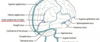
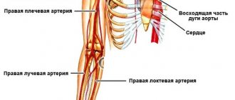
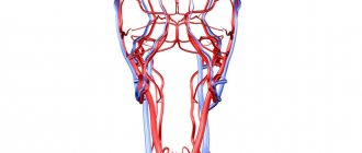
The arterial system of the body consists of many sections responsible for the blood supply to individual organs and structures. The main, most important and largest branches of the system are called trunks and are divided into several mains. At the exit from the left ventricle there is a trunk of large arteries, the beginning of which is the aorta. It continues with an ascending vessel and forms an arch from which the common subclavian and brachiocephalic trunks branch. The latter, in turn, branches into paired carotid and subclavian arteries on the right. From this root portion of the aorta (aortic bulb) the coronary network branches off.
As they move upward, the vessels divide into paired carotid arteries, one of which is responsible for supplying the outer membranes of the head (face, skull, neck), and the other for supplying the brain and eyes. The subclavian branches are divided into paired vertebrates, which are responsible for the blood supply to the chest and diaphragm, the upper part of the sternum. The subclavian tube located at the top of the chest gradually passes into the shoulder areas, which are responsible for the blood supply to the upper extremities. This system is represented by the brachial, radial, ulnar, superficial and deep arteries.
The descending part of the aorta is the beginning of the vessels responsible for the blood supply to the abdominal organs, the vessels supplying the anterior abdominal wall, external genitalia and lower extremities. Several trunks extend from the descending arc:
- multiple paired external intercostal arteries and internal branches that deliver blood to structures and organs located in the chest;
- the abdominal aorta, from which arise many paired (renal, ovarian) and unpaired (gastric, hepatic, etc.) large arteries that supply blood to the abdominal organs;
- as it descends, the main arteries, called iliac, depart from one tube: the internal one supplies the organs of the genitourinary system with blood, and the external one passes into the femoral part of the circulatory system;
- The femoral tubes, as they move down, pass into the popliteal, then into the tibial, peroneal and plantar vessels.
Most of the vessels of the extremities are represented by mixed-type arteries. Only the aorta and the main trunks of the thoracic and abdominal aorta are classified as elastic. Almost all systems have arterial anastomoses - “side” ducts connecting the vessels of one part of the circulatory system. They play the role of bypass channels, which are activated if the conductivity of the main highways deteriorates.
Small arterial branches gradually narrow and branch, forming arterioles and then precapillaries. The diameter of these tubes rarely exceeds 2 mm, and their walls are dominated by the muscle layer.
Definition
Vessels of the legs - arteries, arterioles, capillaries, veins and venules that supply blood to the tissues of the lower extremities.
Healthy leg blood vessels are an important component of human health. They supply the legs with blood, oxygen and nutrients, allowing a person to lead a full, active life. When the vessels of the legs are exposed to diseases, without proper and timely treatment this leads to serious consequences. The arteries of the lower extremities are paired. Here are the main ones:
- femoral artery (continuation of the iliac artery)
- popliteal artery (divides into tibial arteries)
- artery of the foot.
The veins of the lower extremities are divided into superficial and deep. The deep veins are paired; they accompany the arteries and participate in the process of blood drainage. Excessively dilated blood vessels in the legs are not only an aesthetic problem, but also a sign of pathological processes in the body.
Pathologies
The arterial network is characterized by congenital and acquired pathologies of a local and systemic nature. The most common and dangerous are acquired arterial diseases:
- aortic dissection;
- vascular aneurysms;
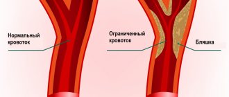
- sclerotic changes;
- deposits of lipoproteins with the formation of plaques;
- arterial stenosis, etc.
Almost all of the listed arterial diseases are the result of a violation of the internal environment of the body. These include imbalances of hormones, metabolism, and metabolic processes. For example, aortic dissection, stenosis and aneurysms are typical consequences of increased stress on the circulatory system due to hypertension, which develops in older people. Numerous age-related changes occur in their body, which are based on a slowdown in metabolic and metabolic processes and a decline in the synthesis of sex hormones.
The most common pathology of the arterial system is considered to be atherosclerosis, caused by the accumulation of lipids (cholesterol) in the blood and its deposition on the walls.
An imbalance of lipid metabolism plays a major role in this disease. Congenital arterial diseases are represented by an extensive list of anomalies in the structure of blood vessels. These include arteriovenous fistulas - ducts between arteries and veins that provoke hemodynamic disturbances. Such diseases can manifest themselves as local symptoms (atypical vascular pattern on the skin), regional signs (hypertrophy or atrophy of organs, body parts) or general disorders (heart failure, brain pathologies, etc.).