The Innovative Vascular Center has all the necessary technologies for the treatment of acute and chronic occlusion of the inferior vena cava. Our specialists have successful experience in solving this complex problem.
Acute thrombosis of the inferior vena cava requires treatment in a specialized vascular surgery hospital. The goal of treatment is to restore the patency of the IVC. This problem is successfully solved using endovascular surgery methods. There are modern thrombolytic drugs and endovascular probes for removing thrombotic masses.
Causes of SVPV
The main cause of superior vena cava syndrome is obstruction, which is based on pathological processes such as:
- germination of the vein wall by a cancerous tumor;
- compression of the vein from the outside;
- thrombosis of a valveless vessel.
In 90% of cases, the cause of obstruction is the presence of a malignant process in the body. At the same time, lung cancer accounts for 85% of tumors (squamous or small cell cancer). In more rare cases, the cause is lymphoma and lymphogranulomatosis (tumor disease of the lymphatic system). Metastases of other malignant neoplasms (breast, testicle) contribute to the development of SVPV.
Among the causes of Kava syndrome, the following should also be highlighted:
- benign tumors;
- aortic aneurysm;
- enlargement of the thyroid gland;
- fibrous mediastinitis (inflammatory process in the mediastinal tissue).
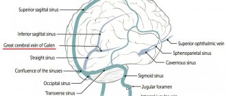
Blockage of blood outflow from the valveless vessel and the head is fraught with the development of a number of pathophysiological effects. Venous return to the right ventricle is impaired and cardiac output is reduced. Systemic hypotension (decreased arterial pressure) and increased venous pressure in the SVC system are also observed, which is fraught with the development of cerebral vascular thrombosis. Due to a decrease in the arterial-venous gradient, irreversible changes in the brain region may develop.
In lung cancer, the patency of a valveless vessel, as a rule, is preserved, despite the fact that invasion has already occurred. Only 10-20% of patients with tumor SVPV live more than 2 years. The life expectancy of such patients, according to statistics, does not exceed 10 months.
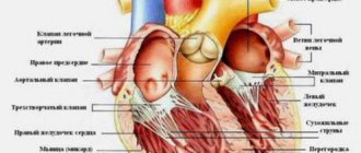
Anatomy of the superior and inferior vena cava
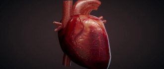
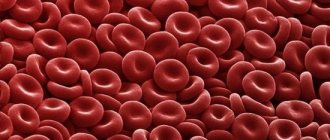
Many people remember from high school anatomy course that both vena cavae carry blood to the heart. They have a fairly large lumen in diameter, where all the venous blood flowing from the tissues and organs of our body is placed. Heading towards the heart from both halves of the body, the veins connect into the so-called sinus, through which the blood enters the heart, and then goes to the pulmonary circle for oxygenation.
System of the inferior and superior vena cava, portal vein - lecture
Superior vena cava
superior vena cava system
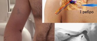
The superior vena cava (SVC) is a large vessel about two centimeters wide and approximately 5-7 cm long, carrying blood away from the head and upper half of the body and located in the anterior part of the mediastinum. It lacks a valve apparatus and is formed by connecting the two brachiocephalic veins posterior to the place where the first rib is connected to the sternum on the right. The vessel goes almost vertically down to the cartilage of the second rib, where it enters the cardiac sac, and then, in the projection of the third rib, enters the right atrium.
Anterior to the SVC is the thymus and parts of the right lung; on the right it is covered with the mediastinal layer of the serous membrane, on the left it is adjacent to the aorta. Its posterior part is located anterior to the root of the lung; the trachea is located behind and slightly to the left. The vagus nerve passes through the tissue behind the vessel.
The SVC collects blood flow from the tissues of the head, neck, arms, chest and abdominal cavity, esophagus, intercostal veins, and mediastinum. The azygos vein from behind and the vessels carrying blood from the mediastinum and pericardium flow into it.
Video: superior vena cava - formation, topography, inflow
Inferior vena cava
The inferior vena cava (IVC) lacks a valve apparatus and has the largest diameter of all venous vessels. It begins by uniting two common iliac veins, its mouth is located to the right than the zone of branching of the aorta into the iliac arteries. Topographically, the beginning of the vessel is located in the projection of the intervertebral disc of the 4-5 lumbar vertebrae.
The IVC is directed vertically upward to the right of the abdominal aorta; behind it actually lies on the psoas major muscle of the right half of the body; in front it is covered with a layer of serous membrane.
Going to the right atrium, the IVC is located behind the duodenum, the root of the mesentery and the head of the pancreas, enters the liver groove of the same name, where it connects with the hepatic venous vessels. Further on the path of the vein lies the diaphragm, which has its own opening for the inferior vena cava, through which the latter goes up and goes in the posterior mediastinum, reaches the cardiac sac and connects with the heart.
The IVC collects blood from the veins of the lower back, the lower phrenic and visceral branches coming from the internal organs - ovarian in women and testicular in men (the right ones flow directly into the vena cava, the left ones into the renal vena cava on the left), renal (run horizontally from the hilum of the kidneys), right adrenal vein (the left one is connected directly to the renal vein), hepatic.
The inferior vena cava takes blood from the legs, pelvic organs, abdomen, and diaphragm. The fluid moves through it from bottom to top; to the left of the vessel, almost its entire length lies the aorta. At the entrance to the right atrium, the inferior vena cava is covered by the epicardium.
Video: inferior vena cava - formation, topography, inflow
Pathology of the vena cava
Changes in the vena cava are most often secondary in nature and associated with diseases of other organs, therefore they are called superior or inferior vena cava syndrome, indicating that the pathology is not independent.
Symptoms of SVC syndrome
Symptoms of superior vena cava syndrome develop against the background of venous hypertension. Signs of pathological disorders directly depend on the progression of obstruction, its degree and area of localization. The degree of progression of collaterals (side or bypass tracts of blood flow) is also important. It is also a component of SVPV.
The course of the disease can be either slowly progressive or acute. Complaints can be quite varied, ranging from changes in appearance and slight dizziness to convulsions and fainting. On physical examination, veins in the neck, arms, and chest wall are swollen and dilated. The picture of the disease in many patients is blurred. Physical diagnosis includes the Pemberton test - raising your arms up. With occlusion of a valveless vessel, cyanosis of the skin of the neck and face is observed, as well as injection of conjunctival vessels.
Clinical signs of superior vena cava syndrome in cancer are conventionally divided into several groups. The result of stagnation in superficial and deep structures:
- swelling in the arms, face and body;
- asphyxia (suffocation) – when swelling moves to the vocal cords;
- when the capillaries narrow, cyanosis develops, which is characterized by an earthy-pale skin color (occurs against the background of lymphostasis - a pathological accumulation of protein-rich fluid).
Congestion in the brain area is accompanied by general cerebral symptoms: characteristic shortness of breath, constant headaches, and frequent attacks of suffocation. With prolonged disturbances, worsening laryngeal edema is noted.
As a result of ongoing disorders, dramatic changes occur in the brain area, which are fraught with the development of emotional fatigue and drowsiness. Attacks of suffocation are accompanied by loss of consciousness, which is a sign of cerebral hypoxia, which develops against the background of ongoing circulatory disorders. Among the most common manifestations of the pathological process, one should highlight the pronounced occurrence of auditory hallucinations and severe confusion.
Among the manifestations of cranial nerve disorders, diplopia (double vision) and a pronounced deterioration in hearing qualities should be highlighted. Such signs are caused by nerve disorder and are accompanied by decreased visual function and tearfulness. It is possible that intraocular pressure may increase.
In order to more fully characterize the patient’s condition with SVPV, the third group of clinical signs caused by the underlying disease should be studied. They manifest themselves in the form of cough, cachexia and hemoptysis. Superior vena cava syndrome in oncology is accompanied by nasal and esophageal bleeding, which occurs due to thinning of the venous walls. During physical activity, hand fatigue occurs quite quickly. It becomes difficult to perform even minor physical work, which is associated with a rush of venous blood to the head. Heart pain, increased heart rate and compression behind the chest are the consequences of disruption of the blood supply to the heart muscle and swelling of the mediastinal tissue.
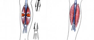
Clinical manifestations of May-Turner syndrome
Unfortunately, the syndrome is not well studied because it is rare. Experts distinguish three stages of its development:
- First, there are no clinical manifestations;
- Second, the lumen of the iliac vein narrows, symptoms of venous insufficiency appear;
- Third, thrombosis develops.
The symptoms of the syndrome are as follows:
- Swelling of the left leg, often with a change in the color of the skin to bluish or purple;
- Intense pain symptoms in the affected area that cannot be eliminated with painkillers;
- Varicose veins visible to the naked eye on the thigh, testicle (in men) or labia (in women) on the affected side;
- Increased discomfort after physical activity, including minor ones;
- The appearance of hemorrhoids.
Both men and women suffering from the syndrome have problems with sexual intercourse. So, in the first case, pulling pains appear in the scrotum, which radiate to the urethra and head of the penis and become more intense after sexual intercourse. In the second case, sexual contacts become completely impossible due to the pain of the node.
Complications
Often, with SVPV, there is the development of bleeding (esophageal, nasal or pulmonary), which develops as a result of rupture of the vascular walls. Due to impaired venous outflow in the head area, cerebral symptoms develop:
- confusion and fainting;
- cramps and weakness of the limbs;
- frequent pain in the head area.
When the nerves are damaged, exophthalmos develops (mono- or binocular anterior displacement of the eyeball), diplopia (double vision), lacrimation, blurred vision and rapid eye fatigue. Hearing impairment, tinnitus and auditory hallucinations are possible.
Diagnosis of Cava syndrome
There are two main stages in diagnosis. This is an initial examination in non-core clinics. The basis is the classic clinical picture and the result of radiography in lateral and direct projection. If there are pathological abnormalities, there is additional darkening in the image. If SVPV is suspected, the patient is sent to a specialized medical facility.
Diagnosis of superior vena cava syndrome includes:
- FBS (fibre-optic bronchoscopy)
– is prescribed for topographic-anatomical localization of the pathological process. - An incisional or puncture biopsy
is performed when peripheral lymph nodes are affected. - Angiography
is used in rare cases and is aimed at determining the extent of the obstruction.
The effectiveness of diagnostic measures at the second stage of the examination is determined by the proportion of morphologically verified diagnoses. Conducted studies prove that the ability to perform certain diagnostic procedures is associated with the degree of progression.
Treatment of superior vena cava syndrome in cancer patients
In case of serious condition of patients, emergency care is required. In this case, there is no time left to conduct a full examination and determine the morphology of the tumor. Treatment measures include taking steps to improve venous outflow through the valveless vessel. There is a need to eliminate respiratory failure, restore cerebral circulation and relieve possible pulmonary edema.
In severe cases, treatment is prescribed aimed at combating dangerous symptoms - the doctor prescribes antitumor drugs that are effective against most malignant neoplasms. With the right approach, the patient's condition quickly normalizes.
Features of treatment of superior vena cava syndrome:
- Symptomatic
. Includes taking measures aimed at increasing the functional reserves of the body. The doctor prescribes a low-salt diet, diuretics, oxygen inhalations and glucocorticoids. - Pathogenetic
. It is prescribed after stopping the main cause of the pathological process. For lymphogranulomatosis, lymphoma and lung cancer, radiation and polychemotherapy are performed. In case of thrombosis, thrombectomy is performed, thrombolytic therapy is prescribed, or a segment of the SVC is resected.
In the case of extravasal compression, radical surgery is performed, which involves excision of a mediastinal tumor, lymphoma, cysts, benign neoplasms, etc. If radical surgery is not possible, then palliative procedures are resorted to (bypass surgery, SVC stenting, percutaneous balloon angioplasty).
At the Medscan clinic in Moscow, treatment of superior vena cava syndrome is carried out on the basis of international protocols. The medical center uses the latest expert-class equipment. Doctors with extensive practical experience are constantly retrained to improve their level of competence. The results of treatment for SVPV primarily depend on the effectiveness of radical therapy for the underlying disease. Superior vena cava syndrome is unfavorable in pediatric oncology. Eliminating the causes of Kava syndrome allows you to stop its consequences and prevent the development of possible complications.
Treatment of pathology
The goal of treatment is to relieve the patient of the symptoms of the pathology. But there is another, primary goal - saving lives. This is exactly what needs to be done at the first stage of treatment. The situation of a patient with primary pathology is extremely serious; he requires oncological resuscitation.
If pathology is the first symptom of cancer, then treatment prospects are good, since a number of forms of oncology are cured under the influence of chemotherapy.
If after surgery and therapeutic treatment are completed, but the pathology is not defeated due to the fact that the oncology is progressing, there are practically no prospects for a cure. Here we need to strive to improve the quality of life and prolong it.
At the Onco.Rehab integrative oncology clinic, all conditions have been created for the treatment of various pathologies and their symptoms. We help our patients.