Veins of the systemic circulation. Lymphatic system
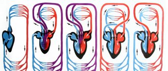
All venous blood from the organs of the human body flows to the right, venous system of the heart through the two largest trunks - the superior and inferior vena cava. Only the heart's own veins drain directly into the right atrium, bypassing the vena cava.
The veins of the systemic circulation are divided into three systems:
1. Superior vena cava.
2. Inferior vena cava.
3. Portal vein.
The superior vena cava (v. Cava superior) is a vessel 5-6 cm long and 25 mm in diameter. It is located to the right of the aorta and is formed by the confluence of the right and left brachiocephalic veins. It is formed by vessels that collect venous blood from the head, neck, upper limb, chest and abdominal cavities.
The main venous collector of the head and neck organs is the internal jugular vein and partially the external jugular vein.
The internal jugular vein carries blood from the cranial cavity and from the organs of the neck. All tributary veins are divided into intracranial and extracranial.
To intracranial
These include veins that collect blood from the cerebral hemispheres.
To extracranial
include
facial, retromaxillary, pharyngeal, lingual, thyroid.
External jugular vein
begins behind the auricle.
The occipital, posterior auricular, and anterior jugular veins
flow into it . N.i.v. flows into the confluence of the subclavian and internal jugular veins or directly into the subclavian vein.
The subclavian vein is a continuation of the axillary vein, merges with the internal jugular vein, and collects blood from the upper limb.
The veins of the upper limb are divided into superficial and deep.
The superficial (saphenous) veins widely anastomose with each other, form a wide looped network, which forms two large trunks: the lateral saphenous vein
- located on the side of the radius and flows into the axillary vein and
medial saphenous vein
- located on the side of the ulna and flows into the brachial vein.
In the elbow flexure, the lateral and medial veins are connected by the intermediate vein of the elbow.
The deep veins are accompanied by arteries of the same name, two each, and have the same names. Their roots are the digital veins, which flow into the palmar arches, moving to the forearm, two ulnar veins
and two
radial
veins.
In the area of the cubital fossa they merge to form two brachial veins, then form the axillary vein. At the outer edge of the 1st rib, the axillary vein passes into the subclavian vein.
The confluence of the subclavian vein and the internal jugular vein is called the venous angle.
As a result of this connection,
the brachiocephalic veins are formed. The veins of the thymus, mediastinum, esophagus, trachea, pericardium, neck muscles, etc. flow into them. The brachiocephalic veins form the superior vena cava , which does not have valves, which at the level of the 2nd rib flows into the right atrium.
Inferior vena cava. (v. Cava inferior) Thick trunk, lies in the abdominal cavity next to the aorta. It is formed at the level of the 4th lumbar vertebra from the confluence of two common iliac veins. The parietal (parietal) and visceral (visceral) veins flow into it.
The common iliac veins are formed from the confluence of the external and internal iliac veins.
The internal iliac vein is located behind the arteries of the same name and is formed from the confluence of the sacral veins, rectal veins, and bladder veins.
External iliac vein
- continuation of the femoral vein. There are veins on the lower limb:
1. Deep; 2. Superficial;
Deep –
double and accompany the arteries of the same name.
Superficial
– form two large trunks:
the great and small saphenous veins.
Big –
originates on the foot along the medial side, rises to the thigh and flows into the femoral vein.
Small –
It rises from the foot along the lateral side, on the lower leg it passes to the back surface and in the popliteal fossa it flows into
the popliteal vein,
which then passes into the femoral vein.
Portal vein (v. Portae)
Collects blood from all unpaired organs of the abdominal cavity with the exception of the liver.
From the entire gastrointestinal tract, where nutrients are absorbed, they enter the portal vein into the liver for neutralization and deposition of glycogen.
This is a thick venous trunk that is formed from the confluence of the splenic vein, superior and inferior mesenteric veins.
On the way to the liver, it takes the gastric vein and at the porta hepatis it divides into two branches, which in the liver parenchyma (in the tissue) break up into small branches and then gather into the central vein
, which leaves the liver and flows into the inferior vena cava.
content .. 150 151 152 153 154 155 156 157 158 159 ..Veins (human anatomy)
Veins of the systemic circulation (human anatomy)
Veins of the systemic circulation
connect to the final large collectors - the superior vena cava, the inferior vena cava and the coronary sinus of the heart, which flow into the right atrium.
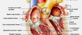
The veins of the heart, venae cordis, are mainly collected in the coronary sinus of the heart, which is formed from the veins of the heart wall (see section Veins of the heart, this edition).
Superior vena cava system (human anatomy)
Superior vena cava
, v. cava superior, formed by the brachiocephalic veins (right and left), vv. brachiocephalicae dextra et sinistra, which are formed from the fusion of the right and left internal jugular veins, vv. jugulares internae, and right and left subclavian veins, vv. subclaviae dextra et sinistra (Fig. 169). The azygos vein, v., also flows into the superior vena cava. azygos.
Rice. 169. Superior vena cava, brachiocephalic veins and their tributaries. 1 - a. facialis; 2, 3 - v. facialis; 4 - v. jugularis interna; 5 - v. jugularis externa; 6 - v. jugularis anterior; 7 - arcus venosus juguli; 8 - v. brachiocephalica sinistra; 9 - a. subclavia; 10 - v. subclavia; 11 - v. thoracica interna; 12 - arcus aortae; 13 - v. cava superior; 14 - v. thyreoidea ima; 15 - v. cephalica; 16 - v. transversa colli
For better assimilation, the structure of the veins is considered according to the blood flow, starting from small veins and ending with large vessels.
Internal jugular vein
, v. jugularis interna, steam room, is formed from the veins of the brain, its membranes and the veins of the face.
Veins of the cerebrum, vv. cerebri, are divided into superficial, formed in the cerebral cortex, and deep, located in the central parts of the hemispheres.
The superficial veins of the brain include the following.
1. Superior cerebral veins, vv. cerebri superiores, collect blood from the cortex of the dorsolateral surface of the cerebral hemispheres, forming a network of veins in the pia mater. Large venous vessels are located mainly in the cortical grooves, vv. cerebri superiores, pierce the arachnoid membrane and flow into the sinus sagittalis superior.
2. Superficial middle cerebral vein, v. cerebri media superficialis, a paired, large vein, passes in the central groove and connects the sinus sagittalis superior and sinus cavernosus.
3. Anterior cerebral vein, v. cerebri anterior, originates on the medial surface of the cerebral hemispheres, extends to the base and connects the great cerebral vein with the inferior sagittal sinus.
4. Inferior cerebral veins, vv. cerebri inferiores, originate in the basal cortex of the brain, flow into the sinus cavernosus, inter cavernosus.
5. Main vein, v. basalis, forms in the area of the substantia perforata anterior, then accompanies the optic nerve tract. Having gone around the cerebral peduncles, it flows over the pineal gland into v. cerebri magna.
6. Superior veins of the cerebellum, vv. cerebelli superiores, begin on the superior surface of the cerebellar hemispheres, flow into the sinus rectus and v. cerebri magna.
7. Inferior veins of the cerebellum, vv. cerebelli inferiores, located on the lower surface of the cerebellum, anastomose with the previous ones. They flow into sinus transversus and sinus petrosus inferior.
The deep veins of the cerebral hemispheres begin in the basal ganglia and white matter. They are represented by the following trunks.
1. Internal veins of the cerebrum, vv. cerebri internae, collect blood from the white matter of the cerebral hemispheres, the walls of the ventricles, the optic thalamus and the basal ganglia. In the transverse fissure of the brain near the quadrigeminal, all branches of the veins merge into the large vein of the brain, v. cerebri magna.
2. Vein of the choroid plexus, v. chorioidea, is formed from the veins of the choroid plexus of the lateral ventricle, penetrating through the foramen interventricularis into the central part of the lateral ventricle and in the transverse sulcus of the brain flows into v. cerebri magna.
3. Veins of the transparent septum, vv. septi pellucidi, begin in the substance of the brain, forming the anterior horn of the lateral ventricle. They flow into v. chorioidea.
4. Great vein of the brain, v. cerebri magna, single, represents a short trunk, 0.5-1 cm long. It is formed from the fusion of the listed branches of the deep veins. In the transverse groove of the brain above the quadrigeminal, it flows into the sinus rectus.
content .. 150 151 152 153 154 155 156 157 158 159 ..
Topic 9.3 Veins of the systemic circulation Lymphatic system
Topic 9.3 Veins of the systemic circulation
Lymphatic system
Venous vessels of the systemic circulation are represented by: the superior vena cava system, the inferior vena cava system and the portal vein system
To the superior vena cava system
These include all venous vessels that collect blood from the head, brain, neck, upper limbs, from the walls of the chest cavity, organs of the chest cavity, and also partially from the abdominal cavity. They are represented by the brachiocephalic veins, internal jugular, subclavian, veins of the upper extremities, azygos and semi-gypsy veins.
Inferior vena cava
collects blood from the lower extremities, walls and organs of the pelvis and abdominal cavity.
Portal vein
collects blood from unpaired abdominal organs; spleen, stomach, gall bladder, pancreas, small and large intestines. This is a short thick trunk that runs deep into the hepatoduodenal ligament.
| Vein | Main branches | Area, organ from which blood is collected |
| I. Superior vena cava system | Head, neck, upper limbs, upper body | |
| 1. Veins head and neck | Superficial veins: external jugular vein | Temporal, parietal and occipital areas of the head, ears, anterior and lateral areas of the neck |
| Deep veins: internal jugular vein | The brain and its membranes; anterior and lateral areas of the face, tongue, pharynx, larynx, thyroid gland | |
| 2. Veins of the upper limb | Superficial veins | |
| Lateral saphenous vein | Skin, subcutaneous tissue of the lateral parts of the upper limb | |
| Medial saphenous vein | Skin, subcutaneous tissue of the medial parts of the upper limb | |
| Deep veins | ||
| Radial vein - steam room | Muscles, ligaments, bones of the lateral sides of the hand and forearm | |
| Ulnar vein - steam room | Muscles, ligaments, bones of the medial sides of the hand and forearm | |
| The brachial vein is initially a steam vein, then the two veins merge into one trunk | Free part of the upper limb (skin, ligaments, muscles, hand bones, forearm, shoulder) | |
| Axillary vein | Free part of the upper limb, skin, subcutaneous tissue of the lateral sections of the chest wall | |
| Subclavian vein | Upper limb, upper anterior and lateral chest wall | |
| 3. Veins of the chest | Azygos vein | Posterior wall of the abdomen and chest cavity, mediastinal organs |
| Hemizygos vein | Posterior wall of the abdomen and left half of the chest cavity, mediastinal organs | |
| Brachiocephalic vein | The anterior wall of the abdomen and chest cavity, mediastinal organs, thyroid gland, thymus, larynx, cervical spinal cord and its membranes, deep muscles of the neck, head, neck, upper limbs | |
| II. Inferior vena cava system | Lower limbs, walls and organs of the pelvis, diaphragm (partially), posterior, lateral and part of the anterior wall of the abdominal cavity, paired abdominal organs, unpaired abdominal organs (through the venous vascular system of the liver) | |
| Vein | Main branches | Area, organ from which blood is collected |
| 1. Veins of the lower limb | Superficial veins | |
| Great saphenous vein legs | Skin and subcutaneous tissue of the anteromedial parts of the foot, lower leg and thigh, external genitalia, anterior abdominal wall | |
| Small subcutaneous leg vein | Skin and subcutaneous tissue posterolateral sections of the bone and lower leg | |
| Deep veins | ||
| Anterior tibial vein—paired | Muscles, ligaments, bones of the dorsum of the foot and the front of the leg | |
| Posterior tibial vein - steamed | Muscles, ligaments, bones of the sole of the foot and the back of the leg | |
| Popliteal vein | Skin, ligaments, muscles, bones of the foot, leg and knee | |
| Femoral vein | Skin, ligaments, muscles, bones of the foot, lower leg, thigh, skin and subcutaneous tissue of the external genitalia, anterior abdominal wall | |
| 2. Veins of the pelvis | External iliac vein | Free part of the lower limb, anterior abdominal wall, external genitalia |
| Internal iliac vein | Walls and organs of the pelvis, external and internal genitalia | |
| Common iliac vein | Walls and organs of the pelvis, external and internal genitalia, lower limb | |
| III. Portal vein system | Unpaired organs of the abdominal cavity (stomach, small and large intestine, pancreas, spleen) | |
| Splenic vein | Spleen, area of the bottom and posterior wall of the body of the stomach, body and tail of the pancreas, left half of the greater omentum | |
| Superior mesenteric vein | Small intestine and its mesentery, cecum, ascending and right half of the transverse colon, appendix, head and part of the body of the pancreas, right half of the body of the stomach and greater omentum | |
| Inferior mesenteric vein | Upper rectum, sigmoid colon, descending colon and left transverse colon | |
| Portal vein | Receives venous blood from the splenic, superior mesenteric and inferior mesenteric veins | |
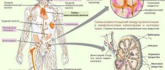
The lymphatic system is a closed part of the vascular system; it complements the venous bed, with which it jointly drains organs through the formation of lymph.
Along with this, the lymphatic system performs specific functions: transport - it transfers metabolic products from tissues into the blood, and into tissues - nutrients and hormones; hematopoietic and lymph-forming - forms lymphocytes; barrier - retains some particles (foreign) and microbial bodies, tumor cells; immune - produces immune bodies responsible for immunity.
The lymphatic system includes: lymph, lymphatic bed (capillaries, intra- and extraorgan vessels, lymphatic trunks and ducts), as well as lymphoid organs (lymph nodes).
Lymphatic system (general diagram)
1 - parotid lymph nodes; 2 - submandibular lymph nodes; 3 - cervical lymph nodes; 4 - superior vena cava; 5 - axillary lymph nodes; 6 - thoracic duct; 7 - cistern of the thoracic duct;
8 - inferior vena cava; 9 - iliac lymph nodes; 10 - superficial lymphatic vessels of the upper limb; 11 - inguinal lymph nodes;
12 - superficial lymphatic vessels of the lower limb; 13 - right lymphatic duct