Historical reference
In the past, when scientists did not yet have informative instruments at hand that could study physiological processes in a living organism, the greatest scientists were forced to search for anatomical features in corpses. Naturally, the heart of a deceased person does not contract, so some nuances had to be figured out on their own, and sometimes simply fantasized. Thus, back in the second century AD, Claudius Galen, studying from the works of Hippocrates himself, assumed that the arteries contained air instead of blood in their lumen. Over the next centuries, many attempts were made to combine and link together the existing anatomical data from the standpoint of physiology. All scientists knew and understood how the circulatory system works, but how does it work?
Miguel Servetus and William Harvey made a tremendous contribution to the systematization of data on the work of the heart in the 16th century. Harvey, the scientist who first described the systemic and pulmonary circulation , in 1616 determined the presence of two circles, but he could not explain in his works how the arterial and venous beds were connected. And only later, in the 17th century, Marcello Malpighi, one of the first to use a microscope in his practice, discovered and described the presence of tiny capillaries, invisible to the naked eye, which serve as a connecting link in the blood circulation.
Phylogeny, or the evolution of blood circulation
Due to the fact that, as animals of the vertebrate class evolved, they became more and more progressive in anatomical and physiological terms, they required a complex structure of the cardiovascular system. Thus, for faster movement of the liquid internal environment in the body of a vertebrate animal, the need for a closed blood circulation system arose. Compared to other classes of the animal kingdom (for example, arthropods or worms), the rudiments of a closed vascular system appear in chordates. And if the lancelet, for example, does not have a heart, but there is an abdominal and dorsal aorta, then in fish, amphibians (amphibians), reptiles (reptiles) a two- and three-chambered heart appears, respectively, and in birds and mammals a four-chambered heart appears, the peculiarity of which is is the focus in it of two circles of blood circulation that do not mix with each other.
Thus, the presence of two separated circulatory circles in birds, mammals and humans, in particular, is nothing more than the evolution of the circulatory system, necessary for better adaptation to environmental conditions.
Major organs
It is believed that in mammals the circulatory system has reached its highest development from the animal kingdom, which is explained by the rate of metabolism, as well as the peculiarities of the biology of animals of this class. It has a closed structure and consists of numerous vessels and a four-chambered heart. The systemic circulation in mammals begins with the heart and passes through the lungs. Blood through the vessels can flow only in one, strictly defined direction.
The main elements of the circulatory system of mammals are:
- heart;
- veins;
- capillaries;
- arteries.
Depending on the specific type of animal, the structure of such internal organs and vessels may vary.
For example, in the smallest creatures the weight of the heart reaches several grams, while in a huge blue whale such a muscle, which pumps blood, weighs about a hundred kilograms and is even larger in size than a person. Vessels and capillaries also differ significantly, including in the thickness of their walls and elasticity indicators.
Scientists in the last century determined how many circles of blood circulation there are in mammals, and established all the features of the heart and blood vessels.
Anatomical features of the blood circulation
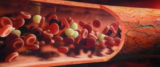
The circulatory system is a set of blood vessels, which is a closed system for the supply of oxygen and nutrients to the internal organs through gas exchange and nutrient exchange, as well as for the removal of carbon dioxide and other metabolic products from cells. The human body is characterized by two circles - the systemic, or large circle, and the pulmonary, also called the small circle.
Video: blood circulation circles, mini-lecture and animation
Systemic circulation
The main function of the large circle is to ensure gas exchange in all internal organs except the lungs. It begins in the cavity of the left ventricle; represented by the aorta and its branches, the arterial bed of the liver, kidneys, brain, skeletal muscles and other organs. Further, this circle continues with the capillary network and venous bed of the listed organs; and through the entry of the vena cava into the cavity of the right atrium it ends in the latter.
So, as already said, the beginning of the great circle is the cavity of the left ventricle. Arterial blood flow, which contains more oxygen than carbon dioxide, is sent here. This flow enters the left ventricle directly from the circulatory system of the lungs, that is, from the small circle. The arterial flow from the left ventricle is pushed through the aortic valve into the largest great vessel - the aorta. The aorta can be figuratively compared to a kind of tree that has many branches, because arteries extend from it to the internal organs (to the liver, kidneys, gastrointestinal tract, to the brain - through the system of carotid arteries, to skeletal muscles, to the subcutaneous fat fiber, etc.) Organ arteries, which also have numerous branches and bear names corresponding to their anatomy, carry oxygen to each organ.
In the tissues of internal organs, arterial vessels are divided into vessels of smaller and smaller diameter, and as a result, a capillary network is formed. Capillaries are the smallest vessels, practically without a middle muscular layer, and are represented by an inner membrane - intima, lined with endothelial cells. The gaps between these cells at the microscopic level are so large compared to other vessels that they allow proteins, gases and even formed elements to easily penetrate into the intercellular fluid of the surrounding tissues. Thus, intense gas exchange and exchange of other substances occurs between the capillary with arterial blood and the liquid intercellular medium in a particular organ. Oxygen penetrates from the capillary, and carbon dioxide, as a product of cell metabolism, enters the capillary. The cellular stage of respiration occurs.
After more oxygen has passed into the tissues and all carbon dioxide has been removed from the tissues, the blood becomes venous. All gas exchange occurs with each new influx of blood, and during the period of time while it moves along the capillary towards the venule - a vessel that collects venous blood. That is, with each cardiac cycle, in one or another part of the body, oxygen enters the tissues and carbon dioxide is removed from them.
These venules unite into larger veins, and a venous bed is formed. Veins, similar to arteries, are named according to the organ in which they are located (renal, cerebral, etc.). From large venous trunks, tributaries of the superior and inferior vena cava are formed, and the latter then flow into the right atrium.
Respiratory system of amphibians
The respiratory system of amphibians is a rather complex complex. Amphibians have lungs in the form of pouches with thin walls and a cellular structure. However, amphibians breathe well through their skin. Unlike fish, they no longer breathe through gills.
The breathing process begins with inhalation, which is carried out through raising and lowering the floor of the mouth. Exhalation occurs through the mouth and nostrils. Underwater, breathing occurs through the skin. Oxygen in water first enters the fluid covering the animal, and then into the blood.
The skin of toads is coated with special mucus secreted from the glands, and the skin itself is equipped with many capillaries. For this reason, the skin participates in gas exchange.
In the cold season, when amphibians are least active, they have to breathe through the skin, but when it gets warmer, skin respiration supplies only 25% to 50% of the total oxygen requirement. In general, only these two types of breathing together will be able to fully ensure gas exchange, despite their different functions.
Mouth breathing is carried out due to the presence of capillary-rich epithelium in the oral cavity. Inhalation is made through the nostrils, the glottis is closed, and the floor of the oral cavity is lowered.
Exhalation occurs due to raising the floor of the oral cavity and contraction of the petrohyoid muscles.
The lungs are hollow organs. The surface of the pulmonary walls is quite small. Almost the entire epithelium of the lungs is supplied with mucus and blood. A bronchus departs from each lung, which then unites into a trachea that communicates with the oral cavity.
In order to ensure pulmonary breathing, the animal has a need to repeat strong swallowing movements. This is what toads and frogs do, for example.
Features of blood flow in the organs of the systemic circle
Some of the internal organs have their own characteristics. So, for example, in the liver there is not only a hepatic vein, which “carries” the venous flow away from it, but also a portal vein, which, on the contrary, brings blood to the liver tissue, where blood purification is performed, and only then the blood collects in the tributaries of the hepatic vein to enter to a big circle. The portal vein brings blood from the stomach and intestines, so everything that a person eats or drinks must undergo a kind of “purification” in the liver.
In addition to the liver, certain nuances exist in other organs, for example, in the tissues of the pituitary gland and kidneys. Thus, in the pituitary gland the presence of a so-called “wonderful” capillary network is noted, because the arteries that bring blood to the pituitary gland from the hypothalamus are divided into capillaries, which then collect into venules. The venules, after the blood with the molecules of releasing hormones are collected, are again divided into capillaries, and then veins are formed that carry the blood from the pituitary gland. In the kidneys, the arterial network is divided twice into capillaries, which is associated with the processes of excretion and reabsorption in the kidney cells - in the nephrons.
Blood pressure readings
It has been established that blood pressure in mammals is similar to that of birds, but at the same time much higher than in amphibians and reptiles. In dogs, this parameter is 112 over 56, in elephant seals - 120 over 90, and in rats - 130 over 90 millimeters of mercury.
Blood pressure in mammals can vary depending on the intensity of blood pumping through the vessels, heart rate, current activity and a number of other factors.
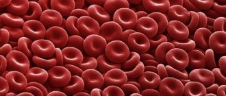
Also, the organs of the circulatory system of mammals are characterized by an increased number of red blood cells and a high hemoglobin content.
Veins and capillaries have a closed system, so the outflow of lymph into cavities and internal organs with tissues does not occur here.
Pulmonary circulation
Its function is to carry out gas exchange processes in the lung tissue in order to saturate the “waste” venous blood with oxygen molecules. It begins in the cavity of the right ventricle, where venous blood flow with an extremely small amount of oxygen and a large content of carbon dioxide enters from the right atrial chamber (from the “end point” of the great circle). This blood moves through the pulmonary valve into one of the large vessels called the pulmonary trunk. Next, the venous flow moves along the arterial bed in the lung tissue, which also breaks up into a network of capillaries. By analogy with capillaries in other tissues, gas exchange occurs in them, only oxygen molecules enter the lumen of the capillary, and carbon dioxide penetrates into the alveolocytes (cells of the alveoli). With each act of breathing, air enters the alveoli from the environment, from which oxygen penetrates through the cell membranes into the blood plasma. When exhaling, the carbon dioxide that enters the alveoli is expelled with the exhaled air.
After being saturated with O2 molecules, the blood acquires the properties of arterial blood, flows through the venules and ultimately reaches the pulmonary veins. The latter, consisting of four or five pieces, open into the cavity of the left atrium. As a result, venous blood flows through the right half of the heart, and arterial blood flows through the left half; and normally these flows should not mix.
Lung tissue has a double network of capillaries. With the help of the first, gas exchange processes are carried out in order to enrich the venous flow with oxygen molecules (relationship directly with the small circle), and in the second, the lung tissue itself is supplied with oxygen and nutrients (relationship with the large circle).