Ultrasound of the vessels of the head and neck is a modern examination technique that allows you to safely obtain data on the state of the arterial network of the brain in a few days, safely for human health. Thanks to ultrasound diagnostics, it is possible to identify pathologies that could cause the patient to complain of headaches and other ailments.
Ultrasound diagnostics includes examination of the veins located inside and outside the skull, as well as the vessels from which the arteries arise (extracranial examination).
Ultrasound of cerebral vessels
Dopplerography of cerebral vessels is prescribed for suspected neurological diseases. The following symptoms may indicate this:
- Panic attacks.
- Difficulty speaking.
- Difficulties in perceiving information.
- Impaired coordination of movements.
- Decreased visual, auditory, kinesthetic sensations.
Based on the results of the ultrasound, the doctor can determine what changes in the structure of the blood vessels could cause these symptoms and prescribe treatment, if necessary.
The procedure is simple and safe and has no age restrictions. It can be carried out at home using a portable scanner.
Contraindications
Ultrasound diagnostics is absolutely safe and cannot harm the body. Therefore, there are no absolute contraindications for its implementation.
Experts may recommend delaying the examination if the following is present on the skin in the examination area:
- rashes;
- burns;
- damage of various types.
Also, a relative contraindication for performing ultrasound scanning of brachiocephalic vessels is the patient’s severe condition. But if necessary, the procedure can also be carried out.
The essence of Doppler ultrasound
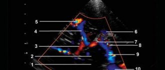
Based on the name of the procedure, it is performed using ultrasonic waves. Their work is based on the Doppler effect. The procedure differs from a standard ultrasound: a vessel with blood passing through it is displayed on the monitor screen. Looking at the resulting image, the specialist assesses the state of blood flow and vascular patency, which affects the quality of blood supply to the brain.
Dopplerography of cerebral vessels makes it possible to study the work of the veins, which “supply” blood to the brain, and the arteries, which are responsible for the outflow of waste products from it. The vessels being examined are located in the cervical and cranial regions. The type of ultrasound will depend on their location:
- Dopplerography of extracranial vessels located on the surface of the neck (jugular veins, carotid, vertebral and subclavian arteries). Any disturbances in their structure affect the state of the brain, which is why these vessels are called “maternal” vessels.
- Dopplerography of transcranial, or “daughter” vessels. The doctor installs an ultrasound sensor in the thinnest parts of the skull and evaluates the functioning of the veins and arteries that supply the brain.
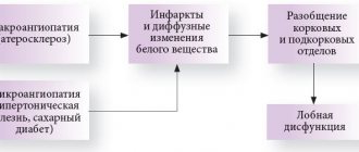
If you are worried about dizziness, tinnitus, ringing in the head, headaches, heaviness, weather dependence, etc. - this may be a sign of impaired, insufficient blood circulation to the brain.
In such a situation, it is necessary to study the blood flow in the vessels of the brain. The most accessible and informative method for assessing the state of cerebral circulation is ultrasound examination of blood vessels, which is aimed at identifying obstacles to blood flow to the brain as the cause of vascular diseases of the brain.
Previously, ultrasound equipment made it possible to study vessels only “blindly” - without visualizing the vessel itself, only by the nature of the blood flow in it (using the Doppler effect, the speed and direction of blood flow was determined. This method is called “USDG” - “Ultrasound Dopplerography of Vessels”) .
Currently, the Doppler ultrasound method is not used independently; it is part of a more modern ultrasound examination of blood vessels, which is called “Vascular Duplex Scanning” (vascular DS). Some doctors continue to call this method in the old way: “USDG”. Therefore, there is confusion in the name of this method of ultrasound examination of blood vessels.
The term “ Duplex scanning of vessels ” means “double scanning of vessels” and allows not only to determine the nature of blood movement through the vessels using the Doppler effect (this is ultrasound scanning), but also to examine the vessels in detail: determine their size, the structure of the vascular wall (which becomes denser and thickens with atherosclerosis, thickens with inflammation of the vessel, etc.); determine the course of blood vessels (which can be smooth or tortuous, up to “loop-shaped”), identify intravascular formations - atherosclerotic plaques, blood clots that create an obstacle to blood flow.
Modern ultrasound machines add to these two components of the study the ability to color vessels depending on the direction of blood flow . Therefore, the term “ Triplex vascular scanning ” (that is, triple scanning) appeared.
The terms “Duplex scanning of vessels”, “DS of vessels”, “Triplex scanning of vessels”, “USDG of vessels” mean one thing - ultrasound examination of blood vessels, which is carried out in triplex mode on modern ultrasound machines.
When performing an ultrasound examination of the vessels carrying blood from the heart to the brain, it is customary to distinguish two levels: the vessels of the neck and the vessels of the brain.
For more reliable information about the state of cerebral circulation, it is necessary to examine both levels of blood flow to the brain, since the “neck and head” are one whole: blood flow in the arteries of the neck is an intermediate stage in the movement of blood from the heart to the brain, and blood flow in the cerebral vessels - this is the final result. Blood flow disturbances can occur at any of these levels.
The first stage of ultrasound diagnostics is the examination of the arteries and veins of the neck :
- common carotid arteries and their branches (internal carotid arteries - carry blood to the brain, external carotid arteries - supply blood to the face)
- vertebral arteries, which at the level of the neck pass through the transverse processes of the cervical vertebrae and carry blood to the posterior parts of the brain.
Obstacles to the flow of blood to the brain at the level of the neck can be tortuosity of blood vessels - congenital (up to the formation of a “loop” by the vessel), or acquired (for example, an uneven tortuous course of the vertebral arteries, formed against the background of spinal osteochondrosis); or there may be narrowing of the arteries - congenital (narrow diameter of the artery), or acquired (narrowing of the lumen of the arteries due to inflammation of the vascular wall, atherosclerotic plaques, blood clots, etc., up to complete blockage of the lumen of the artery). The veins of the neck - jugular and vertebral - are also examined to identify disturbances in the outflow of venous blood from the brain to the heart.
The second stage of ultrasound diagnostics is a study of the arteries and veins of the brain , which is performed through the bones of the skull (transcranial). The arteries of the brain are a continuation of the arteries of the neck on the surface of the brain inside the skull, and form two pools of blood circulation in the brain: “Carotid pool” - from the terminal branches of the internal carotid artery (ultrasound examination is carried out through the temporal bone); and the “Vertebro-basilar basin” - consisting of the terminal segments of the vertebral arteries, which, after entering the cranial cavity, merge with each other into the main (or basilar) artery of the brain (ultrasound examination is carried out through the foramen magnum).
Ultrasound examination of cerebral vessels reveals the resulting indicators of cerebral blood flow: either the blood supply to the brain is not impaired, or there are disturbances in arterial blood flow in one or another circulatory system of the brain (arterial spasm, insufficient blood flow to the brain, etc.) or there are signs of obstructed outflow venous blood from the brain (which leads to increased intracranial pressure). This study is called “Transcranial ultrasound examination of cerebral vessels”, its synonyms: “TCDG” “Transcranial Dopplerography”.
Therefore, it is impossible to study, for example, only transcranial blood flow through the vessels of the brain, without knowing the characteristics of blood movement through the arteries of the neck, where there may be various obstacles to the blood flow to the brain.
And vice versa - the study of only the vessels of the neck will be incomplete in its information content without taking into account the resulting indicators of blood flow in the cerebral vessels.
Carrying out an ultrasound examination of the neck arteries separately is only possible to track the growth dynamics of atherosclerotic plaques identified earlier in them (for timely referral of the patient to vascular surgeons).
There are cases when doctors prescribe a blood flow study in only one of the levels of blood flow to the brain: either in the vessels of the neck or in the vessels of the brain. It is not right.
This may be due to terminological confusion in the name of the method of ultrasound examination of blood vessels (out of habit, they continue to call it “USDG of the neck”).
Or it may be due to mistrust in the results of transcranial examination of cerebral blood flow, which developed in the era of “blind” Doppler examination of cerebral vessels. But those days are already gone. Modern ultrasound equipment allows you to clearly locate cerebral vessels and the movement of blood through them.
The method of ultrasound examination of blood vessels is fast, simple, informative and safe for the patient’s health.
To clarify the state of cerebral blood flow, it is necessary to conduct an ultrasound examination of the vessels of the neck and brain for such complaints as:
- headache
- dizziness
- tinnitus, ringing in the head
- decreased vision and hearing
- memory loss
- sleep disorders
- emotional disorders, psycho-emotional overload, etc.
To prevent the development of stroke, thanks to the identification of atherosclerotic plaques in the arteries, ultrasound examination of the vessels of the neck and brain is indicated for all patients:
- over 40 years of age
- with obliterating atherosclerosis of the arteries of the lower extremities
- for coronary heart disease, heart rhythm disturbances
- for arterial hypertension
- diabetes mellitus
- kidney diseases
- patients who have had a stroke or myocardial infarction.
No other studies other than Duplex Vascular Scanning provide information about the functional state of the vessels of the head and neck and the movement of blood through them. Including this study is not replaced by MRI or CT scan of the brain (since they characterize the structure and condition of the brain tissue itself, and do not characterize cerebral blood flow). Ultrasound examinations of cerebral vessels, MRI or CT scans of the brain complement each other in their information content.
In our department, ultrasound diagnostics of blood vessels is carried out by very experienced and responsible specialists using modern ultrasound equipment. In addition to studies of the vessels of the neck and brain, studies are carried out of the arteries and veins of the upper and lower extremities, arteries of the kidneys, and vessels of the abdominal cavity.
| Name of service | Price |
| Functional diagnostics | |
| Code: A04.12.001.002 (5.067) Duplex scanning of the arteries and veins of the kidneys, | 1050.00 |
| Code: A04.12.003 (5.069) Duplex scanning of the abdominal aorta | 1050.00 |
| Code: A04.12.003.002 (5.070) Duplex scanning of arteries and veins of the iliac segment | 1600.00 |
| Code: A04.12.005.002 (5.042) Duplex scanning of the arteries of the upper extremities | 1500.00 |
| Code: A04.12.005.003 (5.003) DS of brachiocephalic arteries and veins | 1700.00 |
| Code: A04.12.005.004 (5.040) Duplex scanning of the veins of the upper extremities | 1500.00 |
| Code: A04.12.006.001 (5.043) Duplex scanning of the arteries of the lower extremities | 1500.00 |
| Code: A04.12.006.002 (5.041) Duplex scanning of the veins of the lower extremities | 1500.00 |
| Code: A04.12.018 (5.038) DS transcranial arteries and veins | 1100.00 |
In what cases is ultrasound ultrasound prescribed?
The main symptoms signaling a cerebrovascular accident are:
- Impaired coordination of movements.
- Deterioration of hearing and memory.
- Frequent attacks of headache.
- Impairment or partial loss of taste.
- Insomnia, difficulty falling asleep.
- Noise, whistling and ringing in the ears.
- Deterioration of visual function, manifested in loss of visual fields.
- Difficulty concentrating.
- Decreased sensitivity and motor activity of the arms and legs.
A doctor may prescribe a procedure in the following cases:
- The patient has diabetes mellitus.
- For arrhythmia, as this increases the risk of cardiac clot rupture and artery blockage.
- The patient has suffered a heart attack or stroke.
- Osteochondrosis of the cervical spine.
- Before planned heart surgery.
- If the patient is a smoker with many years of experience.
- An area with strong pulsation is visible on the neck (in this case, an ultrasound of the neck is prescribed).
Based on the above symptoms, the doctor can determine the location and name of the vessel in which the patency is impaired. Several pronounced symptoms indicate poor blood flow in a large vessel. In this case, extracranial Doppler ultrasound is prescribed. Violation of only one function will be the reason for conducting transcranial Doppler ultrasound.
Indications
The main indications are the following conditions:
- general lethargy, accompanied by headache, memory and thinking disorders, ringing or noise in the ears, dark circles before the eyes;
- disturbances in facial sensitivity that are predominantly transient in nature;
- pathological changes in heart rhythm (arrhythmias).
Patients with a history of myocardial infarction or stroke are also subject to routine examination using ultrasound. Ultrasound is also used in the event of upcoming cardiac surgery. In addition, such screening is often part of a comprehensive examination and makes it possible to recognize abnormalities in the brain substance visible from the results of computer and magnetic resonance imaging.
Rules for preparing for research
On the day of the procedure, you must refrain from:
- Taking antispasmodics such as “Riabal”, “No-shpa”, “Drotaverine”, “Papaverine”, “Baralgin”, “Cinnarizine”.
- Smoking.
- Drinking black tea and drinks containing caffeine.
- Staying in unventilated, stuffy rooms with large crowds of people, which can have a detrimental effect on vascular tone.
If the patient is undergoing treatment with cardiovascular medications, then the advisability of discontinuing them before the procedure must be agreed with a neurologist. In any case, the sonologist must be informed about the treatment being performed before the ultrasound.
Stages of extracranial Dopplerography:
1. In the office, remove all jewelry from your neck and head.
2. Remove clothing from those parts of the body through which the sensor will be passed: neck, collarbones, shoulder blades.
3. Lie on the couch with your head facing the monitor. The doctor will apply acoustic gel to open areas of the body and begin to move the device over them, placing it in areas where medium and large arteries and veins are located. To obtain more accurate data on the state of vascular tone, the patient will need to hold his breath at the doctor’s request, take special medications and change body position.
Doppler ultrasound of vessels located in the cranial area has a different specificity. Acoustic gel is applied to the temples, the back of the head and the area above the eyes. The device is installed there because in these places the bones of the skull are the thinnest and are able to transmit ultrasonic waves.
By what principle is the received data decrypted?
Each vessel has its own parameters, on the basis of which calculations are made and the degree of their discrepancy with the norm is assessed.
Transcranial and extracranial Dopplerography of arteries compares digital data in each vessel segment:
- Artery wall thickness.
- Vessel diameter.
- Phases of blood flow, its features.
- The degree of symmetry of blood flow through identical arteries located on different sides.
- Diastolic blood velocity.
- Maximum systolic blood flow velocity.
- Pulsation and resistive indices, systole and diastole ratio.
- The degree of pathological narrowing of blood vessels (stenosis), the condition of the artery beyond its limits.
Dopplerography of veins evaluates the following parameters:
- Condition of the venous wall of the vessel.
- The nature of blood movement through the studied vessels.
- Vein diameter
Data obtained at rest and after functional tests are compared with normal values. Any deviations are marked with a special abbreviation of letters and numbers. Based on them, the neurologist prescribes further studies or forms a course of treatment.
Where can I get Doppler ultrasound of cerebral vessels?
The study is carried out in municipal clinics and public hospitals, specialized clinics, and general medical centers. Depending on the location of the ultrasound, the cost of each type of Doppler ultrasound will be in the range of 500-6000 rubles. The procedure has many advantages, including painlessness and the absence of side effects. Within 20 minutes, a sonologist will be able to determine the presence of anomalies in venous or arterial inflow and assess the degree of development of danger to blood vessels.
However, the procedure will not help determine the cause of compression of the thin arteries, since the capabilities of the device do not allow them to be seen from the outside.