1 General rules for applying electrodes 1.1 Features of disposable electrodes
2 Where and how to apply electrodes? 2.1 Features of installing electrodes on the limbs
2.2 Nuances of the placement of electrodes on the chest
The performance of the heart muscle largely determines the health of the body as a whole. An electrocardiogram is a simple and informative research method that allows you to quickly and accurately assess the contraction and relaxation of the heart and even make some dangerous diagnoses.
When recording an electrocardiogram, the serviceability of the equipment is important: the device itself, electrodes and cables. In case of failure, each of the elements can be replaced using the services of an online store that sells medical consumables. In addition, the nurse (or doctor) must place the electrodes correctly, otherwise the test result will be incorrect. Each patient should know how to correctly apply electrodes to be confident in the reliability of the ECG.
What does ECG mean? Types of electrocardiogram
An ECG involves monitoring heart rate, blood circulation, heart rhythm, and activity.
Electrodes are attached to the patient's chest, arms and nails. The doctor records the picture of health in the form of a curve that repeats the work of the heart. A cardiologist can decipher it. If he detects serious deviations from the norm or dangerous diseases, he will prescribe urgent therapy. ECG is an absolutely safe research method that does not cause discomfort to the patient. You can get a cardiogram even during pregnancy.
Let's look at several types of ECG.
Where and how to apply electrodes?
There are 12 leads in the electrocardiogram: 3 main, 3 enhancing and 6 chest. To take data, 10 electrodes are installed: on all limbs and the chest. For ease of use, they often differ in appearance and color.
Features of installing electrodes on the limbs
Applying electrodes to the limbs implies the well-known color order of the traffic light. The terminal installation is as follows:
- A red electrode is applied to the right hand;
- A yellow electrode is attached to the left hand;
- A green electrode is placed on the left leg;
- The right leg is meant to be grounded and a black electrode is attached to it.
Electrodes are placed on the proximal limbs: wrists and ankles, which are pre-treated and gelled. If a person is missing one or another limb, then the terminal is placed on the stump. Sometimes the electrodes are additionally secured with rubber bands.
The nuances of the location of electrodes on the chest
Chest electrodes may look different. Most often they are rubber suction cups. Sometimes the electrodes look like ordinary rectangular plates, then they are additionally secured with an elastic band.
There are 6 chest leads in total. Depending on the equipment of the ECG room, all electrodes are applied at once or the nurse has only one branch, which she installs one by one and records each lead separately.
The procedure for installing chest electrodes:
- The first is the fourth intercostal space to the right of the sternum;
- The second is the fourth intercostal space to the left of the sternum;
- Third - fifth rib along the left parasternal line;
- Fourth - fifth intercostal space along the left midclavicular line (or exactly in the place where the apical impulse is projected, that is, 1.5 centimeters inward from the midclavicular line normally);
- Fifth - fifth intercostal space along the anterior axillary line;
- Sixth - fifth intercostal space in the midaxillary line.
Each chest lead is responsible for a specific part of the heart, so their correct application is of great importance.
Holter ECG monitoring
This method is the most complex and involves round-the-clock recording of heart parameters. A Holter study allows you to identify pathologies that appear during sleep, during periods of physical stress, or after taking certain medications.
Sensors are also attached to the patient and connected to a portable cardiograph. In this situation, a person may not change his lifestyle and do normal things. The only thing that is required of the patient is to record all his activity for the day and refrain from water procedures so as not to damage the sensors.
Our clinics in St. Petersburg
Structural subdivision of Polikarpov Alley Polikarpov 6k2 Primorsky district
- Pionerskaya
- Specific
- Commandant's
Structural subdivision of Zhukov Marshal Zhukov Ave. 28k2 Kirovsky district
- Avtovo
- Avenue of Veterans
- Leninsky Prospekt
Structural subdivision Devyatkino Okhtinskaya alley 18 Vsevolozhsk district
- Devyatkino
- Civil Prospect
- Academic
For detailed information and to make an appointment, you can call +7 (812) 640-55-25
Make an appointment
Indications for the procedure
ECG is a fairly popular procedure that is prescribed to everyone, from the youngest patients to adults. The reasons for the study can be either a routine examination or complaints of ailments. The procedure is required:
- for admission to first grade;
- passing the driver's license exam;
- teenagers who are undergoing changes in their body;
- admission to secondary and higher educational institutions after school.
ECG is also indicated for patients who have the following health problems:
- shortness of breath while walking or other activities, as well as at rest;
- high blood pressure;
- pain in the chest, neck, back and abdomen;
- fainting;
- swelling of the limbs;
- general weakness;
- heart murmurs and so on.
An ECG is also done before surgery, when registering for a sanatorium, and for issuing medical documents. A cardiogram should be done every year for people over 40 years of age.
Where to do an ECG of the heart
In the problem of competently taking and interpreting an ECG, the main role is played by the choice of a specialist and a clinic. This measurement is quite common - the device is available in all free public clinics, but there are many arguments in favor of private ones.
Reasons to have an electrocardiogram done at the Medicenter:
No queues . By making an appointment at a time convenient for you, you will quickly receive results and a professional transcript and go about your business.
Professional doctors. Medicenter employs highly qualified cardiologists with extensive experience. They have knowledge and specificity in taking cardiograms in children and adults.
Possibility to conduct research at home. Not many medical centers offer such services to patients. This offer is especially relevant for elderly patients and children.
The latest equipment. To take an ECG, the Medicenter uses a 3-channel ECG machine “Cardiomax” (Japan), which has the full range of capabilities of modern cardiac equipment.
Low price.
Preparation rules
- Before having an electrocardiogram, you need to get a good night's sleep. Also, you need to go to a medical facility in a calm state. Please know that you will not be given any injections before your appointment. During the examination you will not experience any discomfort at all. Often before the procedure the patient is allowed to lie quietly for 5-10 minutes;
- The ECG room should be at normal room temperature so that the patient feels comfortable. Otherwise, trembling in the body from the cold can distort the data of the organ’s functioning;
- Patients who experience severe shortness of breath are advised to monitor while sitting in a chair. In such patients, the heart rate is recorded better in this position;
- Women should not use greasy creams on the breast area before monitoring. This may reduce skin conductivity, resulting in distorted measurement data;
- Try to wear clothing that can be easily removed, as the electrodes must be attached to bare skin during the procedure. It is better for women not to wear tights, since ECG electrodes are also attached to the ankles;
- On the day of the electrocardiogram, you are allowed to eat two hours before the procedure. You can drink as much as you want.
If you complain about suspicious symptoms in the chest area, you can contact a doctor at the ProfMedPomosh clinic, who will write you a referral for an ECG. The ECG procedure itself is performed by a functional diagnostics doctor or nurse.
What ECG indicators are considered normal?
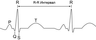
The record looks like a graph. Along the vertical lines you can see a curve that rises and falls. The horizontal line indicates over what period of time the indicators change. This is a special interval, it is measured in seconds.
A normal electrocardiogram will look like this:
- The P wave indicates sinus rhythm. It can be positive or negative, it all depends on the chest leads. If we talk about the norm, then the width will be 0.1 seconds;
- PQ interval. He says how long it took for the sinus impulse to travel through the atrioventricular node. If the heart is working normally, then this interval is 0.1 seconds;
- QRS complex. Demonstrates how excited the muscle tissue of the ventricles is when they contract. This can normally last 0.3 seconds;
- wave T. It shows all the processes that take place during the restoration of muscle tissue before the next contraction.
As you can see, you need to clearly know how to properly take an ECG and apply electrodes, then you can get reliable readings.
How to read a cardiogram correctly?
How to correctly take an ECG, correctly apply electrodes and analyze the results of the study? Decoding should begin with checking the registration technique. That is, the specialist checks to see if there are any interferences. They usually occur due to poor contact between the skin and the electrodes or muscle tremors.
Afterwards, the doctor examines the millivolt amplitude, which should be 10 mm. It is also worth paying attention to the speed of the paper, how it moved during the ECG recording. Most often, this speed should be 50 or 25 mm/s. In both cases, the data will be different, so it is important to pay attention to the tape and its speed.
To correctly take an ECG and analyze it, it is necessary to correctly apply the electrodes, as well as perform several manipulations:
- check whether the heartbeat is stable;
- count how many contractions were made per minute;
- evaluate the P wave;
- analyze the width and shape of the QRS complex;
- determine how the electrical axis of the heart is located.
Why do you need a holter?
ECG monitoring is prescribed by cardiologists to diagnose diseases and select the most effective therapy. You can also undergo an examination to prevent various types of heart pathologies. Indications for a Holter study include:
- complaints of chest pain;
- shortness of breath, arrhythmia and general weakness;
- regular presyncope;
- dizziness;
- interruptions in heart function;
- suspicion of myocardial ischemia, etc.
Holter is the only method of its kind for assessing the effectiveness of treatment for arrhythmia and coronary heart disease. Accurate data obtained from sensors allows us to reconsider the adequacy of the prescribed therapy. Using the information obtained, you can adjust your medication intake.
What the heart holter will show:
- heart rate;
- heart rhythm disturbances;
- changes in PQ and QT intervals;
- ST segment response to physical activity;
- confirmation of arrhythmia;
- quality of conduction through the heart muscle, etc.
Since carrying a child increases the load on the cardiovascular system, a holter during pregnancy allows you to very accurately determine possible risks. It is especially important to consult a cardiologist if a woman has high blood pressure, chest pain, or arrhythmia at rest.
Request a call
Performing an ECG procedure
How to properly take an ECG and place electrodes, steps:
- Initially, the person lies down on the couch, and then exposes his chest, ankles, and wrists (removes his clothes).
- The doctor is always obliged to analyze the patient’s emotional state before the procedure. If you are not feeling well, it is better to do an ECG later. Otherwise, the results of the study may be distorted.
- The doctor uses a special composition to lubricate the places where the electrodes will be fixed. This composition is needed for better conductivity. Then the electrodes are applied.
- An ECG recording is in progress. As soon as the procedure is completed, the specialist removes the electrodes and wipes dry the areas of the body that were lubricated with the composition.
Characteristic
The technique for taking an ECG at the Profmedpomoshch clinic in Moscow is not complicated. Its price is affordable (indicated in the price list). You do not undergo any punctures or radiation. You are simply connected to a special device that will provide information about the work of your heart. The data will indicate the frequency of contractions of the heart muscle, and will also identify possible diseases of the organ. In addition, using this device, diseases such as pulmonary embolism can be detected.
The device for taking an ECG was created at the beginning of the 20th century. Cardiologists all over the world still use it. The device consists of a galvanometer, an amplification mechanism and a device for recording electrical currents arising in the heart.
The monitoring procedure does not pose any danger to the body, even if used frequently. Doctors recommend performing an ECG as soon as pain and discomfort in the heart area occurs. The reason for frequent visits to the doctor may also be shortness of breath, extra pounds, stress, high or low blood pressure, rapid rhythm, etc.
People aged 40 years and above are recommended to undergo annual heart screening, and in some cases up to 4 times a year. In this way, the patient can be correctly diagnosed on time and receive timely medical care. Don't forget that preventive measures help prevent diseases.
Indications for ECG
Everyone needs to check their heart function from time to time. Data on the condition of the heart muscle is necessary in the following cases:
- arterial hypertension;
- age over 40 years;
- smoking;
- increased cholesterol levels in the blood;
- pregnancy;
- past infections;
- upcoming surgery;
- other diseases with suspected involvement of the heart in the pathology;
- deterioration of the condition of patients with “heart disease” (pain, arrhythmia, shortness of breath);
- occupational risk factors (shift work at different times of the day, nervous overstrain, occupational hazards);
- expert assessment of the working capacity of drivers, sailors, pilots.
Make an appointment for an ECG or consultation with a cardiologist.