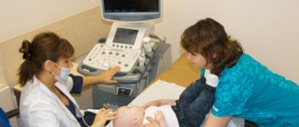
Ultrasound diagnostics is one of the safest and most effective ways to detect various diseases of internal organs. This technique is used in various fields of medicine, including assessment of the condition and possible pathologies of blood vessels. In this case, Doppler ultrasound or ultrasound is performed, which is used to examine the neck and head. Early diagnosis allows you to choose gentle and effective treatment methods and prevent the development of complications. First you need to understand what an ultrasound scan of the vessels of the head and neck shows, why this method is effective in diagnosing many diseases.
The Doppler ultrasound effect is based on the ability of ultrasound waves to be reflected from blood cells. As a result, electronic impulses appear that appear on the device screen in the form of images of veins and arteries with blood flow.
This ultrasound examination allows you to assess the condition of the large arteries that supply the brain, carotid and vertebral arteries, determine the intensity of blood flow, and also identify traces of obstruction.
How is Dopplerography performed?
This examination is carried out in the presence of certain symptoms. Preliminary preparation is necessary so that the results of the ultrasound examination give an accurate picture of the patient’s condition.
There are two types of procedure:
- Transcranial. The sensor is installed on the skull bones in those places where they are thinnest. The procedure gives an accurate picture of the vessels located directly in the brain.
- Extracranial. The technique is used to examine large vessels passing through the neck (carotid arteries, jugular veins, etc.). The sensor is located at each site separately.
Contraindications
There are no contraindications for ultrasound examination of the vessels of the lower extremities. The study can be carried out even on children and pregnant women. Restrictions apply solely to test results. For example, if the patient smoked before the procedure, drank coffee, or took vascular medications, there is a risk of distorting the results.
Relative contraindications to the procedure may include:
- mental disorders;
- open lesions on the skin in the affected area;
- in the presence of allergic reactions to hydrogel.
Indications for the procedure
Doppler sonography is indicated in the following cases:
- Problems with coordination and other symptoms indicating a malnutrition of the brain (the patient is dizzy, has tinnitus, etc.).
- Unstable heart rhythm.
- Hypertonic disease.
- Transient ischemic attacks.
- IOP.
- Previous stroke or heart attack.
- There are suspicions of atherosclerosis.
- Diabetes.
- Fatigue quickly for no apparent reason.
- Cervical osteochondrosis.
This procedure is also indicated if heart surgery is to be performed, if there are pulsating formations in the neck, or if thrombosis is suspected. In children, it is performed in case of serious postural problems and delayed development of the child.
How is it carried out?
Doppler ultrasound is prescribed for suspected vascular pathology. The study is carried out in an office equipped with a modern ultrasound scanner with special sensors. The procedure is completely painless and does not cause discomfort. The doctor applies a special gel to the area of study and uses a sensor to process control points proportional to the projections of the vessel being examined.
The sensor processes the signals and sends them to a computer, which converts them into graphic images and displays them on the monitor. The procedure can last 30-60 minutes, depending on the volume and area of research. The following indicators are assessed:
- wall thickness and diameter of the vessel;
- consistency of vascular valves;
- nature of blood flow;
- resistive and ripple index;
- maximum and minimum speed of blood flow.
15-20 minutes after the ultrasound examination, the patient receives a medical report on a special form. It is also possible to record the result to a flash drive or disk.
Are there any contraindications?
Ultrasound waves are absolutely safe for humans, so there are no special contraindications to this type of ultrasound. The only exception is a violation of the integrity of the skin. The procedure involves contact of sensors with the head or neck, so the examination is postponed until the wound heals.
However, there are a number of special cases that make duplex examination of blood vessels difficult:
- cardiac arrhythmia, which changes the speed of blood circulation through healthy arteries and veins;
- the area under study is hidden under bone tissue;
- slow blood flow in the patient.
These are not contraindications, but in this case changes in the location of the sensors are required. It will be necessary to navigate non-standard conditions, which requires professionalism on the part of the doctor.
Main indications for prescribing an examination
Depending on the age of the patient, the list of indications for diagnosis will differ.
For examination of cerebral vessels in infants, there are the following recommendations:
- if you suspect or accurately determine the location of bleeding in the tissues of the brain or ventricles;
- when it is necessary to assess the degree of ventricular dilatation;
- if there is a suspicion that the white matter of the tissue that surrounds the ventricles of the brain has been damaged;
- in order to identify the presence of abnormalities in the development or structure of the vascular system of the brain;
- for the diagnosis of infectious lesions or tumor processes.
During the treatment of patients in the older age group, scanning of the brain vessels will allow one to accurately determine the location, size and condition of the tumor process (often used during surgery for dynamic control, which allows surgical intervention to remove the tumor to be performed with maximum safety for the patient’s life).
When a patient is diagnosed with sickle cell anemia, he is shown transcranial Doppler ultrasound of the vessels and arteries of the brain. The examination will help determine your likelihood of having a stroke.
Features of ultrasound examination
As a rule, Doppler sonography is planned in advance, but it can be performed urgently. The procedure itself is painless, so patients tolerate it calmly.
The examination is carried out according to the following scheme:
- The patient lies on his back. The head should be relaxed (a flat pillow is placed under it).
- You need to remain calm, breathe evenly and, if possible, not move.
- A special gel is applied to the area under study to prevent the penetration of air between the skin and the sensor.
- The movement of the sensors is controlled by a specialist. As a result, an image of the vessels appears on the monitor screen.
The study lasts no more than 1 hour. During the study, the doctor may ask the patient to do some action: turn his head, squeeze a vein in a certain place, breathe less often, etc.
Dopplerography of extracranial vessels begins with examination of the right lower segment of the carotid artery. The sensor is then moved up the neck. Next, the color mode is activated, which makes it possible to identify a number of pathologies: deformations of the walls of blood vessels, impaired blood flow in certain areas, etc.
What diseases does it detect?
Doppler ultrasound is a highly informative method used to diagnose various vascular pathologies:
- cerebrovascular accidents;
- phlebeurysm;
- vascular malformations;
- deep vein thrombosis;
- hypoplasia of the vertebral arteries;
- atherosclerosis;
- temporal arteritis;
- postthrombotic syndrome;
- vasculitis;
- aneurysms and other vascular pathologies.
The examination allows you to identify the presence of formations that impede or change blood flow (sclerotic plaques, blood clots), check the homogeneity and rhythm of blood flow, assess the compensatory capabilities of blood flow, identify defects in the structure and flow of blood vessels - narrowing, kinks, tortuosity, aneurysms, compression by scars, vertebrae or spasmodic muscles.
What can she show?
Duplex scanning allows you to determine areas of narrowing of the lumen of blood vessels, the direction of blood flow, its speed, as well as the condition of the vascular walls. This study can show the location of cholesterol plaques and the presence of blood clots, which is very important when diagnosing atherosclerosis.
A decrease in the elasticity of the walls of the arteries, as well as their thickening, indicate that the patient has hypertension (high blood pressure). If a change in blood flow is diagnosed, this indicates the presence of obstacles. For example, an aneurysm is a sac-like protrusion of a vessel.
Why is Doppler ultrasound prescribed?
There are many indications for Doppler ultrasound. This can be any disease (or just a suspicion of it) associated with impaired blood flow in the vessels. Among the main pathologies in the diagnosis of which ultrasound plays an important role:
- cerebrovascular accident in acute or chronic form;
- heart failure;
- venous insufficiency and impaired blood flow in the extremities;
- renal failure;
- endocrine diseases.
Examination of the vessels of the head and neck
Doppler ultrasound is most often used to examine the vessels of the head and neck. The doctor prescribes an appropriate prescription if the patient complains of periodic headaches, flickering spots in the eyes, dizziness, or even loss of consciousness. Signals that it is necessary to undergo an examination may also include fluctuations in blood pressure or general weakness in the body for no apparent reason.
If ultrasound examination is prescribed for relatively young patients to diagnose pathologies with severe symptoms, then people aged (after 50-55 years) are recommended to undergo this procedure to prevent stroke.
Doppler of the veins of the lower extremities
Doppler is widely used to assess the condition of the deep and superficial venous systems of the lower extremities. With a probability of 90-100%, such an examination will reveal pathologies of valves, vein walls, and detect varicose transformations of blood vessels.
The most common indications for ultrasound examination of the lower extremities
:
- feeling of heaviness in the legs;
- swelling of the legs;
- leg pain;
- cramps of the calf muscles;
- enlargement, bulging of veins (most often observed in the popliteal fossa).
Doppler ultrasound is used both to diagnose diseases of the veins of the lower extremities and to determine the scope of upcoming surgical intervention. In the second case, during the examination, which is carried out in a standing position, specialists mark with a marker those places on the legs where it will be necessary to ligate and remove the vein.
Examination of heart vessels
Duplex scanning allows specialists to study the structure and evaluate the functional development of the cardiovascular system. Indications for this procedure are as follows:
- pain or discomfort in the chest area;
- high blood pressure;
- congenital heart defects;
- myocardial infarction;
- suspicion of the presence of tumors in the cardiovascular system.
Ultrasound with Doppler is prescribed as an additional study if, for example, a cardiogram or simple echocardiography revealed heart murmurs, angina pectoris, metabolic syndrome and other pathologies.
The use of color Dopplerography allows specialists to study quantitative and qualitative indicators of blood flow in real time, to analyze the state of blood flow not only in the vessels of the heart, but also inside the organ itself.
Doppler testing of the fetus and diagnosis of placental vessels
Today, with the help of Dopplerometry, it is possible to identify pregnancy complications associated with impaired blood flow in the mother-placenta-fetus system in the early stages. Perhaps a pregnant woman does not yet feel any problems with her health, while the fetus in her womb is already beginning to experience a lack of nutrients and oxygen. An ultrasound with Doppler will definitely show this, and the attending physician will be able to prescribe effective therapy in a timely manner.
In addition to scheduled diagnostics with Doppler, a pregnant woman may also be prescribed unscheduled diagnostics if the following indications exist:
- on the mother’s side – gestosis, kidney disease, high blood pressure, diabetes mellitus, Rh conflict;
- on the part of the fetus - developmental delay, oligohydramnios, congenital malformations of organs, asynchronous development of fetuses in multiple pregnancies.
Examination of the vessels of the peritoneal cavity
Ultrasound scanning of the abdominal cavity is prescribed when there is a need to determine the condition of the vessels supplying the kidneys, adrenal glands, and pancreas. Doppler ultrasound allows you to examine the abdominal aorta, inferior vena cava and its branches, paired and semi-gyzygos veins, thoracic lymphatic duct and lymph nodes.
Indications for Doppler ultrasound may include: poor urinalysis, kidney tumors or cysts (can be detected on a simple ultrasound), injuries to the abdominal organs. For preventive purposes, ultrasound examination should be performed if there is a history of chronic or acute inflammatory processes in the pyelocaliceal system (for example, chronic pyelonephritis), in the postoperative period, or with hypertension.
What pathologies can be diagnosed?
Doppler ultrasound has certain limitations in diagnosing diseases. For example, she will not be able to study small veins - for this she needs to perform angiography. But there are a number of pathologies that can be identified:
- Atherosclerosis. By examining the head and neck using ultrasound, it is most often possible to diagnose atherosclerotic disorders. This condition is indicated by echogenicity, the presence of plaques, and thickening of the vascular walls. Moreover, if the lumen of the artery narrows by more than 20%, then the patient is diagnosed with “stenosis.”
- Thrombosis. Thrombosis is characterized by blocking of the arterial lumen by a blood clot. On Doppler ultrasound, inflammation of the vessel wall with a homogeneous formation inside is clearly visible.
- Compression of blood vessels. The examination allows you to see areas where blood flow is impaired due to the impact of swollen tissue on the channels. This allows you to diagnose cervical osteochondrosis and identify problems with blood circulation after injuries.
In addition, these ultrasound examinations can detect a number of other diseases, including vasculitis, post-traumatic dissections of the arterial walls and other pathologies.
Doppler ultrasound is a safe diagnostic method that can be performed in any case if there is a medical need for it. Timely implementation of the procedure allows you to identify the disease at an early stage of its development.
Ultrasound and Doppler Ultrasound
Alexander Pavlovich Rechmedin
Ultrasound Doctor – Expert Highest category Experience over 20 years. Read reviews
Shmarin Alexey Nikolaevich
Ultrasound Doctor, Expert with over 20 years of experience. Go to the doctor's page
Doppler ultrasound and duplex scanning - differences between dopplerography and duplex scanning
The issue of prevention and treatment of vascular diseases remains one of the most pressing today. In the field of angiosurgery, many methods have been developed for the treatment of vascular pathologies, but, as is known, for an angiosurgeon the most important thing in making a diagnosis and prescribing treatment is preliminary ultrasound diagnosis of blood vessels.
Ultrasound is a very common term in the modern world. But, speaking about vascular ultrasound, there are many unclear terms that give rise to misconceptions among patients who are not directly related to them. Several methods are used to diagnose vascular diseases: Doppler ultrasound
(ultrasound Doppler method),
USDS
(ultrasound duplex scanning method) and CDK (color Doppler mapping method). These studies do not harm the body and are considered a safe type of diagnosis.
In this article we will try to figure out how ultrasound ultrasound
from ultrasound, and what is the difference between Doppler ultrasound (
USDG
) and duplex scanning method (
USDS
).
DIAGNOSTICS USING USG METHOD
One of the simple and accessible ways to study venous and arterial patency is Doppler ultrasound. At the heart of ultrasound
lies the Doppler effect, that is, to obtain the necessary data, changes in reflected sound waves from moving blood cells are recorded. The Doppler ultrasound method is prescribed when it is necessary to determine vascular patency, either to obtain an assessment of blood flow, or to identify pathologies of the venous valve.
When performing Doppler ultrasound, veins and arteries are not visible; the blood flow velocity and patency are assessed based on the obtained Doppler effect values. According to the results of the ultrasound
Headaches, hypertension, and varicose veins are successfully diagnosed. But if identifying impaired vascular patency is done with ease, then eliminating the causes of such pathology using Doppler ultrasound is very difficult. This is due to the fact that this diagnostic method does not allow visualization of the vascular walls and their possible curvatures, which affect the speed and quality of blood flow.
Initially, the images shown by the ultrasound device were made in a flat and thin projection of the organ being examined. Modern diagnostic medicine uses equipment that allows obtaining three-dimensional images of organs in real time and in motion.
The Doppler ultrasound method is one of the most common methods for diagnosing diseases of the arteries and veins. As a rule, ultrasound
prescribed to patients with suspected vascular obstruction or venous valve insufficiency. The Doppler effect, which gave rise to Doppler ultrasound, allows us to understand this pathology. With its help, a change in the reflection of a sound wave from the movement of blood cells is recorded, which makes it possible to subsequently diagnose the speed of blood flow, varicose veins, and identify the cause of headaches and hypertension.
Please note: Ultrasound
easily detects blood flow disorders, but determining its cause is not so easy!
As already mentioned, the USDG
does not visualize the walls of blood vessels and their pathological bends, but they affect the speed of blood flow and its quality.
Ultrasound diagnostic method
UZDS
is a duplex scanning and is a “deeper” form of research and, naturally, more effective.
Unlike Doppler ultrasound, ultrasound
combines the Doppler effect with B-mode or “brightness mode”. This combination makes it possible to visualize the walls of veins and arteries, as well as check the condition of nearby tissues. Examination of vascular blood flow is performed using the Doppler method (color mapping) or spectrum analysis technique.
When using the ultrasound
on the monitor screen we can see an image of the vessel being examined, visually assess venous or arterial patency and identify the cause of weak blood flow.
Ultrasound diagnostic method
has a number of undeniable advantages.
With the help of ultrasound scanning
there will be no problem detecting, for example, deformation of blood vessels or their lumens, identifying the formation of cholesterol plaques and the presence of blood clots, and assessing the blood flow of the entire vascular system.
When examining ultrasound scans
of large veins and arteries, pathological conditions can be detected at the earliest stages of development.
What are the advantages of ultrasound
?
The duplex scanning procedure is, of course, absolutely painless. On average, the diagnostic time takes 30-40 minutes, depending on the area of the body being examined, and, as a rule, does not cause discomfort to the patient. After ultrasound examination
the patient can immediately return to their normal lifestyle without restrictions.
The ultrasound duplex scanning method is a highly accurate research method and does not cause any complications after the procedure.
Unlike Dopplerography (
Doppler ultrasound), duplex scanning combines two research functions.
Using Doppler ultrasound, vascular blood flow is characterized, and along with B-mode, this study also makes it possible to visualize the structure of arteries and veins. This gives the right to call ultrasound scanning
a more accurate research method.
Find out the cost
results
Based on the results of ultrasound examination of veins and arteries, a protocol is formed in which the doctor indicates the patency of the veins, the degree of blood flow disturbance, wall parameters, the presence of blood clots and cholesterol plaques, their parameters, peak blood flow velocity in different areas and other data.
The results obtained are compared with established standards. When decoding, the patient's age is taken into account. For young and middle-aged people, the norm is:
- absolute patency of veins and arteries;
- wall thickness without seals;
- absence of pathological reflux and valvular insufficiency;
- normal blood flow velocity with a permissible deviation of 10-15 cm/s: in the femoral artery 75, in the distal artery - 56, in the iliac artery 96, popliteal 53, in the tibial artery 46 cm/s.
If any abnormalities are detected, the doctor will inform the patient and give directions for additional tests and studies.
Pulsation index
PI in the third trimester should be 0.4 -.64. Ultrasound examination results make it possible to understand:
- whether the fetus suffers from intrauterine hypoxia, and if there is a lack of oxygen, then how dangerous the condition is;
- are there any problems due to a conflict regarding the Rh factor;
- the degree of damage to the vessels of the uterus, placenta and fetus due to diabetes mellitus, cardiac pathology or hypertension of the woman;
- Are there any developmental problems during multiple pregnancies, how are the blood vessels connected and developed in twins.
Preparation for ultrasound examination
The study does not require complex preparation. It is enough to take the following measures:
- Before the procedure, you need to take care of foot hygiene;
- stop smoking, alcohol, coffee and energy drinks several hours before the examination: caffeine, ethanol, nicotine cause narrowing or dilation of blood vessels, which can affect the test results;
- It is recommended to stop taking vascular medications or discuss this with your doctor.
You should take a small towel, textile or paper napkins with you to clean the skin of the hydrogel.
Norm of SDO in the umbilical cord artery
| Duration, week | The norm of the systole-diastolic ratio in blood vessels |
| 16-19 | 4, 45 – 4,67 |
| 20-22 | 3,75 -3,95 |
| 23-25 | 3,41-3,6 |
| 26-28 | 3,1-3,27 |
| 29-31 | 2,82-2,94 |
| 32-35 | 2,48-2,52 |
| 35-37 | 2,4-2,45 |
| from 38 | 2,19-2,22 |
The norm of SDO in the arteries of the uterus in the third trimester: 1.3-3.7
Advantages of the method
Doctor Khizriev S.M. conditionally identifies the following advantages of ultrasound of the brain:
the treatment procedure does not imply any age restrictions; Doppler ultrasound can also be performed to assess the effectiveness of treatment and the correctness of the tactics of conservative measures.
Provides several types of access:
- occipital region;
- eyes;
- temporal bone.
Using access through the eyes, the rate of blood flow inside the ophthalmic artery is assessed. Through the temple, the vessels that are located at the base of the brain are examined. As a result, the condition of the veins of Galen and Rosenthal and arteries is assessed. Through the occipital zone, all departments located in the skull are examined. In this case, especially close attention is paid to the vertebral arteries.
Indications
Ultrasound scanning of blood vessels helps diagnose many brain pathologies.
This technique allows you to study the state of the vascular system in dynamics. Thanks to Doppler ultrasound, it is possible to detect a narrowing of the lumen, a decrease in venous tone, a change in the speed of blood flow, and atherosclerotic lesions. Doppler ultrasound is effective in the following indications:
- dizziness;
- pain in the head area;
- fainting;
- swelling of the lower extremities;
- the occurrence of cramps in the calf area;
- overweight;
- the appearance of fatigue;
- hypertension.
It is worth noting that ultrasound cannot be a substitute for angiography. The latter technique is a popular hardware method. It involves x-ray examination of blood vessels, which is used in radiography, fluoroscopy, and computed tomography.