Rheoencephalography (REG) is one of the methods for diagnosing cerebral vessels using weak electrical impulses. Blood has the highest electrical conductivity compared to other media in the body. And since blood circulation is most intense in the brain, this method allows one to evaluate not only the blood vessels, but also the state of the internal structures of the brain. REG is a non-traumatic, absolutely painless and quite informative method of research; it allows the doctor to assess the speed of blood flow, tone, elasticity and blood supply of the vessels of the head. The advantages of this research method are known to every neurologist. Rheoencephalography provides a detailed picture of intracerebral vessels, regardless of their size, can be used in any conditions and allows one to obtain differentiated information about the condition of veins and arteries. At the same time, rheoencephalography is a fairly simple and absolutely comfortable procedure for the patient.
Indications for use
It is necessary to have a child have an REG of the head if he/she has:
- regular headaches of unknown origin;
- hearing or vision impairment;
- hypertension or hypotension;
- tinnitus;
- fainting conditions;
- swelling of the face;
- coordination problems.
The procedure is also prescribed to control blood circulation after operations, neck and head injuries. In some cases, the examination is part of an analysis of the effectiveness of drug therapy and non-drug treatment.
General characteristics of the method
Rheoencephalography is a non-invasive functional diagnosis. Using REG, you can also obtain information about the general functionality of the vascular walls, the magnitude of pulse blood filling, vascular resistance and reactivity of brain vessels. The study evaluates the overall functionality of the venous and arterial systems. The research does not require expensive equipment or specially equipped rooms.
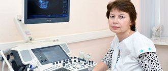
The study is carried out using a device called a rheograph. In the diagnostic room, the doctor places electrodes treated with a special conductive compound on the scalp. They are fixed using an elastic rubber band, after which the device begins to record incoming impulses. The rheograph records them in the form of graphic symbols (curved lines), which are later deciphered by the doctor. During the procedure, the doctor may ask the patient to change his body position, turn his head, stand up and lie down. The fact is that changing the position of the head allows the specialist to draw conclusions about the nature of the disorders - to understand whether they are functional (situational) or organic, associated with certain diseases.
Content:
- General characteristics of the method
- Progress of the study
- Which is better: rheoencephalography (REG) or electroencephalography (EEG)?
It is important to understand that rheoencephalography is not enough for a comprehensive study of veins and arteries. The method describes only the functional state of the elastic walls of blood vessels, and this is not enough to make an accurate diagnosis. REG allows you to detect a problem in a particular part of the brain and concentrate on its further detailed study. Such blood flow parameters are diagnosed as blood viscosity, blood flow speed, vascular tone, the level of blood supply to a particular part of the brain, and the specifics of blood circulation.
Indications/contraindications for REG
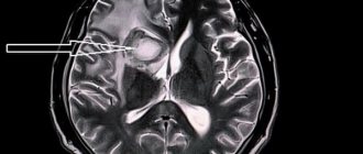
Initially, REG was used to monitor brain function in real time. Currently, the method is used to diagnose atherosclerosis of cerebral vessels, its prevalence and stage. In case of traumatic brain injury, the study provides important information about the size of the injured areas, the presence of intracranial hematomas, their depth and the severity of the condition.
| Indications | Contraindications |
| Acute/chronic vascular pathologies of the brain (for example, vertebrobasilar insufficiency) | Mechanical head injuries |
| Atherosclerosis (chronic pathology of the arteries, in which cholesterol accumulates in the lumen of blood vessels) | Chronic/severe headache |
| Migraine (a neurological disease characterized by frequent, unbearable headaches) | Dizziness |
| Stenosis (persistent narrowing of the lumen of blood vessels) | Abrasions/wounds on the diagnosed area |
| Impaired circulatory functionality | The study is not recommended for newborns and infants |
| Stroke (acute cerebrovascular accident) | |
| General diagnosis of vascular condition after traumatic brain injury | |
| Evaluation of the effectiveness of therapy or individual drugs | |
| Intracranial hypertension (increased pressure in the cranial cavity) |
Preparation for the event
Before the examination, the patient must eat 1.5 hours before the appointment. Light, familiar food is recommended. Breastfed children are examined immediately after feeding. On the day of the test, you do not need to give your child drinks with caffeine and taurine: coffee, tea, cola, kvass.
You need to tell your child that a completely painless procedure awaits him. He must understand that during the doctor’s manipulations he needs to relax and not worry. The more calmly he behaves during the analysis, the more accurate data the diagnosticians will be able to obtain.
Electroencephalography (EEG)
Electroencephalography (EEG) is an accessible and safe method of studying the brain by recording brain biocurrents.
Neurons - the main elements of the central nervous system (and the brain, including) - are capable of generating and conducting electrical impulses that are recorded by an electroencephalograph. This method is of great importance for the early detection of injuries, tumors, vascular and inflammatory diseases of the brain, and epilepsy. In addition, this is the only neurological outpatient study that is performed during attacks of loss of consciousness.
In our clinic, electroencephalography is performed by highly qualified neurologists-neurophysiologists with extensive practical experience in working with patients of various age groups.
1 Electroencephalography (EEG)
2 Electroencephalography (EEG)
3 Electroencephalography (EEG)
EEG is absolutely harmless, has no contraindications, and therefore is used for patients of any age, both children and the elderly.
Electroencephalography is a highly informative study that reflects the functional state of the cortex, subcortical structures of the brain, as well as complex cortical-subcortical interactions, including the hidden pathology of diseases that have not yet manifested against the background of the complete clinical health of the subject.
EEG allows you to monitor the course of the disease over time, adjust treatment if necessary, and evaluate the effect of drug therapy (overdose or withdrawal of antipsychotics, tranquilizers, barbiturates, antidepressants) on brain activity. Unlike CT and MRI studies, EEG reveals structural and functional (reversible) changes that persist for a long time in the brain, for example, after a mild traumatic brain injury.
The role of EEG in the diagnosis of epilepsy
EEG is the most important diagnostic method for epilepsy. Every year, from 20 to 120,000 new cases of epilepsy are registered per year (on average - 70 - 100,000). In the CIS countries alone, about 2.5 million people suffer from this disease. Epilepsy is often combined with other diseases, such as cerebral palsy, chromosomal syndromes, and hereditary metabolic diseases. The incidence of epilepsy, for example, in patients with cerebral palsy is up to 33%.
An experienced neurologist/neurophysiologist can confirm the diagnosis of epilepsy based on the results of an EEG. In addition, diseases that occur without clinical manifestations have recently become widespread, but are characterized by pathological activity in the brain that significantly impairs its functioning (for example, epileptic encephalopathies). In such cases, EEG is the leading research method.
Electroencephalography technique
EEG is completely harmless and painless. During the examination, the patient sits comfortably in a chair. Using a special helmet, small electrodes are attached to his head, connected by wires to an electroencephalograph. The device amplifies the biopotentials received from the sensors hundreds of thousands of times and records them in the computer memory.
1 Electroencephalography (EEG) in MedicCity
2 Electroencephalography (EEG) in MedicCity
3 Electroencephalography (EEG) in MedicCity
No special preparation is required to conduct an EEG, but there are several recommendations. It is important that the patient is not hungry during the examination, as this may cause changes in the EEG. And you should wash your hair the day before the test - this will allow for better contact of the electrodes with the scalp and, accordingly, the results will be more reliable. You should not give up your usual medication intake, as this can provoke seizures and even epistatus. The interpretation of EEG results may depend on the patient’s age, medications he is taking, the presence of tremor of the head and limbs, visual impairment, skull defects, etc.
1 Electroencephalography (EEG) in MedicCity
2 Electroencephalography (EEG) in MedicCity
3 Electroencephalography (EEG) in MedicCity
Diagnostic capabilities of EEG
With EEG you can:
- distinguish epileptic seizures from non-epileptic ones and classify them;
- identify areas of the brain responsible for triggering seizures;
- track the dynamics of the action of drugs;
- assess the functional state of the brain (even in the absence of changes on a CT scan of the brain);
- resolve the issue of professional suitability (the detection of epileptiform phenomena serves as the basis for the selection of professions related to driving, requiring constant attention and quick response to sudden situations and stimuli in conditions of increased risk).
Indications for EEG:
- epilepsy and other types of paroxysms;
- brain tumors;
- traumatic brain injuries;
- vascular diseases;
- inflammatory diseases;
- degenerative brain lesions;
- headache;
- dysontogenetic diseases;
- hereditary diseases of the central nervous system;
- functional disorders of nervous activity (neuroses, neurasthenia, obsessive movement neurosis, somnambulism, etc.)
- psychiatric pathology;
- encephalopathy of various origins (vascular, post-traumatic, toxic);
- post-resuscitation conditions due to somatic pathology.
Our clinic uses the latest biofeedback-EEG training technique, which includes taking an electroencephalogram, which records the main rhythms of the brain (alpha, beta, delta, tetarhythms). An EEG (electroencephalogram) assessment is carried out by an experienced neurologist-neurophysiologist, and a conclusion is given about the characteristics of brain rhythms and the distribution of biopotentials in various areas of the cerebral cortex. Depending on the indications, the necessary course of biofeedback-EEG training is selected (relaxing, activating, etc.).
How the research is carried out
Before the examination begins, the child lies on his back or sits on a chair in a comfortable position. If the child is worried, parents are allowed to be present.
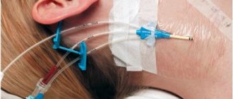
After this, the specialist attaches electrodes to the areas of the head being examined. For diagnostics, a rheographic attachment and a recording device are used. The equipment records the indicators on a special tape. During the examination, the specialist may ask the child to follow simple commands: turn or tilt his head. A functional test allows you to obtain the most complete information. The procedure lasts no more than 10-20 minutes. A transcript of the results is provided 30 minutes after completion of the manipulations.
Liver radiography (REG)
Posted at 23:58h in Services by doctor
Rheography is a diagnostic method that examines blood flow both in specific organs and tissues and in the entire body as a whole.
The essence of rheography is to graphically record, using a special device - a rheograph - changes in the electrical conductivity of an organ caused by pulse fluctuations in blood flow.
Among all the structures of our body, blood has the highest electrical conductivity.
This means that during systolic contraction of the heart, when blood flows into nearby organs, the electrical conductivity of these parts of the body will be high, and at the moment of relaxation of the heart muscle (diastole), on the contrary, it will be low.
Based on the rheograph's readings, a pulse oscillation curve, called a rheogram, is derived.
Rheography literal translation: “reo” - flow, flow and its graphical representation. The rheography technique is a study of blood circulation based on the measurement of a pulse wave activated by the resistance of the vessel wall when an electric current is passed.
When diagnosing all kinds of vascular pathologies of the brain, limbs, lungs, heart, liver, etc., rheography is prescribed.
Rheography of the extremities is used for diseases of peripheral vessels, accompanied by changes in their tone, elasticity, narrowing or complete blockage of the arteries. The rheogram is recorded from symmetrical areas of both limbs, onto which electrodes of the same area, 10-20 mm wide, are applied. To find out the adaptive capabilities of the vascular system, tests with nitroglycerin, physical activity, and cold are used.
Rheohepatography
Rheohepatography is a study of liver blood flow. By recording fluctuations in the electrical resistance of its tissues, rheohepatography allows one to judge the processes occurring in the vascular system of the liver : blood supply, lesions, especially in acute and chronic hepatitis and cirrhosis.
It is carried out on an empty stomach, with the patient lying on his back, in a number of episodes after a pharmacological load (papaverine, aminophylline, nosh-pa).
Vascular rheography
Vascular rheography or rheovasography allows you to assess blood flow in the vessels in the periphery, that is, in the extremities. The main “targets” of rheovasography are the vessels of the shoulder, forearm, hand (upper limbs), thighs, legs, feet (lower limbs).
Vascular rheography is carried out in the same way as described above: rectangular or strip electrodes are used, the skin under them is treated with a solution of sodium chloride or a special electrically conductive gel.
To study blood flow in a specific area (shoulder, lower leg, etc.), one electrode is placed at the beginning of this area, and the other, respectively, at its end.
For example, if we talk about the lower leg, then these points will be the area of the ankle joint and the popliteal fossa. A wave on a normal rheogram has a steep ascending part, a round crown and a gentle descent with possible additional waves.
With the help of vascular rheography, it is possible, for example, to make a diagnosis such as obliterating endarteritis, or, as it is also called, “smoker’s foot”: a chronic disease that affects the arteries of the leg and foot. On the rheogram this is reflected in a decrease in the height of the curve, flattening of the apex, and the absence of additional waves.
Thus, if there are prerequisites or suspicions of problems with peripheral vessels (loss of their tone, elasticity, narrowing of the lumen or even blockage), then vascular rheography will be able to answer questions of concern.
Prices
| Name of service (price list incomplete) | Price |
| Appointment (examination, consultation) with a neurologist, primary, therapeutic and diagnostic, outpatient | 1750 rub. |
| Consultation (interpretation) with analyzes from third parties | 2250 rub. |
| Prescription of treatment regimen (for up to 1 month) | 1800 rub. |
| Prescription of treatment regimen (for a period of 1 month) | 2700 rub. |
| Consultation with a candidate of medical sciences | 2500 rub. |
| Transcranial duplex scanning (TCDS) of cerebral vessels | 3600 rub. |
What is Rheoencephalography (REG)?
Rheoencephalography is a diagnostic method based on graphic recording of electrical resistance of tissues. Along with ultrasound, REG is a safe method that has long been used in medicine. To carry out the procedure, electrodes are used that send impulses to the body tissues, the reaction to which is assessed by the device. REG allows you to evaluate the tone and elasticity of blood vessels, as well as nearby tissues. In addition, this method provides information about the filling of brain vessels with blood.
When is REG prescribed?
Typically, REG is prescribed to study large and small vessels of the brain in addition to ultrasound. Rheoencephalography is also indicated for an already diagnosed vascular problem to monitor the treatment of the disease.
How is the REG procedure performed?
Before the procedure, you must refrain from smoking, drinks and drugs that affect vascular tone. The patient must be at rest: the study is carried out in a lying or sitting position. Electrodes are attached to previously degreased areas of the head. The patient’s task is to remain still and calm, if necessary following the doctor’s instructions to carry out functional tests that increase the effectiveness of the study. The duration of the REG procedure is 15-30 minutes.
Hypoplasia of the ACA
The cause of sudden blockage of a cerebral vessel is often an abnormal decrease in its lumen. The reason for this is not cholesterol plaques, but hypoplasia of the cerebral artery - a pathological narrowing of the vessel. The disease is observed in 80% of older people. In addition to their congenital defect, age-related changes in blood vessels are added.
PMA hypoplasia – what is it and how does it manifest? ACA hypoplasia manifests itself in insufficient development of the right cerebral artery. The vessel has an abnormal shape. With this pathology, the structure of the blood vessels supplying intracranial structures may be disrupted. ACA hypoplasia is a congenital pathology that occurs in utero. With hypoplasia of the ACA, the nutrition of the cerebellum, brainstem and occipital lobes is disrupted. As a result of the pathology, the risk of aneurysm formation or stroke increases. Hypoplasia of the ACA is a dangerous condition, to which neurologists at the Yusupov Hospital and neurosurgeons at partner clinics pay special attention.