In the process of bearing a child, a woman's body inevitably changes. Since impaired blood flow during pregnancy occupies one of the leading positions among all pathologies of the gestational period, assessment of the state of blood circulation between the expectant mother and baby is included in the mandatory examination program for pregnant patients.
Why does disruption of the uteroplacental blood flow (UPB) occur? What types of this pathological process exist? What is the difference between grades 1a and 1b? How dangerous is this phenomenon for a child? What to do if blood flow is impaired? In what ways is its condition checked?
Degrees of disturbance of uteroplacental blood flow
The uteroplacental blood flow is an anatomically complex system consisting of the placenta, blood vessels of the expectant mother and the child. Disorders of uteroplacental blood flow (UPF) are common pathologies caused by dysfunction of the placenta and umbilical cord.
When diagnosing this pathological phenomenon, grades 1, 2 and 3 are distinguished. In this case, the first degree is divided into 2 types. Information about each of them is presented in the table.
| NPMK degrees | Characteristic | Possible consequences | |
| 1 | 1a | Poor communication between the uterus and the placenta when the latter is fully connected to the embryo. | Deviations in the development of a child in mild forms, manifested in the form of underweight and disturbances in general physical characteristics. |
| 1b | The state of the uteroplacental blood flow is normal, but the embryo-placenta blood circulation pattern has deviations. | Developmental delay. | |
| 2 | Placental insufficiency is present at each level. It is almost impossible to compensate for the lack of oxygen, because The embryonic aorta, uterine artery and umbilical cord vessel are not able to fully pass blood. | In 85% of cases the child dies. | |
| 3 | Characterized by centralization of blood flow. | The condition of the fetus becomes critical due to dysfunction of intracardiac hemodynamics. During Doppler measurements, reverse diastolic blood flow is often recorded. The fetus has developmental abnormalities. This degree can rarely be cured. | |
Pathologies are also classified according to other criteria. The table shows the types of disease.
| Sign | View | Description |
| By time of occurrence | Primary placental insufficiency | Develops before 16 weeks of pregnancy. It manifests itself in the form of disruption of the process of embryo attachment and further abnormal formation of the placenta. |
| Secondary placental insufficiency | It is detected by the time the placenta is already fully formed - after 16 weeks of gestation. Pathology occurs under the negative influence of external factors. | |
| According to symptoms | Compensation | Metabolic disturbances occur in the functioning of the placenta, but the blood flow between this organ and the uterus or fetus functions without interruption. |
| Subcompensation | The female body is not able to restore the blood supply to the embryo necessary for its full growth, because all elements of the blood flow system are not working properly. | |
| Decompensation | There is a disruption of blood flow at all levels, which is difficult to treat. |
Kasabulatov N.M.
It is currently known that the most common cause of disturbances in the condition of the fetus during pregnancy is placental insufficiency (PI), which is the main cause of intrauterine hypoxia, delayed growth and development of the fetus, and its injuries during childbirth [4,14,8].
Placental dysfunction can occur under the influence of various damaging factors - endocrine, hypoxic, toxic, infectious; due to obstetric and extragenital pathology of the mother, environmental and pharmacological influences during pregnancy [1].
In clinical practice, primary and secondary PN are distinguished
Primary PN is characterized by changes in the structure, location, attachment of the placenta and maturation of chorionic villi. Secondary PN is characterized by involutional-dystrophic and inflammatory changes in the placenta, developing in the II–III trimesters of pregnancy.
PN can have both an acute and chronic course. Acute PN is determined by disturbances of the uteroplacental circulation. It often develops against the background of extensive infarctions and premature abruption of a normally located placenta, resulting in fetal death and termination of pregnancy. Chronic PN occurs as a result of a violation of compensatory-adaptive mechanisms in combination with circulatory disorders and involutionary-dystrophic processes [13,15].
PN is also divided into relative (compensated) and absolute (decompensated). Compensated PN does not require treatment, and pregnancy in these cases can result in timely birth of a viable and healthy child. Failure of one of the links in the physiological adaptation of the maternal body to pregnancy can manifest itself as hypertensive disorders, acute (placental abruption) or chronic PN, intrauterine fetal growth restriction (IUGR). The nature of the adaptive-homeostatic reactions of the placenta depends on the cause of PN and differs in extragenital and obstetric complications [13]. Absolute placental insufficiency occurs in severe form and is accompanied by IUGR and fetal hypoxia (up to its intrauterine death). Pregnancy in such women often occurs against the background of the threat of miscarriage.
There are two main forms of chronic placental insufficiency
1) a violation of nutritional function (trophic insufficiency), in which the absorption and assimilation of nutritious products, as well as the synthesis of the fetus’s own metabolic products, are impaired;
2) respiratory failure due to impaired transport of oxygen and carbon dioxide. Both forms of PN can exist independently or in combination. In the pathogenesis of PN, disorders of the uteroplacental and fetal placental circulation, metabolism, synthetic function and state of the cell membranes of the placenta are distinguished. With pathology of the uteroplacental circulation, there is a disturbance in the flow of blood into the intervillous space, difficulty in the outflow of blood from it and a change in the rheological and coagulation properties of the uterine blood. The most important pathophysiological changes in the body during chronic PN in a pregnant woman include hypovolemia and decreased organ perfusion. At the same time, the sensitivity of vascular elements to circulating pressor agents increases and the coagulation cascade is activated, which leads to a simultaneous decrease in the perfusion of the intervillous space [13,15,17,19].
Reocoagulation disorders play an important role in disrupting the hemodynamics of the placenta. Hypercoagulation occupies a special place in the pathogenesis of PN. The initial physiological hypercoagulation of the blood, which reaches its maximum development by the end of the third trimester, provides local hemostasis in the uterus after childbirth. During pregnancy, the body develops hypervolemia and decreases peripheral vascular resistance. These mechanisms are adaptive and protective in nature in healthy pregnant women. In the case of pathology leading to activation of the hemostatic system, they lose their protective function and contribute to the worsening of PN [17,19]. During pregnancy complicated by intrauterine growth retardation, the number of trophoblastic elements penetrating the myometrium decreases. Incomplete trophoblast invasion into the maternal spinal arteries causes insufficient trophoblast perfusion and changes in the secretion of humoral factors. The development of modern methods for studying the state of the fetoplacental complex in the dynamics of pregnancy and childbirth allows for timely diagnosis and treatment of the main clinical forms of fetal suffering - intrauterine growth retardation (hypotrophy) and/or its chronic hypoxia.
Prenatal diagnosis of these conditions is established on the basis of echography and fetometry of the placenta, cardiotocography, Doppler flowmetry in the vessels of the “mother-placenta-fetus” system, cytology, amnioscopy, hormonal methods (determination of free estriol, human chorionic gonadotropin, placental lactogen). Postpartum diagnosis of the placenta is carried out mainly using morphometric and morphological methods. The Doppler ultrasound method, which uses direct measurements of blood flow in various vascular zones of the mother-placenta-fetus system in dynamics, allows one to assess the state of uteroplacental blood flow and therefore has important diagnostic and prognostic significance in the group of pregnant women at high perinatal risk. A directly proportional relationship with a high correlation coefficient was noted between the degree of hemodynamic disturbances in the mother-placenta-fetus system and the incidence of fetal growth retardation, intrauterine hypoxia, surgical delivery by cesarean section, severe condition of the newborn and perinatal losses. The final diagnosis is established taking into account the complementary data of a comprehensive study: echography, CTG and Doppler measurements [9].
Prevention and treatment of PN and intrauterine growth retardation
include measures aimed at improving uteroplacental blood circulation and microcirculation, normalizing gas exchange in the mother-fetus system, improving the metabolic function of the placenta, and restoring impaired function of cell membranes.
In recent years, the arsenal of drugs used for PN has expanded significantly. The effect of the drugs used is realized mainly at the level of the microvasculature of the placenta. In this case, agents are conventionally identified that act on the vascular component of the blood flow and on the rheological properties and anticoagulant potential of the blood. Medicines that affect the vascular component of the uteroplacental vascular system include adrenergic agonists. Their effect is due to a decrease in the tone and amplitude of uterine contractions, general peripheral vascular resistance, diastolic blood pressure, and an increase in heart rate without a significant decrease in blood pressure (BP). Under their influence, pulse blood pressure and cardiac output significantly increase, and hemoperfusion of the uterus and feto-placental area increases. This group includes fenoterol, terbutaline, hexopenaline. With high activity of the uterine muscles and acute fetal hypoxia, intravenous administration of adrenergic agonists to the mother helps improve uteroplacental blood flow. The best effect from their use is achieved with a “fluid load” and cardiotonic agents. This treatment method is complemented by metabolic therapy, laser exposure, etc. [5,10,11,12]. To improve oxygen transport to the fetus, vasodilators and drugs that normalize microcirculation processes in the placenta and uterus (theophylline, xanthinol nicotinate) can be used. In recent years, drugs such as Actovegin, Instenon, and Troxerutin have found widespread use in the treatment of placental insufficiency. These drugs act on the pathogenetic links in the development of placental insufficiency , being involved in metabolic processes, participating in the regulation of energy supply, correcting the impaired compensatory capabilities of the mother and fetus [15].
To improve uteroplacental circulation, intravenous infusions of rheopolyglucin, dextrans, as well as drugs that improve the rheological properties of blood (heparin, fraxiparin) are also used. In recent years, efferent treatment methods (plasmapheresis) have begun to be used to treat PN. It is recommended to use ultraviolet blood irradiation, medical ozone, and laser therapy. Prevention and treatment of this pathology are most promising in early manifestations of PN. Improvement of ultrasound diagnostic equipment makes it possible to identify early, preclinical stages of placental dysfunction. Currently, the emphasis of perinatal care is shifting towards preventing the development of placental insufficiency. Therefore, it is especially important to find safe and effective means of preventing this pathology in women with complicated pregnancy. Treatment of pregnant women at high risk of PN is justified. First of all, these are pregnant women with intrauterine infection, gestosis, and autoimmune pathology. According to the literature, the most effective method for treating the initial manifestations of PN is the use of the cellular metabolism activator Actovegin a in combination with ß-mimetics [18]. Actovegin activates tissue metabolism, which improves trophism, and stimulates the regeneration process. The active substance is a deproteinized hemoderivative from calf blood with low molecular weight peptides and nucleic acid derivatives. Actovegin improves cellular metabolism by increasing the transport and accumulation of glucose and oxygen, enhancing intracellular utilization. These processes lead to an acceleration of ATP metabolism and an increase in the energy resources of the cell. Under conditions that limit the normal functions of energy metabolism (hypoxia, lack of substrate), and with increased energy consumption (healing, regeneration), Actovegin stimulates the energy processes of functional metabolism and anabolism. The secondary effect is increased blood supply. In cases of metabolic and blood supply disorders to the brain, for example, in cerebral insufficiency syndrome (dementia), the transport of glucose across the BBB and its utilization by cells deteriorate. The activity of pyruvate dehydrogenase and the concentration of acetylcholine also decrease. The use of Actovegin improves the transport and utilization of glucose, while an increase in oxygen consumption is observed. Under the influence of Actovegin infusions, central hemodynamic parameters significantly improve in pregnant women who had a hypokinetic type of blood circulation. At the same time, the total peripheral vascular resistance is significantly reduced and fetal-placental blood flow improves [6]. The use of Actovegin significantly improves the indicators of arterial and venous blood flow in the mother-placenta-fetus system, which makes it possible to reduce the frequency of early delivery in PN and adverse perinatal outcomes [2]. Infusion therapy with Actovegin has a pronounced therapeutic effect on the condition of the fetus developing under conditions of placental insufficiency, which is expressed in a significant improvement in blood flow in the fetal-placental vessels and the dynamics of its intrauterine growth. In addition, treatment with Actovegin promotes better tolerance of labor to the fetus. The positive effect of Actovegin on fetal-placental circulation is associated primarily with improved oxygen delivery, increased glucose perfusion and restoration of aerobic metabolism in the placenta [6]. Complex treatment of PN developing in the third trimester with Actovegin and Ginipral allows one to achieve satisfactory perinatal outcomes, despite concomitant extragenital pathology in the mother [3]. Falyants A.G., Zakharov I.V. [16] treated PN in 53 pregnant women with uterine fibroids. A complex of drugs was proposed that improved uteroplacental blood flow, each of which normalized individual parts of the impaired placental circulation and, in general, created a system for protecting the condition of the fetus. Treatment was carried out at gestational ages of 22–26 weeks and at 32–36 weeks. The complex included dipyridamole (tablet) - 25 mg 3 times a day. pentoxifylline 400 mg 1 dr. 2 times a day and Actovegin (tablet) – 0.2 g 3 times a day. In case of decompensation of PN, infusions of Actovegin and rheopolyglucin with pentoxifylline were carried out. Significant positive changes were obtained during control CTG and ultrasound examinations. There were no perinatal losses. Demidovich E.O., Ignatko I.V. [7] used Actovegin in the form of tablets 200 mg three times a day for three weeks to correct identified hemodynamic disorders in fetuses with growth retardation syndrome. For pregnant women with PN on the background of extragenital pathology, long-term gestosis, long-term threat of miscarriage and stage II fetal growth restriction syndrome, this drug was prescribed intravenously in the form of a 10% solution with sodium chloride (250 ml for a course of 7–10 procedures). The recommended regimen for using Actovegin for therapeutic purposes includes starting its use with intravenous drip administration of the drug every other day 5 times at a dose of 5 ml in 250 ml of 5% glucose or saline solution. Then continue taking the drug orally, 1 tablet 3 times a day for 3–4 weeks. In patients with recurrent miscarriage, prophylactic use of Actovegin is also possible. In this case, you can limit yourself to oral administration of the drug, 1 tablet 3 times a day for 2–3 weeks. Thus, Actovegin is one of the promising drugs, the use of which can achieve good results in the treatment and prevention of placental insufficiency. However, the problem of treating PN remains not fully resolved and requires further research in this direction.
Literature
1. Aylamazyan E.K. antioxidants in the complex therapy of late toxicosis and associated chronic fetal hypoxia // Obstetrics and Gynecology. 1991, No. 3, pp. 30–34.
2. Afanasyeva N.V., Strizhakov A.N. outcomes of pregnancy and childbirth with fetoplacental insufficiency of varying severity.// Issues of gynecology, obstetrics and perinatology. 2004, T. 3, No. 2, pp. 7–13.
3. Vozovik A.V. Correction of fetoplacental insufficiency in pregnant women with non-toxic nodular goiter. //Materials of the V Russian Forum “Mother and Child”, Moscow, 2003, 44–45.
4. Garmasheva N.L., Konstantinova N.N. Pathophysiological basis for the protection of human intrauterine development. L., 1985., 159 p.
5. Grishchenko V.I., Shcherbina N.A. Improving the diagnosis and therapy of perinatal pathology // Obstetrics and Gynecology. 1990, No. 10, pp. 3–6.
6. Gromyko G.L. Actovegin: Experience of use in obstetric practice./Edited by Ailamazyan E.K./St. Petersburg, 2000, 33–41.
7. Demidovich E.O., Ignatko I.V. Features of fetal renal blood flow in fetoplacental insufficiency. //Materials of the V Russian Forum “Mother and Child”, Moscow, 2003, pp. 56–57.
8. Kosheleva N.G., Arzhanova O.N., Gromyko G.L. et al. New approaches to the treatment of threatened premature birth // Bulletin of the Russian Association of Obstetricians and Gynecologists. 1996, No. 1, pp. 55–60.
9. Medvedev M.V. Clinical guidelines for ultrasound. 1996, Volume II.
10. Ordzhonikidze N.V., Klimenko P.A., Dzhivigelova G.D. et al. New in the treatment of pregnant women with fetal growth retardation syndrome // Obstetrics and Gynecology. 1996, no. 3, pp. 32–36.
11. Parashchuk Yu. S., Grishchenko O. V., Lakhno I. V., Shevchenko O. I. Fetoplacental insufficiency. Tutorial. Kharkov: KhSMU, 1999. 45 p.
12. Pestrikova T. Yu., Chizhova T. V., Petrichko M. I., etc. Management of high-risk pregnancy and childbirth. M.: Souvenir, 1994. 287 p.
13. Radzinsky V.E., Ordiyants I.M. Placental insufficiency in gestosis. // Obstetrics and gynecology, 1999; No. 1, pp. 11–6.
14. Savelyeva G.M., Fedorova M.V., Klimenko P.A. et al. Placental insufficiency. M, 1991, 276 p.
15. Sidelnikova V.M. Current problems of miscarriage. M., 1999, 138 p.
16. Falyants A.G., Zakharov I.V. Correction of placental insufficiency in pregnant women with uterine fibroids. //Materials of the V Russian Forum “Mother and Child”, Moscow, 2003, pp. 244–246.
17. Fedorova M.V. Placental insufficiency. // Obstetrics and gynecology, 1997; 6:40–3. 18. Shapovalenko S.A. Complex diagnosis and treatment of placental insufficiency in pregnant women at different stages of gestation. // Bulletin of the Russian Association of Obstetricians and Gynecologists, 2001, No. 2, pp. 43–7.
19. Salafia CM Placental pathology of fetal growth restriction. // Clin. Obstet. Gynecol, 1997; 40: 740–9.
Causes that can lead to pathology
Many factors contribute to the appearance of disturbances in uterine blood flow. Many of them are capable of influencing the placenta not only at the stage of its formation, but also at later stages. Possible causes of deterioration of uteroplacental circulation:
- Anemia. Due to a decrease in the concentration of hemoglobin in the blood, hemodynamic parameters increase in all blood vessels, including the uterine ones. This is due to the fact that the body seeks to restore the supply of oxygen to tissues by increasing the speed of blood flow, including uterine.
- Incorrect attachment of the placenta. Accompanied by a decrease in blood flow due to thin muscles in the lower segment of the uterus. This problem occurs when the placenta is attached to its scarred area. This zone cannot provide uteroplacental blood circulation, as a result of which the blood supplied to the embryo may not be enough for full intrauterine development.
- Late toxicosis. This condition, during which small blood vessels are affected, often provokes a violation of the uteroplacental-fetal blood flow (UPFB).
- Infectious diseases suffered by a woman during gestation. A number of pathogenic agents negatively affect the condition of the placenta, causing pathological changes in its tissue. Consequence – the IPC was violated.
- Conflict between the Rh factors of the woman and the fetus. This leads to the development of anemia in the baby, which is fraught with a deterioration in the blood supply to his body.
- Pressure surges. They negatively affect blood circulation in the vessels, provoking the development of NMPK.
- Abnormal structure of the uterus. The bicornuate organ has a septum. Pregnancy develops in one of the two cavities formed. The danger in this case lies in the disruption of the child’s full blood supply. Normally, this is provided by two uterine arteries. During gestation, their diameter increases, which leads to the formation of a large number of vessels connecting them, which help normalize blood flow. In a uterus with such an abnormal structure, these processes are absent, so the required volume of blood does not reach the placenta.
- Defects of the umbilical cord vessels. When their number changes, NMPC develops.
- Endometrial pathologies. Their development is caused by inflammation, surgical interventions, and bad habits of the expectant mother.
- Myoma. With the development of neoplasms, their blood supply increases, and the flow of blood to the fetus, on the contrary, decreases.
- Multiple pregnancy. When two or more fertilized eggs are implanted, the placental area increases significantly. In addition, it is possible for a larger volume of blood flow to transfer to one of the embryos. Not only the donor child suffers, but also the recipient fetus, because his heart muscle is not ready for such an amount of incoming blood.
- Diabetes. By affecting the internal walls of the arteries, this pathology triggers the development of placental insufficiency.
Indications for use
Since there are many reasons that become the primary source of abnormalities in blood flow in the placenta and uterus, experts sorted all of them according to three thematic groups. They cover the following categories:
- on the mother's side;
- from the placenta;
- from the side of the fetus.
In the first case, the most common cause is late toxicosis, which in medical terminology is called gestosis. The condition is caused by the natural reaction of the female body to the presence of foreign tissues in the body.
No less common are:
- arterial hypertension;
- obesity;
- anemia.
The first point falls into the chronic disease camp and is persistent high blood pressure. Over time, unnatural blood pressure levels begin to affect the so-called target organs, which include not only the heart muscle, but also the brain, as well as the eyes.
An equally serious problem is obesity, which causes unnecessary pressure on the uterus. Because of this, the inflow and outflow of blood does not occur as intended by nature.
If, when deciphering the results of a duplex analysis, it turns out that the baby suffers from hypoxia, then the reason for this may be the mother’s anemia. It is caused by a decrease in the level of hemoglobin, which is responsible for transporting oxygen throughout the body. If you ignore the disease detected by scanning, the risks of miscarriage, premature birth or impaired development of the child increase.
Women who have previously been diagnosed with uterine fibroids can also be sent for testing. A benign formation of muscle tissue is considered not particularly dangerous, but not during pregnancy. In an interesting position, the neoplasm can compress the placenta and inhibit vascular patency.
Also, diseases on the mother’s side, after detection of which she will be sent for a specific ultrasound, are:
- Chronical bronchitis;
- pneumonia;
- bronchial asthma;
- respiratory failure.
As soon as the supply of oxygen to a new family member is reduced, there is a risk of deviations in child development. By an identical principle, cardiovascular failure poses a danger. The disease may be a consequence of:
- ischemia;
- cardiomyopathy;
- valve defects.
Once the heart's "pump" stops working at full capacity, it causes a drop in blood pressure, which blocks normal blood transport.
In addition to problems on the part of the mother herself, there are also a number of prescriptions that are explained by pathologies of the placenta. Among them are the following:
- changes in tissue homogeneity, which may be a cyst, tumor or previous heart attack;
- premature aging;
- mismatch with gestational age;
- presentation.
The latter option affects situations where the placenta is located in the lower part of the uterus, where blood does not flow particularly actively. This scenario increases the risk of hypoxia several times.
A little less often, the reasons for sending pregnant women for scanning are problems with the fetus itself. Most often, they begin to be suspected after undergoing an initial classical ultrasound, which involves determining developmental delay. This indicates a discrepancy between the growth dynamics of the baby and the established gestational age. A large fruit weighing over 4 kilograms can also become a problem.
Separately, young ladies with diabetes and those who were found to have intrauterine infections during laboratory testing were at risk.
Despite the stereotypes that any examination negatively affects the well-being of the unborn newborn, duplex scanning is not included in this list of risky procedures. The truth is confirmed by the fact that the only significant contraindication for its implementation is the serious condition of the woman. Then the victim requires immediate emergency care first.
How dangerous is a 1st degree violation for a child?
The most common and dangerous consequence of these hemodynamic disorders (HDD) is oxygen starvation. Other complications of poor blood supply to the fetus include:
- decrease in body weight and physical parameters (intrauterine growth retardation);
- acid-base imbalance;
- disorder of the heart in the form of acceleration or deceleration of the pulse, arrhythmia;
- reduction of adipose tissue in the body;
- threat of pathological abortion;
- hormone imbalance;
- antenatal fetal death.
Symptoms of uteroplacental blood flow disorder 1a degree
If this pathology is in the stage of compensation, the expectant mother will not feel any pronounced deviations. In this case, you can find out about the disease only after examination. Obvious signs of the disease accompany the acute form and chronic decompensation. This pathology is accompanied by the following symptoms:
- a sharp increase or cessation of motor activity of the embryo;
- too slow growth of the abdomen (the diameter of its circumference does not correspond to the normative indicators corresponding to the specific gestational age);
- gestosis;
- arterial hypertension;
- strong weight gain of the expectant mother;
- swelling of the legs below the knees;
- proteinuria.
In some cases, bleeding may occur. This symptom most likely indicates placental abruption. If bleeding occurs, you should immediately see a gynecologist.
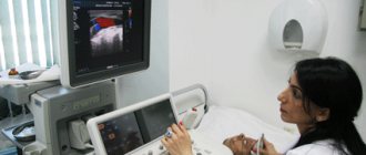
Diagnostic methods
Dopplerography can provide the most reliable and complete information about this pathology. This diagnostic procedure is based on the use of ultrasonic waves and is completely safe for the expectant mother and baby. Using the procedure, signs of circulatory disorders are diagnosed, such as a decrease in diastolic velocity, an increase in the resistance index, and a dicrotic notch in the blood flow curve. The table provides information on how this pathology is diagnosed.
| Diagnostic method | Type of study | Purpose of the event |
| History taking | Analysis of the patient’s complaints, correlation of abdominal circumference with standard indicators corresponding to the gestational age | Making a preliminary diagnosis, developing a plan for further action |
| Physical examination | Auscultation | Determination of fetal heart rate |
| Laboratory research | Blood analysis | Determination of the amount of estrogens, progesterone, human chorionic gonadotropin |
| Instrumental studies | Ultrasound of the pelvic and abdominal organs | Determining the size of the fetus and the condition of the placenta |
| Cardiotocography | Study of the child’s heart function | |
| Dopplerography | Assessment of the intensity of blood flow, determination of the state of intraplacental circulation, flow speed and direction of blood in the vessels of the uterus and umbilical cord |
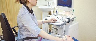
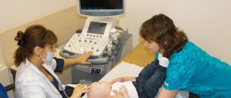
How is the manipulation done?
Diagnostics does not oblige the lady to specially prepare for the appointed date. The examination is carried out in the usual ultrasound room of the clinic. There are no special dietary requirements for the subject before the actual analysis. It can also be carried out at any time of the day without the risk of getting a result with a high percentage of error.
The examination is carried out in a supine position, for which there is a medical couch in the office. First, the diagnostician applies a gel to the sensitive sensor so that the contact with the skin is as dense as possible.
Next, the doctor slowly moves the sensor along the front of the abdominal wall, which makes it possible to assess the blood flow in all the vessels present there. Usually the procedure starts with checking the uterine arteries, and it doesn’t matter which one to start with - right or left. This allows you to identify symmetry problems.
Having completed the uterine part, the specialist switches to examining the structure of the placenta, which is necessary to exclude angiomas - vascular tumors. After this, the umbilical cord and certain problematic vessels such as the middle cerebral artery of the fetus are examined.
You also need to remember that it is not advisable to use the presented method before the 16th week of pregnancy. The reason for this is the unfinished formation of the placenta, which will not allow hemodynamic disturbances to be noticed.
An additional advantage of scanning is the ability, along with studying the clinical picture of the blood flow of the uteroplacental type, to also establish possible anomalies in the baby’s cardiovascular system. In total, the test lasts no more than twenty minutes.
Features of treatment during pregnancy
Therapeutic tactics depend on the degree of the pathological process and the pathogenesis of the disorders. This disease can be treated with medications only in the first degree of circulatory impairment. The second degree is considered borderline. If the pathology has reached the third degree, surgical intervention is indicated. The doctor decides which treatment method to choose on an individual basis.
Conservative methods of therapy
Therapeutic tactics are based on a complex effect on all elements of the hemodynamic process:
- For minor deviations from the norm, Hofitol is used. If symptoms are severe, the patient is prescribed drugs with more active ingredients (Pentoxipharm, Actovegin) (see also: Actovegin: instructions for use during pregnancy).
- When a pregnant woman is diagnosed with a tendency to form blood clots, medications are used that can improve the flow of blood through the blood vessels (Curantil).
- To dilate blood vessels, Drotaverine or No-Shpa is used orally, Eufillin is used as injections.
- For uterine hypertonicity, drip administration of magnesia and enteral use of Magne B6 are indicated.
- The negative consequences of circulatory disorders must be eliminated with the help of ascorbic acid and tocopherol, which have an antioxidant effect.
Medicines are prescribed by the attending physician. Self-medication is strictly prohibited. If the chosen treatment tactics do not improve well-being, the patient is indicated for inpatient treatment. This measure will allow for constant medical monitoring of the condition of the expectant mother and fetus.
Surgical intervention
If signs of pathology are pronounced (grades 2 and 3 MPC), emergency delivery is resorted to. In situations where conservative therapy did not give the expected result, including that which was carried out with diagnosed 1st degree of blood flow impairment, a decision on further actions is made in the next 48 hours. In this case, as a rule, doctors perform a caesarean section. If childbirth in this way is planned to take place before 32 weeks of gestation, the baby’s condition and vital signs must be assessed.
The essence of Doppler measurements of fetoplacental blood flow
During pregnancy, a new structure is formed in the female body, within which the relationship “future mother - placenta - baby” takes place.
Its blood flow system is also separate. Any violation of it can have a negative impact on the fetus. To diagnose such disorders, Doppler measurements of fetoplacental blood flow are used. It allows you to assess the movement of blood through the vessels within this blood flow system, thanks to which the doctor is able to identify any violations that pose a threat to the health and life of the mother and child.
Dopplermetry (abbreviated as “DPM”) is an ultrasound examination technique that allows you to determine the indicators of blood movement in the vessel being examined. It is based on the Doppler effect, which causes a frequency shift when acoustic ultrasonic waves are reflected from objects in motion. In this case, these objects are blood cells that move through the vessels, forming blood flow.
During the procedure, blood flow in the uterus and umbilical cord arteries is assessed in pregnant women.
Preventive measures
Patients who are at risk of developing this disease need to carefully prepare for pregnancy. Spontaneous conception in this case can turn into a tragedy for the couple.
For those women who are already pregnant, to prevent deterioration of uteroplacental blood flow, it is necessary to follow a number of rules:
- Avoid stress.
- Eat properly. Food must contain the required amount of proteins and vitamins.
- Completely quit smoking, incl. passive, and drinking alcoholic beverages.
- Take walks in the fresh air every day.
- Regularly ventilate the living space.
- Avoid excessive physical activity. Preference should be given to yoga, swimming, and gymnastics for pregnant women.
- Monitor your body weight. Weigh yourself regularly and measure your abdominal diameter.
- Sleep on your left side. If the stomach is on the left for a long time, the load on the inferior vena cava, located to the right of the uterus, is reduced. However, sometimes with renal congestion, sleeping on the right side solves this problem.
- Pass all scheduled examinations.
Advantages over analogues
In the normal course of an “interesting situation,” women are only prescribed a series of standard tests and a traditional ultrasound. This progenitor of the modern duplex analogue is considered a safe method that has no significant contraindications and is designed to determine the exact duration of pregnancy.
Also, classical echography is designed to determine the simplest structural changes in the placenta, including difficulties with its location. If any suspicions arise at this stage, then a scan is prescribed to examine the exact picture of the intrauterine development of the fetus.
An ultrasound, which helps to establish a possible multiple pregnancy, as well as the size of the baby and its possible entanglement with the umbilical cord, is unlikely to provide more accurate information about any pathology. And if cardiotocography was initially used for a more in-depth analysis, today it has been replaced by duplex examination. It combines both conventional ultrasound and testing using the Doppler effect. The latter allows you to better study the reaction of the baby’s cardiovascular system to uterine contractions.
At this stage of the study, the girl may be diagnosed with fetoplacental insufficiency, as evidenced by:
- falling outside the basal heart rate (less than 100 beats or more than 170);
- rhythm deceleration;
- decreased heart rate variability.
A similar non-invasive system, which is aimed at studying dynamic blood circulation according to the “mother to child” scheme, makes it possible to recognize the first alarm bells.
In addition to all of the above, scanning also allows you to determine deviations in the patency of the uterine, umbilical and small arteries with fixation of blood flow speed.
If the suspicions are not confirmed, the previously scheduled amnioscopy may even be cancelled. If the duplex technique revealed reasons for concern, but could not accurately determine the location or nature of the disease, then the intervention of an amnioscope cannot be avoided. It is necessary to obtain an indirect assessment with the analysis of amniotic fluid. The attending physician must provide a decoding of the color of the liquid.
The main advantage of the procedure, which mixes the standards of ultrasound and the Doppler effect, is its fairly high accuracy, as well as the absence of undesirable consequences. Doctors themselves value this type of analysis of uteroplacental blood flow due to its ease of execution and the ability to obtain results immediately after the test.