The brain is responsible for coordinating the work of the entire body. A reliable way to obtain information about the structure and functioning of the baby’s brain structures, to conduct NSG / neurosonography / of the brain with determination of the nature of blood flow in the vessels / Doppler ultrasound /
Currently, almost all children undergo NSG in the maternity hospital as a screening method.
To monitor the dynamics of the baby’s development and brain development disorders, repeated examination is recommended in order to exclude congenital or acquired abnormalities. This is especially true for children from the group of premature, low birth weight, immature for gestational age.
Doppler ultrasound of head and neck vessels for children of all ages
Doppler ultrasound is based on the fact that the ultrasound of diagnostic equipment reacts to moving red blood cells in the patient’s blood and thereby gives an idea of the intensity of blood flow, the lumen of blood vessels and many other parameters. The information content of the examination is quite high, however, if necessary, Dopplerography of the vessels of the child’s head and neck can be supplemented with duplex scanning. Dopplerography of the vessels of the head and neck can be performed on a child at any age, since this study does not require prolonged immobility. The anatomy of an adult and a child has certain differences, so a doctor who performs an ultrasound scan of the vessels of the head and neck of a child must know the characteristics of blood flow in the child’s body.
How is it carried out?
It is possible to perform an ultrasound of the child’s brain only until the age when the fontanelles close. The examination is carried out through the anterior, lateral fontanelles or foramen magnum at the base of the child’s neck.
During an ultrasound, the diagnostician moves a special sensor over the baby's head. The resulting pulses are sent to the device and displayed on the monitor in the form of images. This is a comfortable procedure that can be done in the clinic, even while the child is sleeping.
You can sign up for ultrasound diagnostics by calling the reception at (495) 951 87 63 or
In our center, infants undergo comprehensive diagnostics (NSH, ultrasound of the hip joints, EEG/video-EEG monitoring) and consultations with a neurologist, orthopedist, geneticist, and ophthalmologist.
Indications for Dopplerography of the vessels of the head and neck for children
Dopplerography of cerebral vessels in children is an optional procedure, so it is carried out as prescribed by a doctor. In order to understand whether this study is necessary, the doctor must know the general clinical picture and pay attention to alarming symptoms that may be signs of vascular problems.
Headache
The causes of headaches in a child can be both hypertension and injury. With them, children are at risk of vascular deformation, which will be clearly visible on an ultrasound scan of the head if it occurs.
Restlessness
Restlessness and hyperactivity of a child can also be indications for ultrasound examination of the vessels of the head and neck
, since often these symptoms arise as a result of damage to the central nervous system. Assessing the performance of blood vessels helps to identify possible damage and adjust treatment.
The child gets tired quickly
The cause of increased fatigue, which does not allow the child to study and develop normally, may be increased intracranial pressure. This affects the quality of blood circulation in the brain and neck, so Dopplerography of blood vessels will be the most informative procedure for determining the causes of this condition in the child.
Memory and attention disorders
This is one of the symptoms of attention deficit disorder and other disorders that require in-depth research into the causes. It is possible that memory and attention impairments in a child occur due to insufficient blood supply to the brain, and an ultrasound scan of the vessels of the head and neck will help to exclude or confirm this.
Delays in speech development
The most common cause of delayed speech development in a child is minimal brain dysfunction, which can be caused by both birth and postpartum injuries and hypoxia. In case of organic brain dysfunction in children, ultrasound examination of the vessels of the head and neck will be the most accurate way to determine the degree of damage.
How does a Doppler ultrasound machine work?
Doppler ultrasound uses reflected sound waves to assess blood flow in different parts of your baby's body. It is important that ultrasound machines do not use any other type of radiation other than ordinary sound.
The mechanism for obtaining images during Doppler sonography is in many ways similar to the mechanisms that nature uses: with the help of ultrasonic waves, dolphins and whales search in the seas and oceans for schools of fish that they hunt. The tip of the Doppler ultrasound machine emits sound waves, like a dolphin, which are partly reflected from organs and tissues and partly absorbed by them. By analyzing the difference in reflected echo waves, the computer builds images of the structure of blood vessels. Structures that reflect sound well appear brighter on the screen, while organs and tissues that absorb most of the sound wave appear less bright than their surrounding environment. Body tissues that completely absorb sound waves (anechoic) appear black.
When the doctor sees on the screen a vessel in which he wants to evaluate the blood flow, he presses a button to turn on the Doppler function. To measure blood flow parameters, an ultrasound machine uses the Doppler effect, which consists of changing the length of the echo wave when reflected from a moving object (moving blood). In this case, the computer measures the change in the wavelength of the echo signal and calculates all the necessary parameters of the heart, blood vessels and blood movement, while building a color image of the blood movement in the vessels on the screen.
Preparation for the procedure
Ultrasound scanning of the vessels of the head and neck requires, first of all, psychological preparation of the child: it is necessary to explain to him that this is a painless procedure and tell him in detail how it is carried out. Also, before it, children can be shown videos and reviews about ultrasound scanning of the vessels of the head. If a child takes vascular medications due to illness, they must be discontinued in order for the study results to be reliable.
Dopplerography or duplex ultrasound scanning of blood vessels in children
Vascular ultrasonography is a major diagnostic field that uses duplex ultrasound scanning to see in real time how blood flows through the arteries and veins in different parts of the baby's body and organs.
Ultrasound examinations (ultrasonography) are a safe and informative method for modern diagnostics of the condition of the blood vessels of a newborn or older child. Ultrasound scanning of blood vessels can be performed without special preparation, right at the bedside. Such diagnostic studies are easily tolerated by the child.
How is the research going?
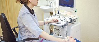
Ultrasound scanning of the vessels of the head and neck for children is done in ultrasound diagnostic rooms. In order to examine the vessels of the head, the child will be asked to lie on the couch, his neck and head should be freed from clothing and jewelry. Transcranial vessels of the head during ultrasound examination are examined with a sensor in the area of the back of the head, temples and eyes, which the child must be told about in advance. In order to examine the vessels of the neck, the patient must lie on his stomach. Doppler ultrasound of head vessels
and the neck of an infant is done with the help of parents who hold the baby in such a way as to ensure his immobility.
Why is this ultrasound examination called duplex?
Duplex ultrasound scanning is used for visualization and differential diagnosis of various conditions and diseases of blood vessels. The term "duplex" means "double". The fact is that this study combines two types of ultrasound diagnostics: traditional sonography and Dopplerography. On the screen of a duplex scanning ultrasound machine, a double layer of image is simultaneously displayed in real time: visualization of the structure of the examined part of the body in shades of gray and a color Doppler image to visualize the movement of blood in the vessels. At the same time, the computer calculates the speed of blood movement, the volume of blood and the diameters of the vessels through which it flows.
Decoding the results
The results are deciphered by the sonologist who conducts the examination. Usually he reports what he sees on the monitor to his parents and enters the findings into the conclusion. Subsequently, the conclusion must be transferred to the attending physician. It is worth noting that deciphering the results of an ultrasound scan of the vessels of the head and neck and making a diagnosis are not the same thing; the final conclusions about the child’s health are made by the attending physician. If necessary, a more in-depth study, such as an MRI, may be prescribed.
Conducting research
The doctor will place the child in a comfortable position on the couch and apply an acoustic gel, warmed to body temperature, to the surface of the area being examined, which helps sound waves penetrate the body tissue. The doctor will move the tip of the ultrasound machine along the gel, which will make pulsed whistling sounds. The doctor may ask the child to lie down in different positions, take a deep breath, and hold his breath. In some cases, a cardiogram can be taken and pressure in the upper and lower extremities (at the ankle and shoulder) can be measured at the same time. The duration of the study is 30-45 minutes.
Causes of vascular problems
Problems with blood vessels in children arise for various reasons:
- birth injuries;
- fast growth;
- shocks, injuries, falls;
- diseases that affect the vascular system.
In babies, the problem arises as a complication after birth trauma, which is associated with the physiology of childbirth. Excessive hyperextension of the head occurs during the provision of obstetric care, which is associated with the peculiarities of the anatomy of the woman in labor. Also, excessive hyperextension occurs during Caesarean section. These processes affect the tortuosity of blood vessels and their tone.
Doppler ultrasound of cerebral vessels in a child
Doppler ultrasound is an abbreviated name for Doppler ultrasound examination of the vessels of the extremities, internal organs, head and neck. This diagnostic technique is highly informative and harmless, even with frequent use.
Considering the safety of the study, it can be used not only for adults, but also for children of all ages. It is also not uncommon to perform ultrasound scanning on newborn children. In this case, Doppler sonography of the cerebral vessels is most often necessary for the newborn. This study helps to supplement the data obtained during neurosonography, namely to verify the absence of vascular pathologies or, if present, to determine their type in order to carry out successful treatment in the future. Since this type of study does not involve radiation exposure, it can be done as many times as necessary, thus observing how the treatment is progressing over time. The non-invasiveness of the technique and the absence of discomfort during the examination do not cause rejection in the child, which undoubtedly facilitates the task of both doctors and parents. For the patient, it is no different from a regular ultrasound.
An ultrasound scan of the cerebral vessels of a child after one year is done without a preliminary NSG examination, that is, as an independent type of diagnosis. Doppler sonography may be prescribed to a child by a doctor if there are complaints of headaches, dizziness, fatigue, delayed speech development, and so on. is also regularly performed on children if there is a history of diseases that cause circulatory problems, such as diabetes mellitus or vasculitis, as well as in the presence of congenital pathologies of cerebral vessels. Sometimes it is necessary to conduct even more detailed studies of the child’s vascular system, which are duplex and triplex scanning. These methods help not only to detect the presence of vascular pathology, but also to determine its cause.
A huge advantage of ultrasound examinations is that they are carried out in real time and on a large scale, which increases the objectivity of assessing the parameters necessary to make a correct diagnosis. Of course, this also requires that Doppler ultrasound for children be performed on modern equipment by competent specialists who have experience working with children and specialize in interpreting the results of Doppler ultrasound in children whose blood flow has its own characteristics.