With age, many people begin to worry about varicose veins and other vascular diseases of the lower extremities. This is not surprising - after all, the main burden of movement falls on the legs. The higher the load when moving, the more difficult it is for the veins and arteries of the legs. That is why most often the pathological condition of the vessels of the lower extremities is observed in women who prefer high heels (even young ones), as well as in overweight people and those who work “on their feet” - stand for a long time or walk a lot.
In order to assess the condition of the blood vessels in the legs, doctors recommend giving preference to modern ultrasound techniques. This procedure does not cause discomfort and has a high diagnostic value.
In what cases is ultrasound of the lower extremities performed?
Ultrasound examination of blood vessels can be carried out for preventive purposes or in the presence of disturbing symptoms. Early diagnosis allows you to begin timely treatment and avoid complications, incl. life-threatening.
Direct indications for the diagnostic procedure are the following:
- Rapid fatigue of the legs;
- Discomfort and pain when walking for a long time;
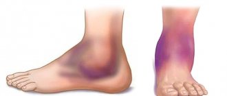
- Pronounced vascular network;
- Periodically occurring feeling of numbness;
- Change in skin tone;
- Constantly cold feet;
- Blue fingers;
- Severe causeless itching;
- Increased sensitivity of the skin;
- Convulsive spasms.
Women who have been using hormonal contraceptives for a long time should be especially careful when diagnosing blood vessels - with long-term use, oral contraceptives can cause an increased risk of blood clots. Vigilance is also necessary for the following factors:
- Recent stroke;
- Diabetes;
- Various bad habits;
- Endocrine diseases;
- Weak blood vessels;
- Unbalanced diet, lack of nutrients.
Equipment in our clinic
In our network of clinics there are more than 20 ultrasound diagnostic devices of a high and expert level, one of them is TOSHIBA VIAMO - a portable Japanese device of expert quality, which allows for a wide range of studies of the vascular system and internal organs. Excellent resolution for diagnosing venous thrombi. Work in Doppler mode and triplex scanning mode.
Mindrey DS 70 is a stationary ultrasound scanner of expert level. Allows you to conduct diagnostic studies of any complexity. Excellent assessment of blood flow in the vessels of the abdominal cavity, arms and legs, neck and brain. All types of sensors are used. Diagnosis of any pathology at an expert level.
Ultrasound scanning of veins is an effective diagnostic method with high clinical value. Correct ultrasound diagnosis of blood vessels is a key factor for correct treatment. For a long time, the diagnosis and treatment of venous pathology was carried out by eye, which led to unsatisfactory treatment results. Venous diseases require the most thorough ultrasound assessment by a specialist with significant clinical experience and working with high-quality diagnostic equipment.
Possible contraindications to the study
There are no direct contraindications to ultrasound examination. However, it is worth understanding that the reliability of the results will decrease significantly if the patient moves his legs during the manipulation. Therefore, people with mental disorders, neurological and other pathologies, in which it is difficult to keep the lower limbs motionless, should consult with a doctor about how best to undergo the study - in some cases, taking sedatives may be recommended.
Indications
In each specific case, the area of localization of the vessels for which ultrasound scanning is required is determined by the doctor. Among the most common indications are:
- thrombosis, phlebitis or thrombophlebitis;
- phlebeurysm;
- congenital anomalies of vascular development;
- diagnostics before and after vascular operations;
- injuries with possible damage to blood vessels;
- swelling, numbness of the limbs, change in skin color;
- pain along deep veins, etc.
Ultrasound diagnostics of the veins of the lower extremities is mandatory for diabetics, people suffering from excess weight and problems with blood pressure. As a preventative measure, it is advisable for smokers to undergo testing.
Do you need preparation for an ultrasound?
Ultrasound examination of the vessels of the lower extremities does not require any serious preparatory measures. But for the information content of the study, it is advisable to adhere to the following recommendations:
If you take any medications (including herbal ones) on an ongoing basis, you must inform your doctor about this. Some of them can affect the condition of blood vessels and pressure. In this case, before the study, it is necessary to limit their intake (unless, of course, these are vital drugs).
From your usual diet, you should exclude foods that can affect the condition of blood vessels and the speed of blood flow. These include tonic drinks, various energy drinks, chocolate, strong coffee and tea.
The day before the upcoming test, you must stop drinking alcohol.
10 hours before the procedure, refrain from smoking.
Before the study, it is necessary to remove hair from the lower extremities.
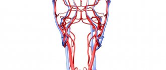
Ultrasound of veins
Ultrasound examination of veins is a method for studying the vascular wall of veins and arteries, which allows one to simultaneously obtain reliable information about the state of blood flow in the vessels. The indisputable advantages of the technique include:
- absolute safety – allows you to carry out the procedure an unlimited number of times, which is ideal for preoperative and postoperative diagnostics, tracking the dynamics of treatment;
- simplicity of execution - does not cause any discomfort to the patient;
- accessibility – the price of the service is low, acceptable for all segments of the population; efficiency – it takes little time, the result is provided within a few hours.
Ultrasound is based on the ability of mobile blood cells to reflect beams of ultrasonic waves with a certain change in their characteristics. A special sensor picks up these reflected signals, and the computer calculates the speed of blood movement, which makes it possible to judge the presence or absence of blood flow disturbances.
The value of ultrasound of the veins of the lower extremities, upper extremities and arteries lies in the possibility of detecting the early stages of vascular diseases, when their clinical manifestations are minor or completely absent.
Ultrasound algorithm
To begin with, a person is asked to prepare the field being studied, that is, to remove clothes below the waist. The patient lies down on the couch. The lower limbs should be shoulder-width apart. The ultrasound doctor applies a thin layer of a special gel, along which the sensor slides. To increase the information content of the study, the doctor may ask the person to change their body position, for example, lie on their stomach, turn on their side, or stand on their feet. An ultrasonic sensor reads the condition of the vessels and displays an image on the monitor in front of the specialist. If necessary, the ultrasound doctor adjusts the frequency of sound radiation and analyzes the data obtained. The study evaluates the vessels of the popliteal, lesser subcutaneous, sural and fibular regions, as well as the posterior surface of the legs.
The duration of the study does not exceed 20-30 minutes.
After research
After the examination, you are wiped off the remaining gel from your skin and allowed to get dressed. After the ultrasound examination, no restrictions on normal activities are required. It will be necessary to wait for a written conclusion from a specialist on the results of the study. No complications after ultrasound examination of veins have been noted so far.
An ultrasound diagnostic doctor or phlebologist will analyze the results of the study and draw up a conclusion, which will be the main document on the diagnosis performed. In most cases, ultrasound of the veins is the final diagnostic method for venous diseases, but sometimes additional research methods such as venography are required.
The conclusion on ultrasound of the veins should answer the following questions:
- Are the venous trunks passable?
- What is the diameter of the trunks of the saphenous veins
- Are there thromboses in the superficial and deep veins?
- Do venous valves work and what is the direction of venous blood flow?
- Are there incompetent perforating veins?
Dopplerography of the vessels of the lower extremities
Often, a comprehensive ultrasound examination with Doppler is performed, which helps to assess the speed of blood flow. As a rule, additional manipulation is indicated in the following cases:
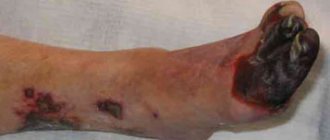
- If the slightest blow causes hematomas and extensive bruises;
- With pallor and cyanosis of the skin;
- If your feet are always cold, they freeze even in warm shoes.
The rules for preparing for the study remain the same as for conventional ultrasound of the vessels of the extremities.
What does ultrasound examination of leg blood vessels evaluate?
The procedure allows you to determine the functioning of the venous collectors, namely:
- The level of patency of deep and superficial veins and the severity of existing pathology.
- The degree of consistency or insufficiency of valves in the venous vessels.
- Detect a blood clot and its level of mobility in the vein.
- Assess the degree of consistency of the perforating veins.
During the study, the level of blood movement through the arteries, vascular patency, the presence or absence of curvatures and aneurysms are checked.
Triplex ultrasound
Today, this method of ultrasound examination of the vessels of the lower extremities is considered the most modern. It allows you to observe a three-dimensional image of the vessel on the screen with detailed visualization. This diagnostic procedure allows us to identify the following pathological processes:
- thrombophlebitis;
- atherosclerosis;
- destruction and structural anomalies of vascular sections;
- angiopathy;
- vasculitis;
- postthrombophlebitic diseases.
The duration of the study is approximately one hour.
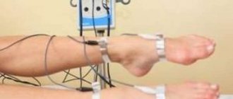
How is the study of veins and arteries performed?
The procedure is practically no different from ultrasound:
- The patient frees the examination area (legs) from clothing. There is no need to remove glasses, contact lenses, dentures or hearing aids.
- Before the procedure, you will be asked to lie on an examination table or couch.
- The doctor applies a special gel to a portable device called a transducer that sends high-frequency sound waves into the arteries or veins being examined.
- The doctor moves the sensor over the patient's skin, examining different areas.
- Information is displayed on a computer monitor when the sensor is pressed against the skin and moved along the legs. The sensor sends sound waves through the dermis and other body tissues into the blood vessels. Sound waves are reflected from the vessels and send information to a computer for processing and recording. The computer creates diagrams that show the flow of blood through arteries and veins.
When examining arteries and veins, the specialist will pay attention to problem areas. First of all, areas with inflammation, pain, and hyperthermia are examined.
The procedure lasts about an hour. After an ultrasound scan of the veins and arteries of the lower extremities, the patient can return to normal activities.
Evaluation of ultrasound data
Ultrasound of the lower extremities is performed during the treatment period to monitor dynamics and as a preventive measure if the development of the disease is suspected. The data obtained is assessed by a specialist using multiple indicators and conclusions are drawn about the need for appropriate treatment.
Evaluated as follows:
- lumen of blood vessels and their permeability;
- blood flow speed;
- thrombus localization and evaluation;
- operation of the valves of the venous channel.
During an ultrasound scan of the leg arteries, the following is assessed:
- thickness of the vascular wall.
- permeability and narrowing of arteries;
- blood flow depending on the phase of the heart (systole or diastole);
- blood flow speed and dynamics of its changes.
The essence and differences of the methods
Dopplerography of the vessels of the brain, neck, upper and lower extremities, as well as their duplex scanning, is a non-invasive diagnostic procedure. Their advantage is their affordable cost, lack of contraindications, and high information content.
The use of the Doppler effect allows one to calculate the speed of blood flow and determine its disturbance in individual vessels. Most often, this data is enough for the doctor to make an accurate diagnosis. In turn, duplex scanning of the vessels of the neck, head and extremities provides information not only about the quality of blood flow, but also about the geometry of the vascular lumen, tortuosity of the bed, the presence of anatomical or postoperative anomalies, wall thickness, the appearance of blood clots and atherosclerotic plaques.
GNICPM offers to take advantage of the capabilities of modern ultrasound diagnostics as part of a comprehensive or routine examination.
Ultrasound equipment
To conduct research, devices are used that are designed for visual diagnostics of the human body. Ultrasound rooms are equipped with multifunctional equipment designed to meet stringent requirements. Modern models of devices have ultra-precise focusing and compact dimensions. They provide high quality images.
To improve performance, the devices are equipped with various modes. For example, two-dimensional visualization, as well as color and power Doppler. Many models are equipped with glare-free monitors that rotate freely around an axis and tilt in the desired direction. It is possible to connect several sensors simultaneously. The received information is recorded on the hard drive and stored in a database.
When developing ultrasound machines, new technologies are being introduced: grain suppression, image detailing, automatic image optimization, wireless information transfer. The scanners are easy to operate. If necessary, a specialist can adjust the settings. The image is instantly transferred to the monitor. The doctor examines the details and assesses the situation.
There are two types of equipment on sale: stationary ultrasound and portable ultrasound. Stationary devices are installed in rooms designed for ultrasound examinations of leg vessels. They are impressive in size and provide reliable diagnostics. Portable devices are portable and small in size. Such devices are designed to conduct research in any place: ward, ambulance, emergency department. They are indispensable in cases where it is necessary to urgently determine a person’s condition and take the right actions.
Contraindications
Despite the apparent safety and gentleness of the main diagnostic methods, they have a number of absolute and relative contraindications. Doctors use in-depth research techniques with caution when:
- Decompensated heart, liver and kidney failure;
- Allergic reactions to the components of the contrast solution;
- Exacerbation of chronic diseases;
- Infections and inflammations;
- Mental disorders.
Studies are carried out with the utmost caution in pregnant and lactating women, elderly patients and patients with disorders of natural coagulation.