In what cases should the blood vessels of the head and neck be checked?
There are categories of patients who, even without symptoms, need to regularly check the condition of their blood vessels. These are elderly people, workers whose specific work involves significant psycho-emotional stress, overweight patients, smokers, and other bad habits.
In addition, there are numerous indications for vascular diagnostics in this area. These include:
•Decreased memory, nausea, vomiting, headaches of unknown etiology, failures in coordination of movements;
•Atherosclerosis, that is, blockage of blood vessels, which has serious consequences for human health, and even life;
•Diabetes mellitus, causing irreversible consequences in many organs and tissues;
•A brain tumor;
•Hypertension;
•Hypotension;
•Cervical osteochondrosis;
•Vertebro-basilar insufficiency.
•Craniocerebral trauma, concussions and spinal bruises;
• Suspicion of ischemic stroke;
•Elective heart surgery.
Initially, the patient should contact a neurologist, who will prescribe the appropriate diagnostic method.
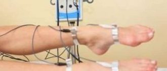
Are your legs bothering you? Check your blood vessels! Who can benefit from ultrasound scanning of lower extremity vessels?
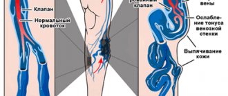
Spider veins, swelling, a feeling of heaviness in the legs - these signs can be, in particular, a manifestation of vascular diseases of the lower extremities. Ruslan Akhyaevich Bidzhiev, ultrasound diagnostics doctor at Clinic Expert Stavropol, talks about in which cases ultrasound scanning of the vessels of the legs should be performed and how this study is performed.
— Ruslan Akhyaevich, Ultrasound scanning of the vessels of the lower extremities - what kind of study is this?
- This is a modern, highly informative method for identifying pathological changes in the vessels of the legs - arteries and veins. The abbreviation “UZDS” stands for “ultrasonic duplex scanning”.
I would like to note that until recently, Doppler ultrasound (USDG) was considered one of the widely used diagnostic procedures for assessing the condition of the vessels of the extremities. However, with its help it was possible to evaluate only one indicator - the patency of the vessel.
Unlike ultrasound doppler scanning, duplex ultrasound scanning of leg vessels allows you to visualize in detail the condition of the vascular wall of both large arteries and veins and small ones, identify the presence of blood clots and atherosclerotic formations, and determine the speed, direction and intensity of blood flow in the vessels. That is, we have the opportunity to obtain more detailed information.
— For what symptoms is ultrasound scanning of the vessels of the lower extremities performed?
— The main complaints of patients for which ultrasound scanning may be prescribed are pain and swelling of the legs. Redness of the skin, spider veins, various compactions (especially in the popliteal region), nodules in the projection of the vessels of the legs can also serve as a reason for prescribing this study.
Typically, ultrasound duplex scanning of the arteries and veins of the lower extremities is prescribed to the patient by the attending physician - a vascular surgeon, cardiologist, neurologist, therapist, traumatologist.
— In the diagnosis of what diseases can ultrasound of the arteries and veins of the lower extremities help?
— Vascular surgeons and phlebologists prescribe it to detect varicose veins, phlebitis (inflammation of a vein), superficial and deep thrombosis (phlebothrombosis and thrombophlebitis).
After operations on the lower extremities, ultrasound scanning is used to monitor the consistency and quality of the performed manipulations. Traumatologists prescribe duplex scanning to prevent thrombosis of the vessels of the lower extremities, associated either with compression of the vessels by a plaster cast or with a blood clotting disorder.
Ultrasound scanning will also be informative in case of some heart pathologies, after chemotherapy, and will also help to distinguish some infectious diseases from vascular pathology (in particular, erysipelas from thrombosis). In addition, this research method makes it possible to detect traumatic injuries and anomalies in the structure of blood vessels, intervascular malformations (connections).
— Do I need any preparation for ultrasound duplex scanning of the vessels of the lower extremities?
- No.
— How is ultrasound scanning of the vessels of the lower extremities performed?
— Traditionally, this study is performed in various projections. Despite the fact that, according to generally accepted recommendations, the patient is examined in a standing position during ultrasound scanning, practice shows that sometimes this is not enough, so our clinic provides a more expanded and thorough approach to this issue.
I would like to emphasize that we carry out diagnostics step by step, relying in our work on the laws of physics. First, the patient is placed on the couch. After normalizing the blood pressure, we perform an ultrasound scan of the femoral vessels, then the patient turns over on his side and the back of the thigh and lower leg are examined.
The next stage is to examine the patient’s limbs in a standing position, that is, under load. If necessary, we perform the examination in a sitting position.
— Does it happen that to clarify a patient’s diagnosis, ultrasound duplex scanning of the vessels of the legs is not enough, and some other research methods are required? And if so, which ones?
— Today, ultrasound scanning is one of the most informative diagnostic methods, which, by the way, is considered the gold standard for studying the blood vessels of the legs. In my opinion, the best method for detecting and assessing pathologies of the veins of the extremities has not yet been invented. To confirm this, I would like to add that, based on our conclusions, traumatologists, vascular surgeons, and cardiologists prescribe treatment and successfully perform operations.
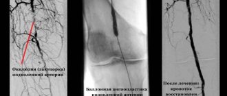
Traditional angiography or CT angiography may also be prescribed to assess the condition of the arteries.
If necessary, the attending physician may additionally recommend that the patient undergo laboratory blood tests. Among them, in particular, assessment of blood clotting indicators (including prothrombin index) and a number of other studies.
You can sign up for ultrasound duplex scanning (USDS) of blood vessels here. ATTENTION: the service is not available in all cities
Interviewed by Sevilya Ibraimova
The editors recommend:
Doppler, duplex, triplex... What types of vascular ultrasound are there? Ultrasound of neck vessels: when is it prescribed? When is ultrasound of cerebral vessels prescribed? When is CT angiography needed?
For reference:
Bidzhiev Ruslan Akhyaevich
Graduate of the Faculty of Medicine of Stavropol State Medical University in 2015.
In 2021, he completed an internship in the specialty “Therapy with a course in dietetics.”
He completed training and advanced training courses in ultrasound diagnostics at the FMBA research center (Moscow).
Currently holds the position of ultrasound diagnostics doctor at the Expert Clinic, Stavropol. Receives at the address: Dovatortsev St., 39A.
What are the purposes of cerebral vascular examination?
Regardless of which specific examination method is prescribed to the patient, his head and neck vessels are always checked for:
•Presence or absence of blockages in vascular narrowings;
•Identification of disturbances in the functioning of blood vessels in asymptomatic disease;
•Checking the effectiveness of treatment already carried out;
•Identification of congenital defects in the structure of blood vessels of the corresponding section;
•Checking the tone of the walls;
•Detection of disruptions in venous blood flow;
•Presence of aneurysms, spasms, deformities.
How is an MRI of soft tissues of the extremities performed?
Magnetic resonance imaging is performed by appointment. At the diagnostic center you will need a passport, a doctor’s referral (if any) and other medical documentation: results of previous MRIs, CT scans, ultrasounds.
If there are implants in the body, you will need a passport or an extract from the clinic where the operation was performed. This will help the specialist assess the possible risks of MRI: metal objects under the influence of a magnetic field can move and heat up.
It is better to wear loose clothes made from natural fabrics during the tomography. Sweatpants and a long sleeve T-shirt will do. It is important that clothing does not restrict movement or cause discomfort.
Synovial sarcoma of the knee joint (indicated by an arrow)
It is better to arrive at the DC 15 minutes earlier than the appointed time to fill out documents and change clothes. If you are planning a contrast-enhanced MRI, you should have a snack before leaving the house. This will help avoid manifestations of vegetative reactions to the administration of the drug. You can eat a small sandwich, yogurt or some cookies.
The sequence of MRI soft tissues of the extremities is as follows:
- The patient undergoes a brief consultation with a radiologist to identify contraindications.
- The person is sent to the locker room, where he needs to remove metal objects (glasses, hairpins, belt, other accessories).
- In the diagnostic room, you should lie down on the tomograph table. The patient's body is secured with soft straps. The equipment is noisy, so it is recommended to use headphones. To urgently stop the study, the patient is given a panic button.
- Upon completion of preparations, the doctor goes into the next room, from where he controls the equipment.
- Perform a survey of the area of interest. Contrast scanning takes place in 2 stages: native tomography, repeated examination after intravenous administration of an amplifier.
- Once the procedure is complete, you can get dressed and wait for the results. The conclusion is taken immediately or at any convenient time. At the patient's request, it can be sent by email.
MR image of the knee joint
Electroencephalography
EEG is also a hardware diagnostic method. Sensors are attached to the patient's head that record brain impulses, and then the recorded curve is deciphered by specialists.
Indications for an EEG include not only obvious disturbances in the functioning of the blood vessels of the head and neck, but also injuries, bruises, and sleep disorders. At the same time, specific areas that experience oxygen starvation are determined.
The procedure requires certain preparation, which consists of stopping taking antispasmodic and anticonvulsant medications. The duration of the EEG is from a quarter of an hour to a couple of hours. This study cannot bring the slightest harm to the body, but it is quite capable of identifying a wide range of abnormalities and ailments.
Definition
Diagnostics and examination of blood vessels is a set of diagnostic methods that allow us to study the condition of blood vessels and determine the pathologies present in them. Vascular examination is carried out to determine the cause of vascular diseases and effective treatment. There are a number of diagnostic procedures that make it possible to identify any deviations from the normal functioning of blood vessels even in the early stages. The examination of blood vessels includes a doctor’s examination, laboratory tests and instrumental techniques for examining blood vessels.
Recommendations before visiting a doctor
At the beginning of the appointment, the patient must voice his complaints to the specialist. To provide your doctor with more complete information, it is recommended to keep a diary of pain attacks. It should note the nature of the pain, its location, strength, frequency of occurrence, and duration. It is also necessary to pay attention to the factors that provoke attacks and, conversely, contribute to their cessation.
If the patient suffers from pressure changes, you need to draw up a table of blood pressure readings in the morning and evening for the last 2-3 days.
The neurologist should be shown a list of medications taken (with exact names and dosage).
It is also advisable to write down questions for yourself that will be asked to the doctor after diagnosis (about the cost and duration of the therapeutic course, prognosis of the disease, the need for consultation with related specialists, etc.).
Before the examination, you should take a shower and put on clean and comfortable clothes.