Description of the phenomenon
Tinnitus or ringing in the ears is a very common condition, affecting approximately one in five adults. Usually it is only an inconvenience, but sometimes it can interfere with the ability to concentrate and sleep well. As a result, the patient experiences constant stress, which has a negative impact on his personal relationships and work.
This condition often accompanies hearing loss, although it does not cause deafness in itself. Many people with tinnitus have excellent hearing. Sometimes they develop increased sensitivity to sounds - hyperacusis, so they are forced to take measures to limit external noise.
In some cases, the pathology disappears after eliminating its cause, for example, otitis media or ear plug. However, often even after treatment, ear noise persists.
Why is there noise in my ears?
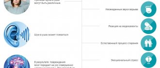
The causes of tinnitus may be associated with damage to parts of the auditory system - the outer, middle, inner ear or brain. There are several theories explaining what happens in the body with this pathology:
- spontaneous electroacoustic emission, that is, the spontaneous generation of electrical signals in the cochlea of the inner ear, which are perceived as tinnitus;
- damage to the organ of Corti with dysfunction of the auditory cells of the cochlea;
- dissonance between healthy and damaged cells of the organ of Corti, which can be caused by the normal aging process;
- increased activity in the posterior cochlear nuclei of the brain caused by exposure to too much external noise;
- auditory plasticity, that is, activation of nerve centers in response to hearing loss due to damage to the cochlea;
- the formation of new connections between the neurons of the auditory nerves when they are damaged, compressed by a tumor or hemorrhage, and the generation of impulses in them in the absence of external sound;
- damage to the trigeminal, facial, glossopharyngeal and other cranial nerves, activating the so-called oto-somatic interaction;
- increased activity of the limbic and autonomic nervous systems due to increased sensitivity to the first episode of tinnitus.
Thus, tinnitus is a complex process that requires accurate diagnosis using modern equipment and competent treatment.
Treatment
Help before diagnosis
You can completely get rid of tinnitus only after treating the underlying disease that caused the discomfort. Therefore, if extraneous sounds appear for no reason, you should not postpone a visit to the doctor. To reduce discomfort until the cause of the disorder is identified, mild herbal sedatives and soothing herbal teas are recommended. In some cases, self-massage of the ears helps: rubbing movements, pressing with your palms on the ears.
Conservative therapy
Since tinnitus is caused by various causes, treatment regimens are selected strictly individually, taking into account the underlying disease and concomitant pathology. A prerequisite for the effectiveness of therapy is the elimination of provoking factors (refusing loud music on headphones, changing jobs, avoiding noisy parties and visiting nightclubs). Etiotropic therapy includes antibiotics, anti-inflammatory and antihistamines, angioprotectors, other groups of drugs, and their combinations. For the symptomatic treatment of tinnitus, use:
- Sedatives
. Herbal and synthetic drugs reduce the excitability of the brain and the speed of nerve impulses. Medicines also normalize the emotional state. - Antidepressants
. Medicines are effective for constant noise, which is caused by chronic diseases, Meniere's disease. The drugs affect centers in the brain and increase the concentration of serotonin. - Tranquilizers
. Indicated for severe ringing or buzzing in the ears, leading to insomnia and reducing performance. The effectiveness of the drugs is due to their sedative and hypnotic effects.
Experimental treatment
Tinnitus retraining therapy (TRT) is an innovative direction that is a type of cognitive behavioral therapy. The program involves individual sessions using psychotherapeutic methods, during which a person is taught ways to relax and techniques for switching attention. A component of the treatment is individually selected sound therapy. The patient is asked to listen to a pleasant noise (the splashing of waves, the rustling of leaves, the sound of rain). Over time, the brain learns to block these sounds, while the perception of pathological noise decreases.
Surgery
If humming, ringing, or tinnitus are associated with purulent inflammatory diseases, it is necessary to open and drain the tympanic cavity, which significantly speeds up the healing process. If tumors of the auditory analyzer are detected as the main cause of the disorder, they are removed with mandatory cytomorphological examination. Treatment of malignant neoplasms involves a combination of surgery with chemotherapy and radiation therapy. In aneurysms, the affected vessel is clipped.
Kinds
There are the following types of noise in the ear:
- subjective: the patient hears noise that does not come from the external environment, it is associated with irritation of the auditory nerve;
- pulsating: the patient hears a buzzing, ringing, clicking or other loud sound that coincides with his heartbeat;
- objective: a rather rare phenomenon associated with the patient’s increased sensitivity to external sounds or vibration of various parts of the body.
Depending on the cause of the noise, it is divided into the following types:
- associated with damage to the vascular system;
- caused by damage to the outer or middle ear;
- muscular;
- neurosensory (peripheral and central), associated with the pathology of auditory cells and pathways.
Additionally, doctors distinguish 3 degrees of noise: with the first, the patient notes sound sensations only upon active questioning, with the second, he considers it not the main problem with hearing, and with the third, tinnitus becomes the main complaint.
Causes of noise not related to hearing aids
If there is noise in the ear, and unbearable pain also occurs, the causes of this condition will not necessarily be related directly to the hearing aid. Often the cause of tinnitus and pain is such diagnoses and conditions as:
- hypertension;
- atherosclerosis;
- metabolic problems;
- diabetes;
- osteochondrosis;
- side effects from taking certain medications;
- poisoning with alcohol or toxic substances.
If unpleasant symptoms do not go away, but only intensify, the best decision would be to consult an ENT doctor to understand the cause of this pathological condition.
Causes of tinnitus
The causes of tinnitus are usually associated with some kind of disease of the hearing organs or nervous system.
- The most common cause of tinnitus is hearing loss. Due to age-related changes, injuries, and the effects of medications, the sensitive cells of the cochlea are damaged. They do not send electrical signals to the brain, and it begins to produce its own impulses, as if compensating for the lack of external stimuli.
- Diseases of the outer and middle ear can cause noise: cerumen plug, otitis media, narrowing of the ear canal, tumor of the tympanic cavity.
- Exposure to loud noise is a very common cause of not only hearing loss, but also tinnitus. Every person should be aware of the damaging effects of loud music and the noise of operating machinery and protect themselves from such influence. Another cause of pathology is barotrauma.
- More than 200 medications can cause this symptom, most often aspirin, aminoglycoside antibiotics (gentamicin, kanamycin) and quinine derivatives. Non-steroidal anti-inflammatory drugs, ethacrynic acid, platinum derivatives, ACE inhibitors and other drugs can cause noise. Exposure to methyl alcohol and benzene is also dangerous.
- Meniere's disease is a disease accompanied by transient dizziness, ringing and congestion in the ears, and temporary hearing loss.
- Acoustic neuroma is a tumor that affects the nerve pathway leading from the cochlea to the brain centers, and causes noise and hearing loss on one side.
- Pulsatile noise is usually associated with pathology of the circulatory system. It is observed during pregnancy, anemia, thyrotoxicosis, arteritis, and also with increased intracranial pressure; In addition, vascular murmur appears with heart defects, vascular development anomalies, and stenosis of the ear arteries.
- The causes of objective tinnitus may be diseases of the temporomandibular joint, pathology of the muscles of the soft palate, middle ear, or gaping of the eustachian tube in the nasopharynx.
- Diseases that can cause tinnitus: hepatitis, diabetes mellitus, atherosclerosis, instability or osteochondrosis of the cervical spine, as well as various hereditary anomalies (Chiari, Gardner-Turner, Klippel-Feil, Pence, Hunt, Konigsmark-Hollender-Berlin syndromes).
The causes and treatment of this pathology are complex, and only a highly qualified otolaryngologist can understand the problem comprehensively.
Tinnitus is a phantom sound perception in the absence of an objective, external acoustic stimulus [1]. Ear noise significantly worsens the patient’s quality of life, as it is often accompanied by hearing loss, hyperacusis, impaired attention, irritability, insomnia, anxiety, and depression.
Ear noise is heterogeneous, and therefore the diagnosis of this pathology requires an integrated interdisciplinary approach. Differential diagnosis should be based on identifying subgroups with established causes of the disease. First of all, it is necessary to distinguish between groups of subjective and objectively auscultated tinnitus.
Subjective ear noise occurs in 5-15% of the population [2, 3], while objective ear noise is much less common.
Typically, objective noise is characterized as a sound that is heard not only by the patient, but also by others. However, it can be viewed from another point of view: objective - as having some additional source of sound generation. Such a source of sound can be pathological blood flow in the vessels located near the middle ear, or myoclonus of the muscles of the middle ear and topographically close areas.
Constant vascular noise is observed with arteriovenous malformations, as well as with vascular tumors of the middle ear [4, 5]. Transient vascular tinnitus may be caused by medications, hypertension, anemia, or intercurrent illnesses such as migraine.
For the differential diagnosis of pulsating tinnitus and the choice of neuroimaging method, examination by an otorhinolaryngologist plays a vital role. Detection of signs of a neoplasm during otoscopy requires first of all to exclude the presence of a vascular tumor in the middle ear. The most common tumor is a paraganglioma.
Paragangliomas (chemodectomas, glomus tumors) are mainly benign tumors of neuroectodermal origin. The source of chemodectome growth is specialized tissue with regulatory functions, concentrated in the paraganglia. Paraganglia are part of the diffuse neuroendocrine system; they are located in close contact with the nerves and vessels of the head and neck: in the bifurcation of the carotid artery, the bulb of the jugular vein, along the vagus nerve, in the tympanic cavity. The areas of primary growth of paraganglioma are called carotid, jugular, vagal and tympanic. The term tympanojugular paraganglioma is used in cases where a tumor that primarily arose in the jugular vein grows into the tympanic cavity, or when it is impossible to determine the site of primary growth of a large neoplasm.
Paragangliomas are characterized by a high degree of vascularization, infiltrative, locally destructive, and slow growth. Clinical manifestations of tympanic paraganglioma (TP) depend on the size and spread of the tumor. Early symptoms include pulsating tinnitus, aggravated by physical activity, and hearing loss [7]. In some cases, TP actively produces catecholamines [8], which is clinically manifested by uncontrolled hypertension [9].
The further development of the symptoms of the disease depends on the size of the tumor and the direction of its growth. Usually T.P. spreads along the path of least resistance: into the auditory tube, mastoid cells, along preformed pathways, i.e. perivascular and perineural. As it grows, jugular paraganglioma destroys the bottom of the tympanic cavity, involving its contents in the process with subsequent destruction of the medial wall, causing dizziness, sensorineural hearing loss, and facial nerve paralysis.
In the absence of middle ear pathology, it is desirable to determine whether the source of tinnitus is a pathology of the arterial or venous segment of the cerebral circulation. Presumably, the topic of the lesion in this case can be judged by the decrease in noise when the ipsilateral carotid artery or ipsilateral jugular vein is clamped [6]. Depending on the results obtained, a visualization method is chosen: CT/MRI arterio- or venography.
Objective muscular tinnitus is based on involuntary, irregular muscle contractions (myoclonus), which are perceived by the patient as clicks in the ear.
The source of muscle tinnitus is most often tremor (myoclonus) of the soft palate or myoclonus of the middle ear. However, in the literature there are isolated descriptions of ear noise associated with myoclonus of the external muscles of the ear and head muscles - m . temporalis
and
m .
occipitalis [10].
Myoclonus of the soft palate is manifested by rhythmic, uncontrolled contractions. In the diagnosis of myoclonus of the soft palate, it is important to differentiate between symptomatic myoclonus, caused by a lesion in the pons or cerebellum, and essential myoclonus, which occurs in patients without intracranial pathology [11].
Clinical manifestations of tremor of the soft palate are the sensation of involuntary movements of the soft palate in combination with rhinolalia or without it - in 20% of patients, clicks in the ear - in 46.7% of patients, or the presence of both symptoms - in 33.3% of patients. In 53.3% of patients, examination reveals movements of the pharyngeal muscles synchronous with the soft palate [12]. Tremor of the soft palate persists during sleep, but disappears when swallowing and with the patient in the supine position. It is believed that objective ear noise in the form of clicks in the ear with tremor of the soft palate is caused by secondary movements of the walls of the auditory tube [13].
Myoclonus of the middle ear muscles is another possible cause of objective ear noise. This term has been proposed to denote tinnitus resulting from dysfunction of one or both intraauricular muscles: m . tensor tympani
and
m.
stapedius .
This type of pathology can be established by characteristic clinical characteristics (feeling of clicking in the ear), as well as on the basis of impedance measurements and otomicroscopy (with myoclonus m . tensor tympani
).
Myoclonus of the muscles of the auricle is extremely rare. In the literature, this phenomenon is characterized by different terms: ear tic, moving ear syndrome, muscular dystonia, ear dyskinesia [14]. Myoclonus of the muscles of the auricle is often accompanied by its visible rhythmic movement; as a rule, it disappears during sleep and when holding the breath.
Treatment of objective muscular tinnitus is very difficult. One of the areas of treatment is pharmacotherapy, but the assessment of its effectiveness is very controversial, and the evidence base is insufficient [3]. Attempts to treat this group of patients by prescribing anxiolytics, antiepileptic drugs, antidepressants, and using masking white noise turned out to be ineffective. Clinically significant results were obtained when botulinum toxin was injected into the soft palate, causing “chemical denervation” of the muscles for several weeks [15, 16, 17]. Botulinum toxin inhibits the release of acetylcholine from presynaptic nerve terminals, causing "chemical denervation" of the muscle for several weeks. When botulinum toxin is injected into the soft palate, side effects are possible in the form of open nasal sounds, velopharyngeal insufficiency leading to ingested food entering the nasal cavity, dysphagia, as well as subjective increased noise in the opposite ear as a result of the unmasking effect. Usually these adverse events disappear within 10-14 days. The severity of side effects can be minimized by targeted administration of botulinum toxin to the area of maximum myoclonic activity under electromyography control [16, 18]. The dose of the drug should be selected individually for each patient using a titration method [16, 17].
The regimen for administering botulinum toxin depends on the patient’s predominant complaint: for tinnitus, the drug is administered transorally into the m. tensor veli palatini
in the lateral part of the soft palate medial to the projection of the hook of the pterygoid process, if the sensation of involuntary movements of the soft palate predominates, the injection is performed on both sides of
the uvula
[17]. The dose and points of subsequent injections depend on the effect of the first injection and the zone of predominance of muscle contractions.
To relieve ear noise caused by myoclonus of the muscles of the auricle, a good effect is achieved by administering botulinum toxin according to the scheme proposed by K. Lee et al. [19]: 10 units. botulinum toxin to the area m. auricularis posterior
and 10 units.
- to area m.
temporalis [20].
If conservative treatment of myoclonus of the intraauricular muscles is ineffective, surgical intervention is possible - selective tenotomy of the affected muscle.
The causes of subjective ear noise are very diverse (Table 2).
Table 2. Causes of subjective tinnitus
Paroxysmal tinnitus can be a manifestation of Meniere's disease [21], dehiscence syndrome of the superior semicircular canal [22], migraine, epilepsy, vasoneural conflict of the auditory nerve. In these cases, differential diagnosis may require MRI, auditory evoked potentials, vestibular tests, and EEG.
Persistent, non-pulsatile ear noise may be associated with conductive or sensorineural hearing loss. In the presence of conductive hearing loss and low-pitched noise, otosclerosis, various forms of otitis, and dysfunction of the auditory tube should be excluded. Sensorineural hearing loss is characterized by high-pitched noise; in these cases, additional examination of the patient is required for unilateral lesions in order to exclude acoustic neuroma and other tumors of the cerebellopontine angle.
If ear noise is accompanied by headache, then the patient’s examination should be aimed at excluding a space-occupying process, benign intracranial hypertension, cerebrospinal fluid circulation disorders, and craniocervical anomalies. In cases of paroxysmal headache of the hemicrania type, combined with the appearance of homolateral noise in the ear, one should remember the presence of Slader's syndrome [23], since the pterygopalatine ganglion plays a leading role in the ventilation function of the auditory tube. With ganglioneuritis of the pterygopalatine ganglion, the function of the auditory tube is observed, which is manifested by a feeling of ear fullness and the appearance of low-pitched tinnitus.
When collecting complaints and anamnesis, it is necessary to note the following characteristics of ear noise: duration of persistence of the symptom; initial appearance of noise (gradual or acute); what the patient associates with the appearance of noise (acoustic, traumatic brain or whiplash injury, hearing loss, dizziness, hearing loss); nature of the noise: constant or intermittent, paroxysmal, pulsating (synchronously with the heartbeat), fluctuating; unilateral, bilateral (symmetrical or asymmetrical), noise inside the head; noise characteristics: low- or high-pitched.
Depending on the answers to these questions, 2 groups of patients can be distinguished. The first group is patients with persistent, long-lasting ear noise, who require a general clinical examination, hearing testing, and treatment as planned.
The second group of patients who exhibit the presence of “red flags” [2] require immediate examination and treatment. These are patients with acute pulsating noise, including after traumatic brain injury; with noise that occurred simultaneously with acute hearing loss; with unbearable noise, accompanied by depression.
A pulsating noise that occurs after a traumatic brain injury may indicate the formation of a carotid-cavernous anastomosis (CCJ). The CCS is a pathological communication between the internal carotid artery or one of its branches and the cavernous sinus, through which arterial blood is discharged into the venous system.
Clinical symptoms of CCS are caused by hemodynamic disturbances. Arterial blood through the formed anastomosis rushes against the blood flow into the cavernous sinus and the veins flowing into it, causing increasing insufficiency of blood supply to the brain and venous stagnation in the orbit. In this case, the cavernous sinus stretches, compressing the cranial nerves passing through it. CCS is manifested by a typical triad of symptoms: proptosis, injection of conjunctival vessels and noise in the head, synchronous with the pulse and decreasing when the ipsilateral carotid artery is clamped [24, 25]. This triad can be supplemented by headache, exophthalmos with visible or palpable pulsation of the eyeball, chemosis, diplopia, ophthalmoplegia, increased intraocular pressure and decreased vision [26]. On auscultation, a pulsating noise is heard in the orbital area, synchronous with the pulse. Occasionally, the carotid-cavernous anastomosis is not accompanied by pulsating exophthalmos, and sometimes there is no vascular murmur.
Natural variations in the structure of the venous system of the skull affect the clinical manifestations of CCS: in patients with a wide superior or inferior petrosal sinus, arterial blood may be discharged into the internal jugular vein, which is manifested by a decrease in the severity or absence of ocular symptoms ( eng.:
"white eye shunt") With a decrease in the functionality of the stony sinuses, blood discharge occurs into the veins of the cerebral cortex, nasal cavity and eyeball, which creates the risk of developing cerebral edema, cerebrovascular hemorrhages and profuse, life-threatening nosebleeds [5, 27].
In the diagnosis of CCS, modern imaging methods are of great importance, revealing the expansion of the cavernous sinus, congestion in the veins of the orbit, pia mater and cerebral cortex. The gold standard for diagnosing CCS is digital subtraction angiography [28]. MRI can detect swelling of the orbital and cerebral tissues, as well as abnormal blood flow in the cavernous sinus [29], and Doppler ultrasound can detect pulsatile or reverse blood flow in the superior orbital artery.
Acute unilateral hearing loss of the sensorineural type is accompanied by the appearance of tinnitus, and in some cases, dizziness. As a rule, this is a manifestation of labyrinthine infarction. The prospect of hearing restoration in this group of patients depends on how quickly treatment is started.
Patients with unbearable ear noise combined with depression also require immediate treatment measures due to the high risk of suicide attempts.
Subjective tinnitus is usually combined with hearing loss (conductive, perceptive, or mixed). Depending on the form of hearing loss and the degree of its severity, a large number of methods for correcting impaired auditory perception and accompanying ear noise have been proposed: pharmacotherapy, acupuncture, hirudotherapy, psychotherapy, physiotherapy, sound and music therapy, hearing aids (traditional, partially implantable hearing aids and cochlear implantation), non-invasive neuromodulation of brain stem structures and others [30], however, all of them can, at best, reduce the intensity of tinnitus, but not relieve the patient from this painful symptom.
The results of modern studies show that with tinnitus, regardless of its original cause, the auditory system is involved in processes that occur in its central parts, and is not accompanied only by morphological changes in the cochlea, as previously thought [31]. The main cause of tinnitus is considered to be increased spontaneous activity of neurons in the auditory cortex of the brain and pathological hypersynchronization of neuronal activity in the auditory system, or a combination of both factors [32].
Based on theoretical concepts according to which subjective tinnitus is a consequence of morphofunctional disorders in the auditory system, research on the treatment of tinnitus in the direction of optimizing the functioning of neural networks and reducing sensory hyperactivity has received new impetus, i.e., taking into account the effects of the implementation of neuroplasticity mechanisms [33] . Such properties are possessed by drugs that have a metabolic and neuroprotective effect on the central nervous system. One such drug is EGb 761 (memoplant).
EGb 761 (memoplant) is a standardized extract of Ginkgo biloba leaves with a strictly defined, stable and precise chemical composition. The main pharmacological properties of this drug are an increase in tissue blood flow (both cerebral and peripheral), a decrease in blood viscosity, replenishment of the deficiency of certain neurotransmitters and growth factors, and elimination of free radicals [34].
The literature contains a large number of clinical studies, including randomized, double-blind, placebo-controlled, which confirmed the effectiveness of EGb 761 in a wide range of diseases of the nervous system: mild cognitive impairment [35, 36], asthenic symptom complex [37], treatment of chronic vascular diseases of the brain [38] and vertebrobasilar insufficiency [39].
The influence of EGb 761 on metabolic processes in nervous tissue was the basis for conducting experimental and clinical studies of the possibility of its use for the treatment and prevention of disorders of the auditory analyzer. It has been established that the neuroprotective effect of EGb 761 may be due to the regulation of phosphorylation of the pAkt protein [40], which affects the adequate expression of proteins-inducers and inhibitors of apoptosis, which ensures greater cell survival in response to damage. In addition, the introduction of EGb 761 limits the release of caspase-3, which is one of the key factors in the induction of apoptosis. The neuroprotective effect of EGb 761 ensures increased resistance of nervous tissue to a variety of damaging factors. Thus, experiments on animals showed a decrease in the ototoxic effect of cisplatin [41] and a decrease in the damaging effect of noise on the auditory analyzer [42, 43] while taking the drug. These effects reflect neuroplasticity processes and help explain the additive therapeutic effect of EGb 761 in patients with sudden sensorineural hearing loss treated with corticosteroids, as was shown in a randomized controlled trial by J. Koo et al. [44]. A similar result was reported in the treatment of blast trauma victims [45].
The obtained data on the effect of EGb 761 on the structural and functional state of the nervous system in the form of stimulation of neuroplasticity processes served as a theoretical basis for studying the possibility of its use for the treatment of chronic subjective tinnitus. In a randomized, controlled, double-blind study, C. Morgenstern and E. Biermann [46] showed the effectiveness of using the drug EGb 761 at a dose of 80 mg 2 times a day. The first significant differences in the level of ear noise between the experimental and control groups were noted after 8 weeks of treatment and reached a maximum 12 weeks after the start of taking the drug. Thus, this study demonstrated a dose-dependent effect of EGb 761.
The problem of the dose-dependent effect of EGb 761 was addressed in a meta-analysis presented in the Cochrane Database, which included studies published before 2006 [47]. It turned out that patients receiving the drug for at least 24 weeks (at least 240 mg per day) had a more significant positive effect compared to patients receiving placebo, while lower doses had a significantly less effect, which confirmed assumption of a dose-dependent effect of the drug. It was noted that improvement in cognitive functions was recorded already at the 12th week of treatment, and when treatment was continued until the 24th week, there was no additional increase in effectiveness. These data were confirmed in a 2015 Cochrane review [48].
Of no less importance is the effect of EGb 761 on emotional and behavioral disorders: a significant decrease in such manifestations as apathy, irritability, dysphoria, dissomnia, depression was noted when taking the drug at a dose of 240 mg per day [49-51], which is extremely important for patients suffering from ear infections. noise. The effect of EGb 761 on emotional and behavioral disorders may be due to its effects on serotonin and glucocorticoid receptors in the hippocampus [52].
Thus, ear noise is a symptom that can signal the presence of serious diseases that threaten the patient’s life. Such patients require careful examination and treatment that can improve their quality of life.
The author declares no conflict of interest.
Symptoms
Manifestations of tinnitus include a variety of sounds heard by the patient. It could be ringing, buzzing, the sound of a roaring aircraft engine, hissing, whistling, clicking. It may be constant or appear only occasionally.
If you experience noise in the ear, you should consult a doctor. This may be associated not only with pathology of the hearing aid, but also be a manifestation of hypertension, hyperfunction of the thyroid gland, anemia and other diseases.
With infectious diseases of the ear, noise may be accompanied by pain and discharge from the ear canal.
When noise is combined with dizziness, urgent medical attention is required, as this may be a manifestation of Meniere's disease or cerebrovascular accident.
With brain tumors, this complaint intensifies at the height of a headache attack. With a tumor located in the posterior cranial fossa, the noise increases with changes in body position.
Our doctors
Novikova Larisa Vaganovna
Neuropathologist, Candidate of Medical Sciences, doctor of the highest category
Experience 39 years
Make an appointment
Pankov Alexander Rostislavovich
Neurologist
40 years of experience
Make an appointment
Diagnostics
To identify the causes of ear noise, the following diagnostic tests are used:
- complete physical examination, medical history, examination of the head and neck, determination of cranial nerve function;
- hearing test (subjective audiography);
- objective audiological examination: method of studying the auditory brainstem response (ABR) and extratympanic electrocochleography;
- computed tomography or magnetic resonance imaging of the skull;
- endoscopic nasopharyngoscopy to identify the causes of muscle noise;
- microotoscopy;
- in some cases - diagnosis of hypertension, hyperthyroidism and other therapeutic diseases;
- according to indications - spinal puncture with determination of intracranial pressure.
High-quality and reliable diagnostics at NEARMEDIC
If tinnitus occurs, the causes must be strictly identified. Only such a diagnosis is complete, because noise is just a symptom of a disease, and the doctor will determine which one. An accurately established diagnosis is the key to targeted and most effective treatment. Therefore, diagnostics should be trusted to professionals. These are the doctors who work at the NEARMEDIC center. Why do patients choose it? Because here:
- Modern diagnostic equipment;
- Highly qualified specialists who regularly improve their knowledge and skills;
- Comprehensive multidisciplinary approach to each patient.
To objectify existing disorders and identify their degree, doctors at the NEARMEDIC center use laboratory and instrumental diagnostic methods. They draw up an individual examination program based on the results of the initial examination. Laboratory methods that may be required include:
- General clinical blood test;
- Determination of lipid profile;
- Hormonal tests (primarily to assess the functional activity of the thyroid gland).
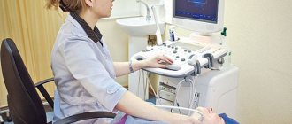
Of the instrumental methods, if there is noise in the left or right ear, the following are shown:
- Ultrasound examination of blood vessels and assessment of the nature of blood flow in them;
- Tone hearing threshold measurement;
- Magnetic resonance examination of the cervical spine and brain;
- Computed tomography of the brain with and without contrast.
When a patient complains of unilateral tinnitus, the doctor always remains oncological alert, because Tumors are particularly dangerous conditions. Their timely identification will help provide the most effective treatment.