Cardiovascular diseases are currently called the “problem of the century,” considering them one of the main causes of disability and mortality, in particular for people of working age. This is due to the steady increase in atherosclerosis and hypertension, leading to local lesions of the fundus.
Retinal vein thrombosis often occurs when:
- hypertension
- vascular atherosclerosis
- blood diseases with bleeding disorders
- diabetes mellitus
- sickle cell anemia
- leukemia
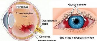
Thrombosis of the central retinal vein is accompanied by a sharp decrease in vision. It appears suddenly. Often the cause of its occurrence is increased blood pressure, physical overexertion and stress. Thrombosis usually occurs in one eye, but occasionally the condition occurs on both sides. With thrombosis of one of the branches of the central retinal vein, changes are limited to the area of the affected vessel. Visual impairment occurs to a lesser extent. Vision after thrombosis of the central retinal vein is usually not significantly restored. In the fundus, swelling of the macular area and optic nerve head is detected - the main cause of decreased central vision; the veins are sharply dilated and tortuous, there are multiple hemorrhages around the vessels. It is believed that the picture of central retinal vein thrombosis is similar to a crushed tomato.
Treatment of retinal vein thrombosis is a long process. The goal of treatment for retinal vein thrombosis is to improve visual acuity and prevent the development of subsequent complications of thrombosis, such as vitreous hemorrhage; increased intraocular pressure, with the development of secondary painful glaucoma; retinal detachment.
Treatment should be carried out in a hospital. In the first hours of the disease, it is possible to perform thrombolysis or resolve the blood clot using fibrinolytic drugs and anticoagulants. In the acute stage of the disease, heparin is injected into the fatty tissue behind the eyeball.
Currently, laser coagulation is used along the branches of the central retinal vein, which allows one to obtain good treatment results.
The patient must remember that the sooner he seeks help from an ophthalmologist, the sooner he will begin to be treated, and accordingly, the prognosis for vision will be better.
Thrombosis of the central retinal vein - what is it?
Central retinal vein thrombosis is a violation of blood flow in the central vein of the eye or its branches.
Due to the obstruction of blood outflow, the veins become overfilled and the vessels become permeable, which leads to hemorrhages in the retina. As a rule, thrombosis occurs in one eye, but can occur in both eyes at once.
There are two types of disease:
- Ischemic - characterized by significant difficulty in blood circulation with many hemorrhages and a strong decrease in visual acuity (up to 0.1 diopters or less). The ischemic type can cause complications and therefore requires careful regular examinations after treatment
- Non-ischemic - this type is characterized by moderate changes in the fundus (visual acuity above 0.1 diopter).
Central retinal artery occlusion
The cause of arterial occlusion of the retina can be hypertension (25%), atherosclerotic changes in the cardiovascular system (35%), rheumatic carditis (7%), and temporal arteritis (3%). In approximately 25-30% of cases, the etiology of the disease cannot be established.
In the mechanism of circulatory disorders in the central retinal artery and its branches, the main role is played by embolism, which can be caused by damage to the heart valve, disintegrating atherosclerotic plaque of the carotid artery or collateral blood flow tracts, as well as blood clots forming in the heart cavity.
Central retinal artery occlusion (CRA) is a predominantly unilateral disease. The age of patients varies from 40 to 70 years. Men are more often affected. The disease begins with a sudden persistent decrease in vision, narrowing or sectoral loss of the visual field, most often in the morning. Sometimes patients note precursors of decreased vision in the form of flickering, the appearance of sparks, short-term and spontaneously disappearing loss of vision.
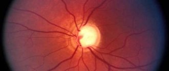
The classic ophthalmoscopic picture of central retinal artery occlusion is widespread ischemic retinal edema with a bright red spot in the macular area (cherry pit sign).
Urgent hospitalization is necessary, since treatment started in the early stages of the disease is most effective. It is usually not possible to determine the exact cause of artery blockage.
Treatment includes massage of the eyeball, administration of active vasodilators (papaverine, compolamine, aminophylline) both systemically and locally in the form of parabulbar injections, dehydration (diacarb, furosemide). Local (parabulbar) corticosteroids are prescribed. To increase perfusion pressure, instillation of beta blockers is indicated. Rheopolyglucin with trental and dexamethasone is administered intravenously. The patient must remember that the effectiveness of treatment is largely determined by the timing of its initiation and is highest in the first minutes and hours of the disease.
The consequences of acute obstruction of the central nervous system are primary atrophy of the optic nerve, central secondary retinal degeneration and a sharp narrowing of retinal vessels. Secondary neovascular glaucoma develops in 1% of patients with central retinal artery occlusion.
Thrombosis of the central retinal vein: treatment
Treatment for central retinal vein thrombosis should begin as quickly as possible, immediately after its onset. With timely, qualified assistance, swelling and hemorrhage resolve within two to three months and vision is completely restored.
To treat thrombosis, intravitreal injections are used (directly into the vitreous body), as well as to treat the underlying disease together with other specialists: hematologists, cardiologists, etc.
If retinal edema persists for more than three months or newly formed vessels appear that cause constant hemorrhages, the patient is prescribed laser treatment.
Doctors at Dr. Belikova's Eye Clinic have extensive experience in treating retinal diseases of varying degrees of complexity using modern drugs and equipment.
Causes
According to statistics, retinal vein thrombosis is initiated by chronic diseases of the cardiovascular or endocrine systems, accompanied by changes in blood viscosity, deterioration of vascular patency, etc. The most common precursors of pathology are:
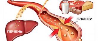
- Vascular atherosclerosis
is a condition in which the level of lipoproteins in the circulatory system increases, depositing on the intima of arteries and veins, forming plaques, and in severe cases completely blocking the lumen of the tubes. Blood clots often form on or near the surface of these plaques, which can break off and float throughout the circulatory system, including the ocular bloodstream. - Diabetes mellitus
is an endocrine disease, the symptom of which is an increase in blood glucose levels. The so-called “sugar” makes blood vessels more fragile and brittle. The body, trying to “patch” the damage, sends blood clotting factors to the damage, resulting in the formation of a blood clot. - Hypertension
is a systemic cardiovascular disease in which the load on small vascular networks, including the eye, increases. As a result, they are damaged, and thrombosis becomes a natural protective reaction (it prevents extensive hemorrhage). - Vasculitis
is an inflammatory disease of the walls of blood vessels of an autoimmune or infectious nature. The formation of blood clots is one of the body's defense reactions. - Tumor processes and metastases occur
in the area where the ocular vessels are located. They create pressure on the vessels and slow down blood flow, resulting in the formation of blood clots. Another scenario is the destruction of the tumor and its fragment entering the bloodstream, followed by blockage of the vessel.
The rarest reason why retinal thrombosis can occur is associated with dysfunction of the thyroid gland - thyrotoxicosis. In the advanced form of this disease, the tissue under the eye grows, causing it to protrude from the eye socket (the so-called thyroid bulging eye). This creates constant tension on the blood vessels, causing them to burst and become clogged with blood clots.
Disease prevention
Compliance with preventive measures will help prevent the development of thrombosis:
- Maintaining and maintaining a healthy lifestyle;
- reduce consumption of foods that increase blood pressure;
- playing sports;
- performing exercises to develop eye muscles;
- high-quality treatment of vascular diseases;
- timely examination by an ophthalmologist.
For the prevention and treatment of pathology, you can make an appointment with a doctor at the Santa Clinic in Chelyabinsk by calling. Operators will select a convenient time for a visit for the patient.
Symptoms
The clinical picture of retinal vein thrombosis depends on the stage of the disease and the location of the blood clot. So, at the first stage, called prethrombosis, symptoms are invisible, although the speed of blood flow in the retina is already reduced. The only sign of the disease is periodic short-term blurring of the field of vision. At the second stage, when a moving blood clot clogs a vessel, the fog before the eyes becomes denser and does not disappear. The third stage is accompanied by post-thrombotic changes and gradual restoration of visual acuity.
The most striking symptoms that make it possible to make a diagnosis are observed at the stage of thrombosis itself.
The intensity of its development depends on the type of occlusion:
- if a blood clot enters an artery of the eye, vision loss occurs sharply, within a few minutes;
- if a blood clot clogs a vein in the visual apparatus, vision gradually decreases over several hours, and sometimes days or even weeks.
The degree of decrease in visual acuity also differs depending on what kind of thrombosis of the retinal vessels occurred - ischemic or non-ischemic. In the first case, a lack of blood supply is observed over a large area of the retina, so vision may become zero, that is, the person does not even see light. In the absence of pronounced ischemic processes, moderate or severe loss of vision is observed, but with preservation of the ability to distinguish the contours of objects or see light.
Important! Painful sensations are not typical for thrombosis of the blood vessels of the eyes. Only in rare cases may a patient complain of dull pain in the orbit.
Treatment methods
Thrombosis therapy begins immediately after diagnosis. The prescribed measures are aimed at resolving the resulting hemorrhages, reducing IOP, improving tissue nutrition and blood circulation.
Sometimes a positive result for the disease can be achieved with the help of drug therapy. The patient is prescribed eye drops that reduce pressure on the retina and accelerate the destruction of thrombotic formations. The use of B vitamins and ascorbic acid is indicated.
If traditional methods of treating the disease do not produce results, the ophthalmologist prescribes laser coagulation. Surgical intervention involves the impact of a laser device on a specific area of the shell.
Laser coagulation is good because it is bloodless, safe and does not require special preparation. The laser has coagulating properties, so it seals blood vessels and accelerates regeneration. After surgery, the rehabilitation period is significantly reduced.
The surgery takes place in a sitting position. The patient is seated on a chair and a drip of anesthesia is injected into the visual organs. With the help of drugs, the pupil is dilated to provide access to hard-to-reach areas of the retina.
A lens is installed on the patient's eye, thanks to which the light radiation will be focused. At this point, the patient may feel tingling or slight discomfort.
The surgeon controls the process of surgical intervention for the disease using a stereomicroscope. The laser beam seals certain areas and glues the membrane to the vascular fundus. Coagulation points keep the retina from further detachment.
Upon completion of the operation, the lens is removed. The patient should sit quietly for some time. At this time, ophthalmologists monitor the patient and monitor his condition. If there are no negative feelings, the patient goes home.
After surgery, you should not overstrain your visual organs. For a while you need to stop watching TV and working on the computer. If the operation was performed on one eye, the patient will not notice any special effects on vision.
Complete rehabilitation takes place in 2 weeks. During this period, it is important to avoid visiting the sauna, swimming pool, and heavy physical activity. It is not advisable to drink alcohol or antidepressants. Do not bend sharply. Since the operated eye is easily susceptible to infectious influences, it is important to promptly treat colds and viral diseases.
Complications after surgical treatment are rare. Sometimes repeated detachment occurs, in which case the operation is repeated. Possible inflammation of the conjunctiva or minor changes in vision.
Reasons for appearance
Thrombosis occurs under the influence of other pathologies in the patient that negatively affect the vascular system of the organs of vision:
- atherosclerosis of arteries;
- hypertension;
- infectious viral and bacterial diseases;
- diabetes;
- sepsis;
- swelling of the optic nerve.
The disease can develop due to the patient's sedentary lifestyle, obesity, and endocrine disorders. Improper therapy for the listed diseases also leads to blockage of blood vessels.
Symptoms of occlusion
As a rule, occlusion of the central artery develops suddenly and completely painlessly. The patient notices an unexpected loss of vision in one eye, which occurs rapidly within just a few seconds. In 10% of cases, episodes of short-term visual impairment occur beforehand. With occlusion of the central nervous system, visual impairment may be preceded by photopsia - light flashes.
Less commonly, occlusion is accompanied by loss of sectors of the visual field. The level of decrease in visual acuity in the victim can vary from distinguishing objects to complete blindness.
Symptoms and clinical diagnosis
Patients with central thrombosis most often complain of sudden, painless unilateral vision loss. Such clinical signs are more typical for the ischemic occlusion scenario. Non-ischemic thrombosis has less pronounced symptoms and is manifested by unilateral clouding and blurred vision. Detecting occlusion of smaller branches is somewhat difficult because it can be asymptomatic. Sometimes there are sectoral opacities and blurred vision, with which patients are in no hurry to see a doctor.
The diagnostic process is based on a detailed study of the patient’s complaints and existing clinical symptoms. General clinical examinations such as general and biochemical blood tests, as well as a coagulogram are performed to determine the state of the blood coagulation system and the risks of recurrent thrombosis. The following instrumental methods are also used to make a diagnosis:
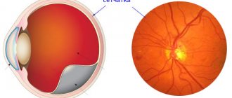
- Ophthalmoscopy to assess the condition of the vessels of the fundus and retina - allows you to see the expansion and blood filling of the venous segment, as well as subsequent swelling of the optic nerve head
- Fluorescein angiography - contrast helps to identify the level of occlusion and assess the functional state of blood vessels
- Optical coherence tomography – if necessary, is performed to visualize the central parts of the retina.
The diagnostic methods described above do not exclude the determination of visual acuity and boundaries.
Treatment of central vein thrombosis
Treatment for retinal vein thrombosis should begin immediately after diagnosis. Typically, several groups of drugs are used in the treatment of the disease:
- Fibrinolytics - to restore blood flow in the vessel. Such drugs are prescribed as an injection into the infraorbital region, after which direct anticoagulants can be used.
- Hormonal agents - to reduce inflammation and swelling. They are prescribed locally and systemically.
- Antihypertensive drugs - to lower blood pressure, since it is often the cause of thrombosis. Most often, intramuscular injections of Lasix are prescribed, as it can also reduce retinal swelling. Reducing intraocular pressure is achieved by using special eye drops.
- Antiplatelet agents - to reduce blood clotting, since there is a risk of a blood clot occurring again. Their use is carried out under mandatory monitoring of coagulation indicators.
Also, medications to improve microcirculation are necessarily prescribed. As a rule, these are intravenous injections. Treatment is not complete without the use of vitamins (necessarily C and B), angioprotectors, and antispasmodics.