In this article we will tell you:
- Types of eye angiopathy
- Causes of angiopathy
- Symptoms and diagnosis of angiopathy
- Angiopathy during pregnancy
- Treatment of angiopathy
- Prevention of angiopathy
- conclusions
Angiopathy of the retina cannot be called an independent diagnosis - rather, it is a pathological condition, a symptom. It occurs as a result of other diseases that specifically affect the blood vessels of the retina. And not only her, but the whole body. Typically, retinal vascular angiopathy in an adult is diagnosed after the age of 30. As soon as nervous regulation is disrupted, the vessels undergo negative changes.
General information
Angiopathy is a pathological process in macro/microcirculatory vessels, which is a manifestation of various diseases, accompanied by damage and disruption of the tone of blood vessels and a disorder of nervous regulation.
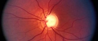
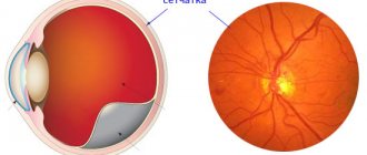
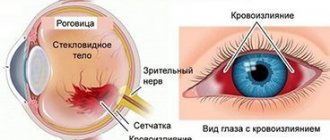
Retinal angiopathy is a change in the microcirculatory vessels of the fundus of the eye, manifested by impaired blood circulation in the retinal tissue, developing under the influence of a primary pathological process. As a result, their narrowing, tortuosity or expansion occurs, hemorrhages into the vitreous body/subretinal space, the formation of microaneurysms, the formation of atherosclerotic plaques, thrombosis of the retinal artery, which leads to a change in the speed of blood flow and disruption of nervous regulation. Thus, angiopathy is a secondary condition, the causes of which can be both ophthalmological and general factors. If left untreated, it leads to irreversible changes in the retina of the eye due to its insufficient blood supply, which can lead to hypoxia of the eye tissue and dystrophic changes in the retina, atrophy of the optic nerve, decreased quality of vision or complete/partial loss. It occurs mainly in adults, but can also occur in children in response to exacerbation of chronic rhinosinusitis or respiratory infection, which is due to the close anatomical connection of the orbit (common innervation, lymphatic/circulatory system) and paranasal sinuses.
Congenital tortuosity of blood vessels in a child is also possible. Since retinal angiopathy is not an independent nosological form, there is no separate ICD-10 code for retinal angiopathy.
Pathogenesis
The pathogenesis of angiopathy is determined by a specific etiological factor.
- Hypertensive angiopathy - persistently elevated blood pressure negatively affects both general hemodynamics and the endothelium of the retinal vessels. High pressure on the vessels leads to pathological narrowing (hypertonicity) of the retinal arterioles and expansion of the retinal veins, uneven caliber and tortuosity of the retinal vessels, destruction of the inner layer (compaction and ruptures), causing local vascular dysfunction and gradually developing disorders of retinal hemodynamics (arterial/venous blood flow ) and the formation of blood clots.
- Hypotonic angiopathy - the tone of blood vessels decreases, which provokes their branching and the formation of blood clots, makes the walls of microvessels permeable and negatively affects the speed of blood flow.
- Diabetic angiopathy of the retina - chronic hyperglycemia , activation of the renin-angiotensin aldosterone system, decreased synthesis of glycosaminoglycans are the main pathogenetic links of diabetic angiopathy. The development of morphological/hemodynamic changes in the vessels of the microvasculature is caused by dystrophic changes in endothelial cells and subsequent disruption of the permeability of the microvascular wall to blood plasma proteins, activation of pericytes, loss of elasticity, hemorrhages and new formation of incompetent vessels.
- Traumatic angiopathy is based on a pronounced increase in intracranial pressure caused by injuries to the eyeballs, skull, cervical spine, prolonged compression of the chest, which provokes ruptures of the walls of microvessels and hemorrhages in the retina.
Frequently asked questions about angiopathy
What is angiopathy in diabetes mellitus?
It is a complication of the underlying disease and aggravates its course. It can affect both small and large vessels, choosing target organs, including the lower limbs, brain, kidneys, and heart.
What is hypertensive angiopathy?
This is a characteristic vascular lesion (swelling, change in wall tone as a result of increased pressure) against the background of constant arterial hypertension.
What is angiopathy of the lower extremities?
The pathological condition of the vessels of the legs (feet) is a severe complication of diabetes mellitus, the manifestations of which can be non-healing ulcerations of the feet (diabetic foot), leading to gangrene and amputation.
Classification
The main factor in the classification of retinal angiopathy is the various diseases that cause its occurrence, according to which they distinguish:
- Diabetic angiopathy - occurs in the presence of diabetes mellitus .
- Hypertensive (hypertensive type) - caused by prolonged and stable hypertension. Hypertensive angiopathy is more common .
- Hypotonic (hypotonic type) - caused by hypotension .
- Traumatic - occurs with traumatic brain injuries, damage to the cervical spine, prolonged compression of the chest.
- Juvenile (Youthful).
- Mixed type angiopathy - occurs when several forms of angiopathy .
conclusions
Angiopathy is a change in the nervous regulation of retinal vessels, leading to their weakness (relaxation) or spasm. Blood supply processes are disrupted, as a result of which vision suffers. The causes of angiopathy in both eyes are usually other diseases: diabetes, hypertension, etc. As well as injuries to the spine and brain. If angiopathy is detected at the initial stage, its further development can be prevented. Lack of timely treatment is fraught with the development of dangerous complications such as retinoschisis. The main methods of treating retinal angiopathy are drugs and surgery. It is necessary to periodically visit an ophthalmologist for a preventive examination, since ocular angiopathy does not manifest itself in the initial stages.
Treatment depends on the type of ocular angiopathy. It should be timely and aimed at treating the underlying disease (diabetes, hypertension, etc.).
Causes of retinal vascular angiopathy
The main etiological factor of retinal vascular angiopathy are various diseases:
- Hypertonic disease.
- Atherosclerosis.
- Diabetes.
- Renal dysfunction.
- Rheumatism.
- Hematological defects.
- Thyroid gland dysfunction.
- Vascular syndromes ( Buerger's , Raynaud's , periphlebitis , periarteritis ).
Physiological conditions that contribute to the development of angiopathy include: pregnancy (early/late toxicosis) and old age.
Exclusively “ocular” causes of angiopathy are various acute circulatory disorders of the retina (embolism, thrombosis), prolonged hypotonic conditions of the central retinal artery. Retinal vascular angiopathy can develop with frequent abuse of alcohol, smoking, radioactive exposure to the body, or working in hazardous industries.
What are the causes of the disease?
Most angiopathy is associated with the development of diabetes mellitus. The pathological process aggravates the course of the underlying disease.
The causes of retinal angiopathy can be both diabetes and blood diseases, hypertension, trauma, smoking, pregnancy, autoimmune diseases, and intoxication.
Cerebral amyloid (CAA) is a consequence of senile dementia or Alzheimer's disease.
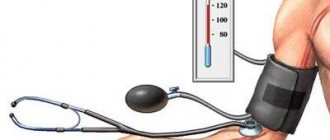
Hypertension causes increased blood pressure.
Striatal angiopathy of newborns is the result of lining of the brain vessels with calcium formations
Symptoms
As a rule, at the initial stage of development of retinal angiopathy, there are practically no symptoms, and patients seek medical help only when vision problems arise. The main symptoms of retinal angiopathy:
- blurred (blurry) vision;
- decreased visual acuity and narrowing of visual fields;
- impaired color sensitivity/reduced dark adaptation;
- the appearance of floating “floaters” in the eyes;
- pain, feeling of pulsation and pressure in the eye;
- the appearance of black blind spots;
- frequent bursting of blood vessels in the eye.
Retinal angiopathy in a child
Changes in the blood supply to the retinal vessels are detected in children under one year of age. Assessing retinal vascular tone is a very subjective method. In the same child, one ophthalmologist will see a violation of tone, and another doctor will regard this as a variant of the norm. In childhood, overdiagnosis most often occurs, when all children are given this diagnosis only on the basis of changes in the blood supply to the vessels. In most cases, children are healthy, since there are no causes or diseases against which angiopathy develops. In children, changes in vascular tone may appear during emotional stress or even with a change in body position, which, of course, cannot indicate pathology.
If the opinions of several doctors coincide and there are changes in behavior or other symptoms, it is necessary to clarify the cause of changes in retinal vascular tone. Most often, fundus pathology is determined in diseases of the central nervous system. If full-blooded veins are detected in the absence of narrowing of the arteries and changes in the optic nerve, the child should be shown to a neurologist.
With increased intracranial pressure, swelling of the optic nerve occurs. At the same time, the veins and arteries of the child’s retina are pinched - the veins become overcrowded, and the arteries narrow and become empty. Diseases of the central nervous system in a child are often associated with perinatal damage - they occur during the period of intrauterine development and childbirth. Perinatal risk factors for the development of any ophthalmopathology are iron deficiency anemia of pregnant women, early toxicosis and acute respiratory infections. In newborns with perinatal pathology of the central nervous system, changes in the organ of vision of varying severity are detected: from angiopathy to strabismus and glaucoma. Epilepsy is one of the diseases associated with perinatal pathology, in which retinal angiopathy .
For rheumatic diseases, retinal angiopathy also appears. Rheumatic diseases have been on the rise in recent years, especially in children. The reason for this is an increase in the aggressiveness of streptococcal infections and a decrease in the sensitivity of streptococci to penicillins. An important role is played by genetic factors - a higher incidence of illness in children if one of the parents suffers from rheumatism. Most often it occurs at school age, and almost never occurs before the age of 3 years. Usually, the first symptoms of rheumatism appear 2-3 weeks after a sore throat - in children, the temperature rises, symptoms of intoxication, pain in the joints and in the heart, shortness of breath, palpitations, weakness. Perhaps an asymptomatic onset - increased fatigue, low-grade fever . But in both cases, changes in the fundus of the eye are noted.
Another disease in which angiopathy is detected is leukemia , which often debuts with changes in the eyes, for example, rapidly increasing unilateral exophthalmos . On examination, in addition to exophthalmos, angiopathy is detected: tortuosity and dilatation of veins, hemorrhages, swelling of the optic nerve head.
In children with secondary hypertension, fundus changes are also detected. Symptomatic hypertension accounts for 40% of the number of children with high blood pressure. Characteristic features of angiopathy with high blood pressure are narrowing of the arteries and dilation of the veins. There is no stage of angiosclerosis in children of primary and school age. Hypertensive retinopathy develops quickly: first, foci of effusion, hemorrhage and swelling of the optic nerve are detected on the eye. With pheochromocytoma or severe kidney pathology, changes in the fundus appear very early and vision quickly decreases.
Moreover, in addition to typical angioretinopathy, maculopathy is present due to the deposition of cholesterol in the nerve fibers of the retina. With this pathology, the tumor is removed and local very long-term absorbable therapy is carried out, which helps improve vision. Children with arterial hypertension should be observed by a pediatrician and ophthalmologist.
Diabetic angiopathy is an inevitable companion to diabetes mellitus cataracts can also develop . For a long time it was believed that children were characterized by type 1 diabetes (insulin-dependent), but more and more often they were diagnosed with type 2 diabetes, which predominates in adults.
In some countries, this type is even more common in children than the first type, which is due to the genetic characteristics of this population. Diabetic angiopathy appears at this age in the late stages of diabetes. The fundus reveals dilation and tortuosity of the veins, minor hemorrhages and retinal edema. Treatment of the underlying disease delays the onset of retinal angiopathy, so it is very important to maintain the required blood sugar level. The traumatic type of angiopathy is also common in children, since they often receive injuries (eye bruises).
The cause of retinal vascular damage in juvenile angiopathy ( Eales disease ) is unknown. The disease progresses rapidly with the appearance of small hemorrhages in the vitreous and retina. In the fundus, dilated and tortuous veins are revealed, which are enveloped in muffs of exudate. New vessels appear on the periphery of the retina (neovascularization), which occurs at the border of the normal retina and the area with poor blood supply.
In severe cases, this disease causes malignant tumors of the retina and its detachment, which leads to complete blindness. The disease progresses slowly over many years. The prognosis for vision is unfavorable due to complications: cataracts , vitreous hemorrhages, glaucoma , retinal detachment . Treatment is systemic (corticosteroids) and surgical. Laser and photocoagulation reduces and prevents vitreous neovascularization and retinal detachment. In case of massive hemorrhages in the vitreous body and the appearance of vitreal cords, vitrectomy (removal of the vitreous body) is indicated.
Treatment of angiopathy
The first task in the treatment of angiopathy is to identify and eliminate the negative factors that served as the background for the development of the disease. As a rule, these are chronic diseases. It is necessary to monitor blood pressure and prevent hypertensive crises. Diabetics must maintain optimal blood sugar levels. These measures are ensured by appropriate treatment regimens prescribed by specialized specialists. The ophthalmologist monitors how the treatment affects the blood vessels of the eyes, and also prescribes treatment aimed at eliminating hypoxia, normalizing the blood supply to the optic nerve head, and strengthening the walls of the blood vessels of the eye. Vitamins, antioxidants, neuroprotectors and mineral complexes can also provide good support to tissues experiencing dystrophy.
Specialists of the Moscow Ophthalmological Center will offer an individual treatment regimen, which may include drugs in the form of:
- tablets;
- injections;
- parabalbar injections into the eye area;
- intravenous drips.
Additionally, hardware treatment programs, physiotherapeutic procedures and massage are prescribed.
Surgical care can be aimed mainly at eliminating complications that have already developed. Most often, laser surgery is used to treat and prevent retinal detachment caused by angiopathy.
During pregnancy
Changes in the condition of the retina during pregnancy occur acutely against the background of toxicosis and go through several stages: angiopathy , retinopathy and neuroretinopathy . First, narrowing and tortuosity of the arteries is detected, then loose spots in the retina (like cotton wool), hemorrhages, swelling of the optic nerve and retina appear. With intense swelling, retinal detachment may develop. Central vein thrombosis is also possible. A pregnant woman's visual acuity is significantly reduced and blindness may occur. All pregnant women with angiopathy due to gestosis are advised to administer Actovegin . At least 5 injections are given in the second half and before birth. Actovegin improves tissue respiration and microcirculation of the retina. This is an effective means of preventing eye complications.
With toxicosis, in contrast to hypertensive angiopathy, there is a non-permanent narrowing of the arteries, which disappears after elimination of toxicosis, sclerosis of the retinal vessels does not occur and obstruction of the lumen of the vessels is not detected, and restoration of blood vessels and vision occurs after childbirth. In pregnant women with gestosis, due to vasospasm and high blood pressure, pronounced changes in the hemodynamics of the eye develop, which have three degrees of severity:
- In the first degree of impairment, the arteries narrow and the veins dilate. There can only be dilation of the veins or narrowing of the arteries with simultaneous dilation of the veins in a ratio of 1:2. If the process progresses, a pronounced narrowing of the arteries is noted. The optic disc has clear contours.
- In the second degree, tortuosity of the arteries and veins appears, unevenness of their caliber, and the ratio of arteries to veins becomes 1:3.
- The third degree is characterized by a change in the color of the disc, the retina becomes pale due to severe vasospasm. The disk is stagnant. Hemorrhages , exudative lesions and “star” formation in the macular area.
As the severity of gestosis worsens, arteriospasm progresses against the background of varicose veins. The severity of the changes depends on the severity of gestosis. The appearance of grade 2-3 angiopathy indicates the progression of toxicosis, and with eclampsia, severe changes in the fundus are noted.
Changes in the fundus are more common with nephropathy in pregnancy. The stage of angioretinopathy is ischemic retinal edema , the appearance of exudative foci and hemorrhages in the retina. Starting from the stage of angioretinopathy, the number of premature births increases. The early stage of neuroretinopathy is accompanied by ischemic disc edema, and the late stage is accompanied by congestive disc. A prognostically unfavorable sign of changes in the fundus is neuroretinopathy , which is manifested by hemorrhages, exudative lesions and the formation of a “star” in the macular area. The outcome of neuroretinopathy is retinal detachment and is often bilateral.
The main danger of angiopathy is that there is a risk of rupture of blood vessels during childbirth and pushing. Therefore, angiopathy is an indication for a cesarean section - this completely eliminates the load on the vessels. Indications for a cesarean section are determined by an ophthalmologist.
Unresolved sharp vasospasm of the retina is an indication for accelerated delivery. Natural childbirth is possible only if the risk of vascular rupture is low. Severe changes in the eye are an indication for termination of pregnancy during gestosis. Absolute indications include:
- retinal disinsertion;
- hypertensive neuroretinopathy ;
- retinopathy with hemorrhages and the presence of cotton wool-like foci;
- central vein thrombosis
- degenerative retinopathy.
Normalization of eye hemodynamics during gestosis occurs only 1.5-2 years after birth.
Pregnant women with mild myopia also have fundus abnormalities, but these are not an indication for cesarean section. With mild myopia, in the first trimester there is a slight increase in the vascular pattern, which disappears by the second trimester. Angiopathy is also observed - a slight narrowing of the arteries and a uniform expansion of the veins. Pregnant women retain vision throughout pregnancy. Unexpressed spasm of arterioles is detected even during physiological pregnancy.
Prevention
Considering that retinal angiopathy is secondary and is a manifestation of various diseases and conditions mentioned above, to maintain eye health you need to monitor your health:
- Have an annual medical examination, including determination of blood sugar and lipids.
- After 50 years of age, an annual fundus examination is required.
- Monitor blood pressure and take timely measures to treat hypertension . For cervical osteochondrosis, regularly do massage, self-massage and physical therapy.
- With increased stress on the organ of vision, it is important to do eye exercises and take special vitamin complexes.
- Active lifestyle (sports, walking, outdoor walks, etc.).
- Rejection of bad habits.
- Balanced diet.
Diagnostics
The disease is diagnosed by an ophthalmologist based on symptoms and after a general examination of the patient. In order to clarify the diagnosis, studies are prescribed, including ultrasound scanning of blood vessels, which provides information on the speed of blood circulation, as well as the condition of the walls of blood vessels, or X-ray examination, which is carried out using a contrast agent, to inspect the patency of blood vessels. Sometimes magnetic resonance imaging may be needed, which allows you to determine the structure of soft tissues and their condition.
Forecast
The prognosis for angiopathy depends on the timeliness of treatment of the underlying disease. Under this condition, processes can be stopped and complications can be prevented. If diabetes mellitus is compensated (that is, the blood glucose level is maintained within the normal range acceptable for a patient with diabetes mellitus), vascular disorders do not progress, or at least do not progress so rapidly.
Stabilization of blood pressure stops the progression of hypertensive angiopathy, but altered and sclerotic vessels do not restore their function. In the traumatic form of vascular pathology, active treatment of the consequences of injury and periodic preventive courses of vascular therapy can improve the condition of the fundus. Vascular changes aggravated by gestosis in pregnant women are gradually eliminated within 2 years after childbirth with adequate treatment, which distinguishes this form of angiopathy from hypertensive one. Juvenile angiopathy of all forms has a poor prognosis and is prone to progression, which ultimately leads to significant vision loss.
Medicines
If the problem of treating the underlying disease is solved, drugs such as pentoxifylline (Pentyline), Trental, Vazonit (help to improve blood flow) are used in treatment.
Xanthinol nicotinate and ginkgo biloba preparations (Bilobat, Tanakan) improve the condition of the vascular wall. To normalize microcirculation, take vitamins C, A, E, B.
ATP-long, actovegin, cocarboxylase drugs have a metabolic effect: they activate blood circulation and improve metabolism.
Soft antiplatelet agents: Magnicor, dipiradamole prevent the formation of blood clots.
Since all such pathologies seriously affect the quality of life, and in some cases are threatening, drug therapy should be agreed with the attending physician.
List of sources
- Egorov E. A. Ophthalmological manifestations of common diseases. Guide for doctors, Moscow, 2006.
- Balashevich L.I. and others//Ocular manifestations of diabetes – pp. 11-85, 90-96, 123-189.
- Katsnelson L.A., Forofonova T.I., Bunin A.Ya. Vascular diseases of the eye. – M.: Medicine, 1990. – 270 p.
- Galileeva V.V., Kiseleva O.M. The use of the antioxidant Mexidol in patients with diabetic retinopathy // VII Congress of Russian Ophthalmologists. Abstract. report Part 2. M., 2000. P.425–426.
- Boroday A.V., Saburova G.Sh., Ishunina A.M. Tanakan in the treatment of diabetic microangiopathies // VII Congress of Russian Ophthalmologists. Abstract. report Part 1. M., 2000. P.304.
Our advantages
"Moscow Eye Clinic" offers comprehensive diagnostics and effective treatment of eye diseases. The use of modern equipment and the experience of specialists working in the clinic eliminate diagnostic errors.
Based on the results of the examination, each visitor will be given recommendations on choosing the most effective methods of treating the eye pathologies identified in them.
The high level of theoretical training and practical experience of our doctors guarantees the achievement of good treatment.
