Author:
Yakovleva Yulia Valerievna 5/5 (1 rating)
Med.
portal: Treatment price Signs Treatment Our advantages
Hypertensive retinal angiopathy is characterized by damage to the vessels of the retina caused by hypertension, which endangers the circulatory system of the entire body.
Arterial hypertension is a chronic disease, the main symptom of which is a persistent, prolonged increase in blood pressure. Hypertension is diagnosed when blood pressure remains within the following ranges for a long time: systolic pressure is above 140 mm, and diastolic pressure is above 90 mm.
Against the background of long-term arterial hypertension without proper compensation, serious complications can arise. And, one of these complications is hypertensive angiopathy. More serious complications include myocardial infarction, stroke, and early mortality.
Nowadays, hypertension is usually called one of the main dangers for humanity.
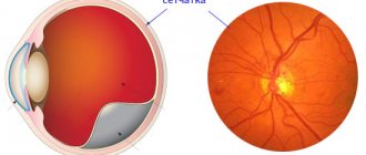
Hypertensive retinal angiopathy, as mentioned above, develops due to a long, uncompensated course of hypertension. With it, due to constantly increased pressure, vascular damage occurs. The defeat gradually and steadily progresses. The development of hypertensive retinal angiopathy is divided into stages:
- The first stage is functional changes. It is characterized by some dilation of the veins and, conversely, narrowing of the arteries, which causes disruption of blood microcirculation. At the first stage, all disorders are difficult to detect and are detected only with a scrupulous study of the fundus (biomicroscopy);
- The second stage is organic changes. At the second stage of hypertensive angiopathy, changes in the structure of the walls of blood vessels begin to occur - their thickening is observed, and later they are replaced by connective tissue. The high density of the vessel walls leads to impaired blood supply in the retina. At this stage, the signs of angiopathy become more and more noticeable: mild swelling of the retina occurs and hemorrhages occur. When performing biomicroscopy, the narrowing of the arteries and the expansion of the venous beds, which become more and more branched, become more noticeable. A characteristic shine of the vessels is revealed due to the compaction of the walls;
- The third stage is angioretinopathy. This stage of hypertensive angiopathy is characterized by a critical change in microcirculation, which leads to the formation of inflammatory exudate in the fundus, released from the affected vessels. All previously appeared pathological changes are aggravated. Vision decreases, and there is a real danger of losing it completely.
- In advanced cases of hypertensive angiopathy, not only the retina, but also the optic nerve undergoes changes (stage of neuroretinopathy).
Our advantages
"Moscow Eye Clinic" offers comprehensive diagnostics and effective treatment of eye diseases. Modern equipment and the high professional level of specialists working in the clinic eliminate diagnostic errors.
Based on the results of the examination, each visitor will be given recommendations on choosing the most effective methods of treating the eye pathologies identified in them.
Causes
Doctors call the main cause of hypertensive retinal angiopathy hypertensive vascular syndrome, in which a high level of tension in the walls of arteries and arterioles remains. As a result of this process, the diameter of their lumen is reduced and, accordingly, the pressure on their walls increases. Arterioles can resist the increased load for some time, and veins stretch and often burst.
The appearance of hypertensive changes in the vessels of the eyes and the body as a whole can be provoked by:
- neurological disorders;
- chronic endocrine diseases - diabetes mellitus, thyroid dysfunction, etc.;
- chronic intoxication of the body due to living in unfavorable environmental conditions, smoking or alcoholism;
- congenital abnormalities in the structure and patency of blood vessels;
- diseases accompanied by increased or unstable intracranial pressure;
- diseases of the blood and hematopoietic system, accompanied by decreased fluidity and increased formation of blood clots;
- chronic diseases of the spine, especially the cervical spine, as well as injuries to the soft tissues of the neck.
Despite the fact that the pathology is caused by various diseases, ophthalmologists cite the patient’s age as the most common cause of hypertensive retinal angiopathy. According to statistics, more than 80% of people with this diagnosis are elderly. Such figures are explained by the fact that a worn-out body is not able to stop the transformation of blood vessels in time, so angiopathy occurs in different parts of the body and organs - weakness of the blood networks.
Important! In most cases, patients with chronic diseases have unilateral angiopathy of the eye vessels. If we are talking about bilateral deterioration of retinal trophism, this may be the consequences of a cervical spine injury.
Our licenses
Symptoms and stages of the disease
The manifestation (the first obvious manifestation) of the symptoms of hypertensive retinal angiopathy occurs at the stages of actively developing vascular insufficiency in the fundus. According to statistics from ophthalmologists, in 90% of cases we are talking about irreversible tissue changes in the retinal area. To identify the disease in the early stages, when it does not manifest itself at all or is accompanied by a slight deterioration in vision, doctors pay attention to the following signs:
- regular stable increase in blood pressure;
- complaints of frequent headaches;
- complaints of inability to concentrate on work that requires prolonged eye strain;
- complaints of temporary visual impairment, for example, the inability to focus in the dark or the need to “get used to” bright light for a long time.
In general, retinal angiopathy of the hypertensive type is characterized by clinical similarities with retinal detachment. In this case, the patient may have difficulty recognizing the text due to its blurriness or many dark spots in the field of vision.
The intensity of symptoms depends on the stage of development of the pathology:
- The initial stage is asymptomatic, although vascular changes are already clearly visible when examining the fundus. Only with significant and prolonged strain on the eyes do signs of “visual fatigue” appear - lacrimation, headache localized in the bridge of the nose and forehead, burning sensation.
- The second stage is accompanied by transient symptoms, a significant increase in the venous pattern, retinal edema and the presence of pinpoint hemorrhages. The patient begins to experience mild discomfort in the eyes, headaches, and difficulty with twilight vision.
- At the third stage, the symptoms characteristic of the second stage of disease development intensify. The discomfort becomes longer lasting. During a hypertensive crisis, retinal detachment may occur, accompanied by loss of part of the visual field or complete blindness.
- The fourth and final stage is accompanied by the release of exudate into the cavities adjacent to the retina and the onset of the inflammatory process. The patient's vision drops sharply or disappears if exudate is released into the space between it and the underlying layer. Additionally, intense headaches may occur; in the most severe cases, fever occurs.
Ophthalmologists note that in the early stages of development, hypertensive retinal angiopathy is easier to correct. The earlier the disease is detected and measures to save the retina begin, the higher the chance of saving vision.
What usually causes retinal angiopathy?
The brain is considered a unique organ that is involved in all processes of the body. Information from various organs is sent to brain cells, where it is analyzed and then transmitted along nerve fibers to various organs of our body. Retinal angiopathy (like most other diseases) appears due to many different factors.
These factors lead to a malfunction of some parts of the brain and disruption of neural connections. In other words, the brain stops giving the “correct” orders for the clear operation of your visual system, which leads to retinal angiopathy, and then to more serious diseases.
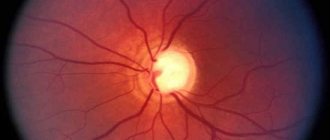
Diagnostics
Hypertensive retinal angiopathy is diagnosed during complex therapy. To identify vascular abnormalities, the ophthalmologist only needs to examine the fundus of the eye using the ophthalmochromoscopy method (a method of examining the retina in red and white light) and visualize the signs of the disease:
- expansion of the venous network;
- narrowing of arteries, arterioles and capillaries;
- pinpoint hemorrhages, etc.
To identify the causes of angiopathy, additional diagnostic methods are used:
- Ultrasound of the eye;
- Doppler scanning of the vessels of the retina, head, and, if necessary, neck;
- radiography with contrast;
- MRI of the head.
If hypertensive retinal angiopathy is suspected, a therapist will refer you for examination, an ophthalmologist will monitor the process, and treatment will be prescribed by a therapist, ophthalmologist, and a neurologist and cardiologist.
How to cure retinal angiopathy as quickly as possible?
We offer an innovative method of color pulse therapy, the discovery of which was awarded the Nobel Prize.
The answer is obvious: in order for the visual system to work correctly, it is necessary to debug the normal functioning of the brain centers responsible for its regulation. This is exactly the problem that the innovative Neurodoctor device solves.
The device is based on the pulse therapy method. This treatment method is considered modern medicine, one of the most progressive and highly effective. Pulse therapy affects certain centers of the brain and activates certain areas of the visual system, eliminating malfunctions in their functioning.
How it works?
Neurodoctor affects the control centers of the brain.
The brain begins to produce dozens of neurohormones and form “corrective” nerve impulses.
Neurohormones are many times more effective than the most powerful drugs in quickly curing the disease. Nerve impulses eliminate pathological processes at the cellular level and correct the functioning of other organs and systems of the body.
LEARN MORE ABOUT THE METHOD
Treatment methods
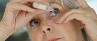
Since ocular angiopathy is not an independent disease, for its treatment it is necessary to use two types of therapeutic measures:
- restoration of vascular tone
, strengthening and normalization of blood circulation; - stabilization of blood pressure.
Groups and names of funds aimed at strengthening and restoring eye blood vessels:
- vasodilators;
- agents that improve blood flow;
- vitamins to strengthen blood vessels.
Additionally, local agents Taufon or Quinax are prescribed, which help reduce the load on the eye vessels, reduce the inflammatory process, and improve metabolic processes in the tissues of the visual organs.
Since hypertensive retinal angiopathy is often provoked by high blood pressure, during therapy it is important to use medications to normalize blood pressure:
- beta blockers to maintain blood pressure at a stable level;
- diuretics for emergency reduction of blood pressure during a hypertensive crisis;
- medications that reduce spasm of vascular walls.
Important! Medicines for complex therapy are selected by a doctor. Self-medication with the medications listed above can cause life-threatening complications.
Prevention of pathology
To prevent the symptoms of retinal angiopathy, it is first of all necessary to undergo regular medical examinations in order to timely identify and treat diseases that cause this dysfunction.
In addition, the following simple rules will help prolong eye health:
- Create correct, sufficient lighting of the workplace.
- Do not read in transport when shaking and at home while lying down in poor lighting.
- When working at a computer for a long time, be sure to take breaks for eye exercises. Ideally, work at the monitor will be forty-five minutes, and rest from it will be fifteen minutes.
- When working visually for a long time, you need to give your eyes a rest - lie on your back, close your eyes and completely relax. If you can't lie down, you can simply lean back in your chair and close your eyes.
- Massaging the eyes with your fingertips is very useful.
- It is worth monitoring your blood pressure, taking timely measures to reduce it. Do physical exercise regularly, walk outside, do breathing exercises, walk more, create a diet rich in foods that strengthen the vascular wall.
- Check your blood sugar levels twice a year.
- Limit the amount of sweet, starchy, fatty, salty and spicy foods in your diet, as well as fried foods and alcohol.
- When planning pregnancy, carry out complete prophylaxis of the body to eliminate foci of chronic infection (caries, tonsillitis and other chronic diseases).
- Annually undergo preventive treatment of diabetes mellitus, hypertension, osteochondrosis with complex therapy, including taking Actovegin, Vinpocetine, Trental, ATP, as well as B vitamins.