Author: Soldatenkov Ilya Vitalievich
, for sindrom.info ©
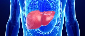
The liver is a vital and multifunctional organ. Impaired liver function affects the condition of the entire body and leads to the development of various diseases. There can be many reasons for such a failure. One of the pathological processes causing liver dysfunction is Budd-Chiari syndrome. This set of symptoms is caused by obstruction of the liver veins, leading to a decrease in the vascular lumen and disruption of the blood supply to the organ.
Budd-Chiari syndrome was discovered at the end of the 19th century by the British surgeon Budd and the Austrian pathologist Chiari. They independently described thrombosis of the large hepatic veins at the level of their junction, which blocked the flow of blood from the hepatic lobules. According to ICD-10, complex pathology of the liver veins was classified in the group “Embolism and thrombosis of other veins” and received code I82.0.
Modern medical scientists distinguish two medical terms: “disease” and “Budd-Chiari syndrome”. In the first case, we are talking about an acute process that is associated with thrombosis of the venous vessels of the liver, or a chronic form of the disease, which is caused by thrombophlebitis and fibrosis of the intrahepatic veins. In the second case, they talk about the clinical manifestation of non-vascular diseases. This division of a pathological process with the same symptoms and manifestations into two different forms is conditional. Oxygen deficiency in the liver tissue leads to its ischemia, which has a negative impact on the vital functions of all organs and is manifested by intoxication syndrome.
Budd-Chiari syndrome is a rare disease that has an acute, subacute, chronic or fulminant course. When the liver veins are occluded, its normal blood supply is disrupted. Patients experience severe pain in the right side of the abdomen, dyspepsia, jaundice, ascites, and hepatomegaly. The acute form of the pathology is considered the most dangerous. It often leads to coma or death of the patient.
This gastroenterological disease most often affects mature women aged 35-50 years suffering from hematological diseases. In men, the disease is much less common. Recently, doctors have sounded the alarm because the syndrome is becoming significantly younger. In Europe it occurs in one person in a million.
All patients with suspected Budd-Chiari syndrome undergo a full medical examination, including Dopplerography of liver vessels, ultrasound, CT and MRI, and liver tissue biopsy. Budd-Chiari syndrome is an incurable disease. Complex drug therapy and timely surgical intervention can only prolong the life of patients and improve its quality. Patients are prescribed diuretics, antiplatelet agents and thrombolytics. In acute forms, even these measures remain powerless. Such patients are indicated for liver transplantation.
Etiology
Budd Chiari disease and syndrome is a rare condition associated with changes in blood flow in the liver due to a decrease in the lumen of the veins adjacent to this organ. In this condition, blood cannot flow out of the liver and return to the heart due to blockage in the hepatic veins. This disorder is also known by other names such as hepatic vein obstruction and hepatic veno-occlusive disease. Clots or congenital membranes occur at the junction of these vessels with the great vein that carries blood from the lower part of the body to the right upper chamber of the heart. The disease may begin gradually or suddenly.
Symptoms associated with Budd Chiari syndrome include pain in the upper right abdomen and an abnormally large liver. As well as the accumulation of fluid in the abdominal cavity between the two layers of the membrane that lines the stomach. Additional conditions that are associated with the disorder include nausea, vomiting, and an abnormally large spleen.
The severity of the disorder depends on the location and number of veins affected. The etiology of the disease is mixed and varied. Most cases result from thrombosis in the hepatic veins. However, 25% is due to external compression, which leads to obstruction. This is a rare disease that can occur in both children and adults.
Note! Most cases tend to affect people between the ages of twenty and forty. Women have a higher risk of the disease compared to men.
Classification
Classic signs of the disease such as abdominal pain, ascites and hepatomegaly are observed in most patients with the disease. If the liver has had time to develop decompression, patients may be asymptomatic or have minor symptoms. However, as the syndrome progresses, it can lead to liver failure and portal hypertension. Budd-Chiari disease can be primary - congenital obstruction by membranes or diaphragms and secondary, caused by several reasons.
The disease has 2 clinical forms associated with the course of the disease:
- Chronic.
- Spicy.
Chronic outflow obstruction, developing over weeks or months, may have no symptoms until it progresses. Causes fatigue, abdominal pain and hepatomegaly. Lower extremity edema and ascites can occur as a result of venous obstruction even in the absence of cirrhosis. Cirrhosis of the liver may develop, leading to variceal bleeding, massive ascites, splenomegaly, or a combination of these.
The cause of the acute condition is thromboembolism of the hepatic or inferior vena cava. Acute obstruction causes fatigue, right upper body pain, nausea, vomiting, mild jaundice, painful hepatomegaly, and ascites. Fluid may accumulate in the abdominal cavity for several days. Often accompanied by thrombosis of the portal vein and, as a consequence, edema of the lower extremities, expansion of the vascular venous network of the abdomen.
Types of Budd-Chiari syndrome, depending on the location of the narrowing of the venous vessel:
- type 1 – blockade of the inferior vena cava and secondary hepatic vein;
- type 2 – blockage of large hepatic veins;
- type 3 – obstruction of small vessels of the liver.
The final stage of development of Budd-Chiari syndrome is the irreversible dilatation of the inferior and portal veins. The condition is accompanied by bleeding, blockage of the peritoneum and blood clots, as well as thrombosis of the blood clot. Patients with ascites may develop peritonitis. If the cause of the syndrome is the fusion of vessel membranes, liver cancer can be expected to occur in 30-45% of cases.
Symptoms of Budd-Chiari syndrome
Clinical manifestations directly depend on the volume of the lesion: if only one of them has pathological narrowing, the disease is asymptomatic. If 2 or more vessels are involved in the process, patients note a deterioration in their well-being, namely:
- pain in the right hypochondrium;
- yellowness of the skin;
- nausea;
- vomiting - may be mixed with blood clots;
- increased liver size;
- swelling in the legs;
- expansion of the saphenous veins.
The difference between a disease and a syndrome
Budd-Chiari syndrome or disease is called a blockage of the veins that supply the liver at its confluence with the inferior vena cava. Such vascular occlusion is caused by primary or secondary causes: congenital anomalies, acquired thrombosis and vascular inflammation and leads to disruption of the normal outflow of blood from the liver. Budd-Chiari syndrome should be considered separately from the disease. Differing in development and symptoms, they occur when blood circulation in the liver is impaired.
The disease is a primary veno-occlusive disease, thrombosis and subsequent occlusion of the liver, as well as abnormalities of the hepatic veins leading to impaired blood flow in the liver. The syndrome is a secondary attack of hepatic blood flow in a number of pathologies not associated with changes in the blood vessels of the liver.
Characteristics of the disease:
- Budd Chiari disease is a characteristic and potentially fatal form of liver injury that occurs secondary to vascular damage due to concomitant pathology. The disease manifests itself in acute, subacute or chronic form, accompanied by abdominal pain and swelling. As well as signs of portal hypertension and varying degrees of increased enzyme levels in the serum and jaundice. This most often occurs in patients with a bleeding disorder, such as those who are pregnant or who have a tumor. As well as chronic inflammatory disease, bleeding disorders and infection.
- Budd-Chiari syndrome is a rare but significant cause of liver dysfunction. A common cause of hepatic venous outflow obstruction worldwide is hepatic vein thrombosis secondary to an inherited or acquired hypercoagulable state.
Causes, development mechanism and classification
The exact cause of approximately 70% of all cases of the disease is unknown. Approximately 10% of people with the syndrome have polycythemia vera. Symptoms develop due to blockage of the veins that carry blood from the liver to the heart. Thrombosis occurs due to the clotting or overgrowth of fibrous tissue in the veins. Other identified causes may include exposure to radiation, arsenic, trauma, blood poisoning, cancer, or certain chemotherapy drugs.
The disease develops when a clot narrows or blocks the hepatic veins that drain blood from the liver. Because blood flow from the liver is obstructed, blood accumulates in the liver, causing it to enlarge. The spleen may also become enlarged. This congestion causes an increase in blood pressure in the portal vein, which carries blood to the liver. This increased pressure, called portal hypertension, can lead to varicose veins in the esophagus. Portal hypertension, plus a swollen and damaged liver, leads to fluid accumulation in the abdominal cavity. The kidneys contribute to ascites by causing salt and water retention.
The clot may expand and block the vena cava or vena cava, which carries blood from the lower parts of the body, including the liver, to the heart. Varicose veins in the abdominal area near the surface of the skin may develop and become visible. As a result, severe scarring of the liver (cirrhosis) occurs.
This is interesting! In 25-30% of cases, the cause of the disease cannot be determined; this pathology is called idiopathic.
Symptoms
Diagnosis can be made in several ways by observing characteristic symptoms in combination with magnetic resonance imaging or liver biopsy, which help rule out the hypothesis of other diseases.
The main signs of the syndrome are:
- hepatomegaly or enlarged liver;
- ascites, or abnormal collection of fluid in the abdomen;
- abdominal pain.
Hepatic vein obstruction makes it difficult for blood to flow out of the liver normally, causing it to become enlarged due to blood accumulation. It can also cause an enlarged spleen and severe bleeding in the esophagus.
Other symptoms of the disease include:
- abdominal swelling;
- fatigue;
- swollen ankles;
- elevated liver enzymes;
- coma;
- jaundice;
- hepatic encephalopathy or brain dysfunction;
- lactic acidosis – excess lactic acid;
- Liver failure due to cirrhosis or scarring of the liver;
- prominent collateral vein;
- nausea;
- vomiting blood;
- skin ulcers;
- serious damage to liver cells.
When the blockage is severe, the onset of the disorder may be sudden and accompanied by severe pain. If the disease is chronic, signs of the disease may be gradual. In some cases, unusual swelling occurs due to abnormal accumulation of fluid in the tissues of the legs.
Diagnostics
Diagnosis of the syndrome is made based on a thorough clinical assessment, detailed medical history, and various medical tests. Signs of liver enlargement may be detected during a physical examination.
Diagnostic tests used for this purpose include:
- Blood tests to determine if the organ is working properly.
- Ultrasound of the liver.
- MRI and CT scan of the abdominal cavity, which are performed to identify any abnormalities in the body.
Liver biopsy is another useful diagnostic procedure. During this process, the doctor takes a small sample of liver tissue for examination. Any tissue damage can be easily recognized by this test.
Hepatic vein catheterization is also performed to determine the presence of abnormalities. In this test, a catheter is inserted into the body to examine the liver veins. A small instrument attached to the tip of the catheter helps doctors measure blood pressure in the hepatic veins.
4.Treatment
Conservative therapy includes the prescription of hepatoprotectors, anticoagulants, diuretics, and hormonal anti-inflammatory drugs. However, all these measures are usually ineffective; The mortality rate with drug treatment alone reaches 90%. Thus, the method of choice is vascular surgery (bypass surgery, angioplasty), but in this case the prognosis and surgical plan vary widely depending on the etiopathogenetic and clinical features of a particular case.
Treatment
Treatment of Budd-Chiari syndrome is complex and depends on clinical findings and the underlying disease. Drug therapy with a blood thinning drug may be sufficient for timely diagnosed Budd-Chiari syndrome. Additionally, this form of therapy can be combined with vascular catheter studies. Hollow tubes are used to open the hepatic veins or create short circuits using special lattice vessel prostheses.
Dilation of the affected veins can relieve pressure on the vessel walls. In some cases, Budd Chiari syndrome can be treated surgically by redirecting blood flow from one vein to another (bypass). In other cases, the blocked vein is cleared and then a thin rod is inserted into it to maintain blood flow. In severe cases, a liver transplant may be required. If left untreated, worsening liver failure due to fibrosis and possible death may occur.
Note! Full recovery from Budd-Chiari syndrome is impossible. Drug therapy and surgical methods in combination prolong the life of a patient with a chronic form up to 10 years, but they are ineffective in acute diseases.
Conservative treatment
The goal of treating the disease is to maintain liver function. Doctors first determine the exact area where blood flow is obstructed. The severity of the syndrome depends on where the clot is located and the number of veins affected. Anti-clotting drugs such as urokinase are sometimes used to treat people who experience sudden onset of clotting in the hepatic veins. Patients should be treated first with heparin and then with warfarin to correct coagulation. These drugs are only used to treat the disease in its early stages, as they do not work once blood clots form.
The use of high doses of prednisolone may also be prescribed. Medicines that prevent blood clotting, such as heparin, may be useful in treating patients with the disease. Doctors may recommend topical thrombolysis, which is useful in dissolving blood clots. Systemic thrombolysis is not generally recommended as it is considered a high-risk treatment.
Radiologists will use X-ray techniques to directly examine the arteries and veins to determine the location of the clot. These procedures involve inserting a catheter into a blood vessel through an easily accessible vein in the arm, neck, or groin. Dye is then injected through the catheter to illuminate the blood vessels so they are easier to see under X-rays. In some cases, the scan shows that only the end of the vein is blocked and that most of the vein remains clear.
In other cases, when the vein is more blocked, doctors will need to access it through a tube inserted into the liver from the abdomen. Once the tube hits the blockage, doctors can remove the clot and open the vein. This is called venoplasty or angioplasty. If the clot in the hepatic veins is recent and difficult to remove, the catheter tube may be left in place for a day or two. This will give the drugs that cause the clot a chance to get rid of it. When venoplasty is successful, it results in an improvement in the patient's general condition.
Surgery
People suffering from this disease often require surgery to restore normal liver function. The specific method to be used is determined depending on the location of the blood clots and various other factors. Many other surgical techniques are also used for this treatment. Liver transplantation is performed to prolong the lives of patients who would otherwise not live beyond six months.
Other radiological interventions performed to treat patients with this type of liver disease include:
- Portosystemic shunts.
- Balloon angioplasty.
- Transjugular, portosystemic shunt.
- Surgical shunts.
However, the success rate of these surgeries differs from one patient to another. These surgical procedures can be effective, but there is a risk that they may create additional problems. Shunts mean that less blood passes through the liver to clear toxins. As a result, there is a risk that these toxins will accumulate and this can cause a condition called hepatic encephalopathy.
A liver transplant is recommended only if other treatments have failed and end-stage disease is life-threatening.
An organ transplant may be required when:
- the liver ceases to perform all its functions adequately;
- Bypass procedures cannot prevent further deterioration of health.
Note! Once a shunt is installed, there is an increased risk of infection. Regular washing is important, especially in the affected area. Moderate outdoor exercise and a healthy diet can promote recovery.
Treatment of Budd-Chiari syndrome
Drug therapy can be used, however, it does not contribute to the complete recovery of the patient and is aimed at temporarily improving the state of health. It involves prescribing the following drugs:
- diuretics - to remove excess fluid from the body;
- glucocorticosteroids - necessary to relieve pain;
- antiplatelet and fibrinolytic agents - used to resolve the formed blood clot;
Activities related to the profile of surgery for Buddy-Chiara syndrome have the following areas:
- anastomosis - involves creating synthetic connections between stenotic veins;
- shunting - to exclude the affected vessel from the general blood flow and create an alternative path for blood movement;
- angioplasty - involves the introduction of special balloons into the cavity of a narrowed vein to increase its lumen, followed by the introduction of a stent;
- liver transplantation.
Indications for surgery are the absence of liver failure and blockage of the veins of the affected organ by blood clots.
Prevention
Budd-Chiari syndrome is an extremely rare and potentially fatal disease. Early diagnosis along with timely treatment is important to ensure a relatively long and normal life for patients. There is no way to ensure prevention of this disease. However, screening and genetic testing of family members of patients with the disease will help understand the risk of developing the syndrome. This process is necessary for those diagnosed with thrombophilic disorder and a family history of Bud Chiari disease.
Proper nutrition is important to manage symptoms and prevent further liver damage. Eating a low-salt diet to maintain a negative sodium balance helps control ascites. The disease can only be prevented conditionally. Patients prone to the syndrome due to a pre-existing condition such as thrombosis, cancer or hepatitis should undergo regular screening. It is important not to put stress on the liver, for example through excessive alcohol consumption or medications.
Forecast
Various studies and studies have tried to predict the survival rate of patients suffering from Budd-Chiari syndrome. However, overall, almost two thirds of patients suffering from the syndrome remain alive for at least ten years from diagnosis. Necessary negative prognostic indicators include encephalopathy, ascites, elevated child Pugh scores, improved prothrombin time, and altered serum levels of various substances such as creatinine, sodium, albumin, and bilirubin. Patient survival depends largely on the underlying causes of Budd-Chiari syndrome. For example, patients with chronic myeloproliferative disorders may progress toward severe leukemia and succumb to death.