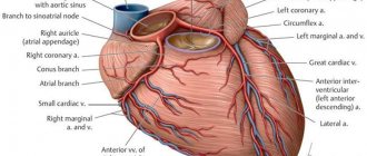
1 The role of the vessel
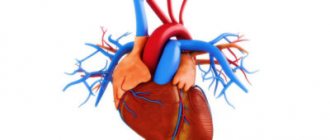
Arteries are blood vessels that carry blood from the heart to the organs. They differ from veins in the reverse process, that is, veins supply blood to the heart.
The common carotid artery transports blood from the heart muscle to the brain and other peripheral organs of the human head. The artery is quite wide. This is explained by the need to transport sufficient levels of oxygen to enrich brain tissue and the presence of a stable but intense blood flow.
The carotid artery is quite “tender”. Compressing it can lead to a sudden loss of consciousness. Those who have ever worn a tight tie or a sweater with a high and narrow collar have noticed a peculiar feeling of discomfort. Such unpleasant sensations are caused by compression of the carotid artery.
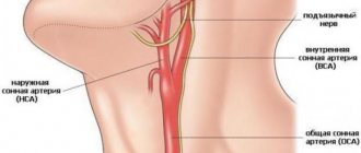
Before answering the question about the location of the carotid artery, it is necessary to make a reservation that there are two of them. One is on the right side of the neck, and the other is on the left. The artery that runs along the left side is slightly longer than the artery that runs along the right, since the first originates at the brachiocephalic trunk, and the second in the aortic arch.
To feel the pulse of the carotid artery in the neck, you need to find a point under the cheekbone in the fossa, on the right or left side of the Adam's apple. In people with highly developed muscles, detecting the pulse in this way may take a little longer than in the average person, since the artery may be blocked by muscles.
Determining the presence of a pulse in the neck is considered optimal in a critical situation. The fact is that not all people feel pulsation in their wrist.
We recommend Fetal heart rate by week, methods of measurement and reasons for deviation
In what cases is diagnostics indicated?
Experts recommend undergoing an ultrasound of the carotid arteries and other vessels of the neck at least once a year, especially after 50 years. Early diagnosis helps prevent heart attack and stroke. An examination may be prescribed in the following cases:
- History of diabetes mellitus;
- Diseases of the heart and blood vessels;
- Long-term smoking;
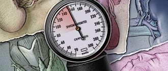
- High blood pressure;
- Dizziness not associated with hunger;
- Frequent headaches;
- Episodes of loss of coordination;
- Noise in ears;
- Sleep disorders: insomnia or constant drowsiness;
- Lack of appetite;
- Decreased concentration.
Complaints and symptoms
Atherosclerosis of the carotid arteries can be asymptomatic or cause complaints associated with impaired cerebral blood flow. Most often, patients may complain of temporary impairment of brain function (transient ischemic attack) or persistent loss of brain function (ischemic stroke).
Transient ischemic attack (TIA)
A TIA occurs when blood flow to the brain is briefly interrupted. This is the initial phase of acute cerebrovascular accident, which is reversible. It has the same symptoms as a stroke, but these symptoms go away within minutes or hours.
A TIA requires emergency medical attention because it is impossible to predict whether it will progress to a stroke. Immediate treatment can save lives and increase the chances of a full recovery.
Modern research has shown that patients who have had a TIA are 10 times more likely to suffer a major stroke than a person who has not had a TIA.
Ischemic stroke has the following symptoms:
- Sudden loss of vision, blurred vision, difficulty in one or both eyes.
- Weakness, tingling, or numbness on one side of the face, one side of the body, or one arm or leg.
- Sudden difficulty walking, loss of balance, lack of coordination.
- Sudden dizziness.
- Difficulty speaking (aphasia).
- Sudden severe headache.
- Sudden memory problems
- Difficulty swallowing (dysphagia)
Ischemic stroke and transient ischemic attack begin in the same way, so any ischemic stroke can be called an ischemic attack if the symptoms completely resolve within 24 hours of the onset of the disease. The presence of a time interval between the onset of stroke symptoms and the death of parts of the brain allows for urgent surgery to restore cerebral blood flow.
How to prepare
If you have ever had to undergo an ultrasound examination, you probably know that this diagnostic procedure requires certain preparation; depending on the area being examined, the requirements may vary.
But if you have to undergo an ultrasound of the carotid arteries, you do not need to take any special preparatory measures. You only need to limit tonic drinks and sedatives - one day before the procedure you should avoid tea, coffee, alcohol, and 5 hours before the procedure you should stop smoking. Otherwise, the data obtained may be unreliable. If you are taking medications, you should inform the doctor who prescribed the test for you.
What are they responsible for?
The main purpose of the carotid artery is to provide the tissues of the head and neck with oxygen and nutrients.
At the same time, the functions of the internal and external carotid arteries are different, since they supply blood to different organs:
| The external carotid artery branches to the front of the head and supplies: | The internal carotid artery ascends to the base of the skull and supplies blood to: |
| Mimic and chewing muscles of the face. | Brain tissue. |
| Facial skin and epidermis under the hair. | |
| Oral cavity. Including the tongue, salivary glands and tooth roots. | Muscles of the forehead and temporal part. |
| Nasal cavity and middle ear tissues. | |
| Thyroid gland, pharynx and larynx. | Ocular tissues. |
The external and internal arteries are additionally connected to each other by numerous arteries, this ensures an uninterrupted flow of blood to the tissues if suddenly a slowdown in blood flow occurs in any part of the artery.
The vessels are also equipped with nerve endings, as a result of which the carotid arteries perform the following additional functions:
- normalization of blood pressure;
- regulation of the number of heart contractions;
- react to a lack of oxygen in the blood.
If you know how to act correctly on the carotid artery, you can lower blood pressure without taking medications. It is important that this procedure is only allowed to be performed by a specialist, since stronger or longer manipulations can lead to death.
How is the carotid artery ultrasound procedure performed?
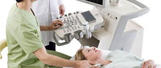
Ultrasound examination of the carotid arteries is a popular technique that allows one to detect possible diseases in the early stages. During the study, the person does not feel any discomfort, either physical or moral.
The specialist asks the patient to remove jewelry or collect long hair, lie down on the couch, and a small cushion may be placed under the head. The area being examined is coated with a gel that improves the operation and movement of the sensor. During an ultrasound, a specialist moves a sensor across the area being examined. He may ask the patient to change the position of the head, turn the head left, right, up, tilt down, or strain the neck.
In total, the diagnostic study does not exceed half an hour. One of the advantages is the quick receipt of a transcript from a specialist. With the transcript, you can go to your doctor.
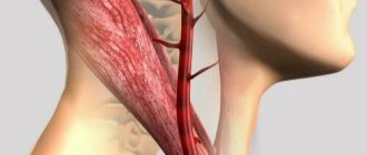
Where are they located?
The carotid artery in humans is located on the sides of the neck near the thyroid gland. The left carotid artery comes from the aorta (the main artery that originates in the left ventricle), and the right one from the brachial artery (which is a branch of the aorta). In this regard, the left carotid artery is longer than the right by about 20-25 cm.
Further, near the Adam's apple (the protruding part of the thyroid cartilage), the right and left carotid arteries branch into external and internal. Above, each artery divides into numerous capillaries that supply all the tissues of the head and neck.
What diseases can be detected by ultrasound of the carotid arteries?
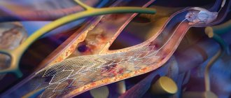
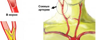
When conducting diagnostics using ultrasound, not only the carotid arteries are assessed, but also the vessels of the neck as a whole. According to statistics, most often during the study the presence of atherosclerotic plaques, narrowing of the lumen of the arteries, and kinks of blood vessels are revealed. These pathological processes contribute to the development of stroke, which can result in death or disability. It is for this reason that experts recommend carrying out preventive diagnostics at least once a year.
Blood clots and aneurysms are also often diagnosed. They can be detected on ultrasound even in the initial stages. Stenosis (narrowing of the vessel), noticeable during diagnostic procedures of the carotid arteries, may explain ischemic attacks, since the blood flow worsened due to the narrowing of the artery does not allow the brain to receive enough oxygen. If pathological processes are detected, a more thorough additional study may be required.
Causes and risk factors
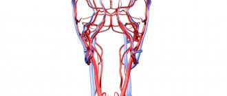
The carotid arteries are paired large arterial vessels that supply blood to the brain in those sections where the centers of thinking, speech, personality, sensory and motor function are located. The carotid arteries run through the neck and enter the brain through openings in the skull.
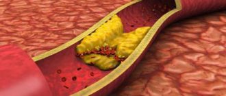
When fatty substances and cholesterol accumulate, an atherosclerotic plaque is formed, which narrows the carotid arteries. This reduces blood flow to the brain and increases the risk of ischemic stroke. A stroke occurs when blood flow does not reach any part of the brain. During a stroke, some brain functions are suddenly lost. If the lack of blood flow lasts more than three to six hours, then these disorders become irreversible.
Why does a stroke develop when the carotid artery narrows?
- Significant narrowing of the carotid artery reduces blood circulation in the brain, and with a sudden drop in pressure (suddenly getting out of bed, flying, overheating in the sun or major surgery), the blood flow suddenly stops, which leads to the death of nerve cells.
- Tearing off a piece of atherosclerotic plaque with its transfer by the bloodstream into small arteries of the brain, which leads to their blockage.
- Acute thrombosis (formation of a blood clot) due to narrowing of the carotid artery with complete cessation of blood flow in certain areas of the brain.
What affects heart rate?
The heart rate is influenced by several factors: - training, - ambient temperature, - body position (standing, sitting, lying), - emotional state: excitement, anger, fear, anxiety lead to an increase in heart rate, - excess weight, - intake medications, alcohol or smoking. If an untrained person's heart beats too slowly - less than 60 beats per minute - this is called bradycardia
.
If, at rest, in an untrained adult, the heart beats faster than 100 beats per minute - this is called tachycardia
.
If you experience similar symptoms, which are accompanied by dizziness, shortness of breath or fainting, consult a doctor immediately
.
How to find it on your neck yourself?
To locate the carotid artery in the neck, it is necessary to visually determine the location of the Adam's apple. Further to the right or left of it, in the area next to the depression under the lower jaw, you need to place your index and middle fingers (these fingers are more sensitive). With proper manipulation, pulsating impulses of blood through the vessel will be felt.
Where is the carotid artery located in the human neck?
The pressure should be gentle, without squeezing the arteries.
It is recommended to independently identify the carotid artery on the right side using the index and middle finger of the right hand. It is important that the pulse on the carotid artery can be felt better than on the wrist, so it is better to measure the number of heart beats per minute on the carotid artery (especially in unconscious people).
BCA segments
The carotid artery (internal part) has a conditional division into segments. The division begins from the bifurcation (the place where the main carotid artery divides into the external and internal) and to the point where the internal carotid artery branches into small vessels and capillaries. Next, we briefly consider where these areas are located and what functions they perform in humans.
Segments of the internal carotid artery
The branching of the main carotid artery into external and internal occurs approximately near the third cervical vertebra.
From this section the internal artery is divided into the following segments:
| Segment name | Start of segment | End of segment | What organs and tissues does it nourish? |
| Cervical | Bifurcation | External opening of the temporal bone canal | The site has no branches, so it does not feed tissue. But in this segment, the carotid artery borders on the laryngeal, hypoglossal and vagus nerves. |
| Rocky | Temporal bone region | Ragged hole (located at the base of the skull) | Located in the carotid canal. Nourishes the tympanic cavity (inner ear). |
| Ragged hole | The segment is located in the torn hole | It is the shortest segment and has no branches, so it does not provide tissue nutrition. | |
| Cavernous | Ragged hole | Proximal ring of dura mater | Surrounded by the cavernous sinus. The section has a C-shaped bend, and is called the siphon of the internal carotid artery (the more detailed purpose of the segment is discussed below). Nourishes brain membrane cells, pituitary gland and nerve fibers. |
| Wedge-shaped | Cavernous sinus | Distal ring. | The segment is short in length and normally has no branches. In extremely rare cases, the presence of a branch of the ophthalmic artery (that is, it nourishes the tissues of the eye) was noted. |
| Ophthalmic | Distal ring | Origin of the posterior communicating artery | The segment nourishes the pituitary gland and the eyeball. |
| Communicative | Branch site of the communicating artery | Branches into small vessels | Is the final segment. Participates in the nutrition of the brain and has connections with the vertebral arteries. |
The internal carotid artery, branched inside the skull, nourishes not only brain tissue, but also the optic nerve, pituitary gland and hypothalamus, thereby ensuring their normal functioning.
What is a normal heart rate?
For a person over 18 years of age, the normal resting heart rate is between 60 and 100 beats per minute
. The more trained the cardiovascular system is, the fewer heartbeats are required for the body to receive the necessary nutrients and oxygen from the blood. Professional athletes have a resting heart rate of about 40 beats per minute. A normal resting heart rate is considered to be: - For a newborn - 120-160 beats per minute, - For a baby from 1 month to a year - 80-140 beats per minute, - For a child aged 2 to 6 years - 75-120 beats per minute minute, - For a child aged 7 to 12 years - 75-110 beats per minute, - For people over 18 years old - 60-100 beats per minute, - For adult athletes - 40-60 beats per minute.
What is maximum heart rate?
This indicator tells you how many beats per minute your heart can make maximum - during physical activity. When playing sports, it allows you to evaluate how intense the load you are receiving. Typically, the maximum heart rate is calculated using a mathematical formula that takes into account the person’s age. For adult men, MHR = 220 – age
. That is, a 25-year-old man would have a maximum heart rate of 195 beats per minute. For adult women, the calculation is the same, but sometimes an adjusted formula is used: MHR = 226 – age. That is, for a 25-year-old woman this figure will be 201 beats per minute.
Tags:
- Workout
- Pulse
- Diagnostics