The section of the venous network, which is commonly called the jugular vein (abbreviated as JV), is a collection of veins that provide the outflow of depleted blood from the cranium and facial part of the head. Not only well-being, but also a person’s life depends on the functioning of this vessel, because the jugular vein is as important an element of the blood supply system to the head as the artery that supplies the brain. Any violation in one of them can provoke serious consequences for human health, including disability or death.
Types and location
The venous vein consists of 3 independent venous channels. Accordingly, their anatomy is separate.
The veins of the head and neck, which are responsible for the correct outflow of blood from the brain cavity, are divided into 3 types. These are the anterior, external and internal jugular veins.
Internal
It has a relatively wide trunk, compared to the other 2. In the process of pushing out blood, it easily expands and contracts, thanks to its thin walls and diameter of 20 mm. The outflow of blood in a certain amount occurs with the help of valves.
As the lumen expands, the superior bulb of the jugular vein is formed. This occurs at the moment when the IJV enters from the hole.
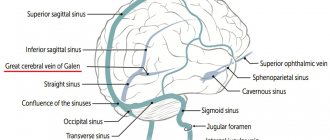
Typical anatomy diagram:
- start – area of the jugular foramen;
- localization - the skull, or rather its base;
- further - its path goes down, the place of localization is in the posterior muscle, the place of attachment is the collarbone and sternum;
- the place of intersection with the posterior muscle is the area of its lower and posterior parts;
- after that the path is laid along the trajectory of the carotid artery;
- a little lower it comes forward and is located in front of the carotid artery;
- then, together with the carotid artery and the vagus nerve, they are directed through the site of expansion;
- as a result, a powerful bundle of arteries is created, which includes the carotid artery and all jugular veins.
Blood enters the IJV from the tributaries of the skull, the location of which is the cranium and outside it.
Comes from vessels: brain, eye, auditory. Also suppliers are the dura mater of the brain, or rather its sinuses.
Outdoor
Location: neck tissue. Blood is directed from the face, head and outer part of the cervical region. Perfectly visible visually when coughing, screaming or stress.
Construction scheme:
- origin - lower angle of the jaw;
- further down the muscle that attaches the sternum and clavicle;
- crosses the outer part of the muscle. The place of intersection is the area of the rear and lower part.
It has only 2 valves located in the initial and middle parts of the neck.
Front
The main task is to carry out drainage from the chin area. Location: neck, middle line.
Anatomical features:
- passes along the muscle of the tongue and jaw (along the front), downwards;
- Then, on both sides, the veins connect with each other, and a venous arch is formed.
Sometimes the arc of collected together as\nals forms a middle one.
External jugular vein
The structure and location of the external jugular vein (abbreviated EJV) makes it easy to visualize it, especially if a person talks passionately and loudly for a long time, sings or screams. It is smaller in diameter than the outer one, which makes its anatomical structure more typical of the venous network: the muscle layer in it is less developed, but there are more valves for directing blood flow, and there are no bulbs at the base.
The external jugular vein has many lateral tributaries that fill it with blood along its entire length. This is what distinguishes it from the IJV, which is filled with venous blood only from one end. The posterior auricular, occipital, suprascapular and transverse branches, as well as the third anterior UR, flow into the MN. The main function of the external jugular vein is to drain blood from the lower jaw and chin, neck and surface of the skull.
It is the NJV that is used to install a catheter when it is necessary to administer long-term intravenous drug solutions.
Diseases and changes
The reasons for the expansion make it known about the dysfunction of the circulatory system. This situation requires an immediate solution. You should know that there are no age restrictions for YV pathologies. They affect both adults and children.
Phlebectasia
A thorough, accurate diagnosis is necessary, the result of which should be the identification of the causes of the pathology, as well as the prescription of comprehensive effective treatment.
Extensions occur:
- in case of stagnation, due to injury to the neck, spine or ribs;
- for osteochondrosis, concussion;
- for ischemia, hypertension, heart failure;
- for endocrine disorders;
- with prolonged sitting at work;
- for malignant and benign tumors.
Phlebectasis can also be caused by stress and nervous tension. With nervous excitement, pressure can increase, resulting in a loss of elasticity of the walls of blood vessels. This can lead to valve dysfunction. Therefore, phlebectasia needs to be detected early.
Circulation can be negatively affected by factors such as: alcohol consumption, smoking, toxins, excessive mental and physical stress.
Thrombosis
It may occur due to the presence of a chronic disease in the body. If they are present, as a rule, blood clots form in the vessels. Once a blood clot has formed, there is a possibility of it breaking off at any time, which entails blocking vital arteries.
Signs of thrombosis:
- sometimes pain occurs in the hand;
- swelling of the face;
- appearance of venous networks on the skin;
- When turning the head, pain occurs in the cervical region and neck.
The result of thrombosis can be rupture of the jugular venous channels, which leads to death.
Phlebitis and thrombophlebitis
Inflammatory changes occurring in the mastoid process or middle ear are called phlebitis. The cause of phlebitis and thrombophlebitis can be:
- bruises, wounds;
- placement of injections and catheters in violation of sterility;
- penetration of medications into the tissues around the vessel. This can often be triggered by calcium chloride when it is injected past an artery;
- infection from the skin.
Phlebitis can be uncomplicated or purulent. The treatment of the 2 pathologies is different.
Aneurysm
A rare pathology is an aneurysm. It can even occur in children at an early age from 2 to 7 years. The pathology has not been fully studied. It is believed that its occurrence occurs from improper development of the base of the venous bed, or rather its connective tissue. It is formed during intrauterine development of the fetus. The anomaly does not manifest itself clinically. It can only be noticed when a child cries or screams.
Symptoms of an aneurysm:
- headache;
- anxiety;
- sleep disturbance;
- fast fatiguability.
Treatment consists of draining venous blood and vascular prosthetics.
Human anatomy is undoubtedly a major core subject for study in medical schools. Despite the fact that normal human anatomy is a discipline that stood at the origins of the development of medicine, a large number of scientific works still appear that make adjustments to modern anatomical atlases.
It would seem that human anatomy cannot change so quickly with the course of evolution, but our understanding of it is constantly improving as new research methods appear - evidence of this is provided by ever new versions of the anatomy atlas.
Atlas of anatomy Sinelnikova R.D. in 4 volumes, this is perhaps the most authoritative and time-tested source of knowledge on this topic. It is constantly republished, delighting us with its visual illustrations and text accessible to everyone. Many students tried to download Sinelnikov’s atlas for study, but the links either did not work, or there was a virus in the folder... We solved this problem by making a website dedicated to this source.
The main goal of studying human anatomy is to create a fundamental knowledge base for students for further study of other medical disciplines. It is difficult to imagine mastering a curriculum in physiology, pathological physiology, pathological and topographic anatomy, operative surgery, and a whole range of clinical disciplines without a thorough study of normal human anatomy.
It is very important for a student to have a visual image of the material studied; for this it is necessary to study human anatomy in pictures. The main feature of this science. of course, is the structuring of its sections and subsections, as well as a clear systematization of the entire nomenclature.
Thus, we can distinguish the following directions that correspond to each system:
- osteology (section on the bones of the human skeleton). Studies the skeleton, both as a whole mechanism and as individual bones. There is also a study of age-related changes in bones.
- syndesmology (joints, ligaments). An extremely important section for future orthopedists and traumatologists.
- myology (muscular system). He studies not only the structure, but also the development and physiology.
- splanchnology (internal organs). Includes the anatomy of the endocrine, digestive, respiratory, excretory and genitourinary systems.
- angiology (vessels and their derivatives). Information is presented on the structure of blood and lymphatic vessels.
- neurology (central and peripheral nervous system). An extremely important section for successful diagnosis of diseases and perhaps the most difficult.
- aesthesiology (the science of the sense organs). Everything about vision and hearing. And also about taste, olfactory and tactile sensitivity. Closely related to neurology.
Who is involved in diagnosis and treatment?
If symptoms of the disease occur, you should consult a physician. After consultation, he can refer you to see a phlebologist.
Based on the patient’s complaints, the phlebologist conducts an initial visual examination, the result of which should be the identification of pronounced symptoms of venous disease.
In addition, all patients suffering from vascular diseases must be registered with a cardiologist. Jugular vein diseases must be identified in the early stages. Be aware of possible serious consequences.
If at least one symptom of a particular disease appears, you must immediately contact a therapist.
What is characteristic of phlebitis?
The most common factor in the progression of phlebitis is inflammation in the middle ear, or the tissues of the mastoid process.
When a blood clot becomes inflamed and embolized, infected particles can circulate throughout the bloodstream, settling in unexpected places.
Also, factors may be:
- Infectious lesion;
- Traumatic situations and bruises;
- Distribution of the drug in the tissues around the vessel.
The main features are:
- Painful sensations;
- Swelling;
- Swelling;
- Signs of damage to the body by toxins;
- Acceleration of heart contractions;
- Rash;
- Fever;
- Hard breath.
Anatomy of veins and arteries
How to study the vessels of the neck
When visiting an ultrasound diagnostician’s office, the doctor usually asks you to remove jewelry from your neck and ears and put your long hair in a ponytail so that nothing interferes with the examination process. Next, the patient lies on a flat couch, with his head towards the doctor. The gel is applied to the skin of the neck and the sensor is moved over it. The head is thrown back or tilted. First, the carotid artery is examined, then all other vessels located in the neck.
The procedure lasts no more than 45 minutes. The doctor may also conduct the examination in a sitting or standing position and ask you to tilt your head. At the end of the procedure, the doctor gives the results of the study to the patient.
What's the forecast?
Prediction is made in each individual case of jugular vein damage. If the vein is affected by ectasia, then no treatment is required, you just need to eliminate the cosmetic defect, in which case the prognosis is favorable.
When the jugular vein thromboses, blood access to certain parts of the head is blocked, which is already a more dangerous situation. Oxygen starvation is possible, which will lead to the death of brain tissue and possible death.
Any defects in the walls of the jugular vein can lead to its rupture, which will lead to severe internal bleeding. In most cases, patients die because they are outside the hospital.
Prevention
Prophylaxis to prevent damage to the jugular vein is general to maintain normal vascular condition.
The main recommendations are:
- Once a year, undergo a routine examination , which will help diagnose possible pathologies in the early stages of development;
- Maintaining water balance . Drink about one and a half liters of clean drinking water per day;
- Proper nutrition . Should contain a large amount of vitamins and nutrients for the elasticity of the walls of blood vessels;
- Carefully study the instructions of medications in order to avoid allergic manifestations that lead to inflammation of blood vessels;
- More active lifestyle . Daily walks in the fresh air are recommended;
- Treat infectious diseases in a timely manner;
- Maintaining a daily routine. The working day should contain sufficient rest and healthy sleep.