Catheterization kit
To insert catheters into venous tubes, the doctor uses standard kits for catheterization of central veins or peripheral vessels.
They differ in the diameter and configuration of the catheter tubes, as well as the presence of additional tools in the kit for inserting and fixing devices on the human body. The standard kit for catheterization of the subclavian and jugular veins (SJV) contains:
- a catheter tube made of polymer material, visible on x-rays, with a diameter of 1.2 to 2.3 mm and a length of 130 to 210 mm, with extensions;
- a round or triangular metal needle with a diameter of 1.1 to 1.6 mm and a length of 57 to 100 mm;
- conductors - straight made of polymer material or J-shaped made of metal;
- expanders;
- fixing elements;
- plug with membrane.
The standard set of catheters for the peripheral parts of the venous system differs from sets for catheterization of central veins in the absence of dilators and guides, as well as the size of the tubes: their thickness varies from 0.62 to 2.1 mm, and length from 19 to 45 mm.
The choice of catheter size depends on many factors, including the age and equipment of the patient, his anatomical and physiological characteristics. For example, the smallest sizes are used for catheterization of children, and larger ones are used for installation in large branches of the circulatory system.
Kinds
The official classification divides catheters into several types depending on the purpose of the devices, the materials from which they are made, sizes and design features. According to their purpose, they are divided into three types:
- CVCs, represented by sets for catheterization of central veins. Suitable for long-term installation in all large veins.
- PVK, represented by kits for catheterization of peripheral veins. Suitable for long-term installation in vessels of the upper and lower extremities.
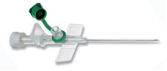
- Butterfly catheters, which are a monolithic structure consisting of a tube and a needle, as well as a fixing element in the form of two round plates. In clinical practice, such a catheter is used for infusion into small veins with procedures lasting no more than an hour.
Butterfly catheter
By size, they are divided into 8 types and are marked with a two-digit number and the letter G (geich). The higher the number on the central venous catheterization kit, the thinner the tubing and needles.
According to their design features, catheters are divided into single-channel and multi-channel. Single-channel ones are used for administering drugs according to Seldinger during emergency care, for long-term administration of solutions and blood components. Multichannel designs are used for the simultaneous administration of drugs that are incompatible with each other.
Polyethylene and polyurethane tubes are considered the most common in kits for catheterization of subclavian veins. The industry also produces catheters made of polyethylene, PVC, silicone and Teflon.
Indications
Conditions requiring long-term administration of medicinal solutions, nutrients and blood components are considered absolute indications for central venous catheterization:
- Venous catheter - what is it? Vein catheterization
- the patient's inability to eat;
- oncological diseases (chemotherapy);
- renal failure requiring hemodialysis;
- administration of drugs that provoke irritation and spasm of peripheral vessels;
- the need to regularly monitor hemodynamics.
Catheterization of peripheral vessels is carried out if it is necessary to administer moderate amounts of medication over 3-5 days.
Preparing for treatment
Before you begin treatment for subclavian artery stenosis, you must first diagnose it and confirm the diagnosis. To do this, we use the following research methods:
- Ultrasound diagnostics
- Computed tomography with vessel contrast;
- X-ray of the lungs.
Additionally, general clinical blood and urine tests and biochemical blood tests are performed. It is necessary to perform endoscopy of the stomach to exclude ulcers, since after surgery antithrombotic drugs are prescribed, which can provoke gastric bleeding in case of ulcers.
Types of jugular vein catheters
For catheterization, a central venous catheter (CVC) is used. There are different types of such medical devices:
- Non-tunneled is a catheter that is fixed at the entrance to the vein through the skin. Its disadvantage is its short (5-7 days) period of use, after which it is removed. A typical type of non-tunneled CVC is the Quinton catheter.
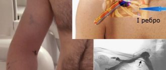
- Tunneled - the instrument is inserted into the skin (for example, on the chest) and driven under it (in a tunnel) to a vein in the neck. This type of catheter is used for a long time. It can stand for up to 30 days. This introduction makes it possible to make the device more invisible, safer and securely fix it. This group includes: Hickman and Groshong catheters.
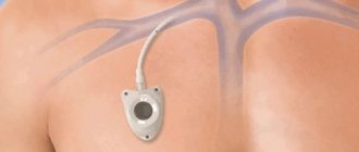
- Implantable Ports - These products are completely implanted under the skin, may have a medication reservoir, and are designed for long-term infusion therapy.
The specialist selects the type of CVC in accordance with the task.
How the treatment method works
Angioplasty and stenting operations are performed in a specialized cath lab. An introducer is installed through an artery on the arm or thigh, through which a special guiding catheter is installed in the affected subclavian artery. Using a guide catheter under X-ray control, a special device is inserted into the subclavian artery - a filter designed to protect the brain from complications of embolism. Then a balloon catheter is inserted into the area of narrowing of the artery and inflated, achieving expansion of the lumen of the vessel. After expansion, the balloon is restored to its original state and removed. In the narrowing zone, a metal structure is installed - a mesh stent, which serves as an internal frame for the artery and presses the loose elements of the deformed atherosclerotic plaque to the vascular wall. After this, carefully remove the elements of the delivery device and the filter itself. Quite often plaque fragments and small blood clots are found in the filter trap. At the end of the operation, a control angiography is performed, reflecting the result of the operation.
Jugular vein catheterization technique
The standard catheterization technique requires appropriate preparation, the position of the patient during manipulation, and the use of a set of instruments. To carry out the procedure, extensive experience of a specialist and the use of visualization techniques for internal structures are required.
Preparation
The patient first undergoes laboratory tests and a coagulogram. Then the features of the upcoming procedure are explained to him:
- target;
- necessity;
- technology;
- possible complications.
The patient signs voluntary informed consent. After this, he is taken to a sterile operating room. The patient is positioned in a supine position on the operating table. The head is turned in the direction opposite to the puncture area. The cervical spine is slightly extended.
If the patient’s condition allows, then to increase the blood supply to the vessels, he is placed in the Trendelenburg position - with the pelvis elevated, the angle of the body relative to the horizontal surface is 45°. Sometimes the legs are also elevated to expand the internal jugular vein, which has a diameter of 7 mm.
The skin on the neck is treated with an antiseptic solution from the angle of the lower jaw to the collarbone. Used as an antiseptic:
- Why do the veins in the arms hurt in the elbow bend?
- 70% alcohol solution;
- 2% chlorhexidine solution;
- 5% povidone iodide solution;
- iodoform;
- tincture of iodine.
A sterile napkin with a hole in the area of manipulation is placed next to the patient. The specialist prepares the CVC by rinsing it with saline and preparing it for installation.
Ultrasound scanning
The insertion point can be determined using the search puncture technique using a needle for intramuscular injection. A hollow needle is inserted into the vessel and a portion of blood is withdrawn. Based on its color and the intensity of blood flow, the type of vessel is determined - vein or artery. But this method has many limitations and can lead to mechanical damage. The use of external anatomical landmarks determined physically does not provide a 100% guarantee of safety, since their relationship with the target vessel can be variable.
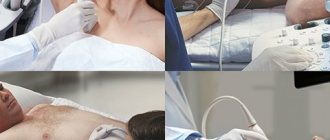
Today, the “gold standard” for diagnosing the location of the jugular vein and adjacent structures is ultrasound. To carry out diagnostics, multi-frequency linear and microconvex sensors are used, the radiation frequency of which ranges from 7 to 10 MHz. They allow detailed visualization of surface structures located at a depth of 6-7 cm. These sensors have special marks on one side.
Of the main veins, the internal jugular vein is most accessible to ultrasound examination. A gel that increases the permeability of radiation is applied to the patient’s side of the neck, and a scan is performed, according to which the position of the vein and access points are marked. The use of color mapping during Doppler scanning facilitates diagnostic measures. The use of ultrasound can reduce the number of unsuccessful punctures and postoperative complications by 50%.
Local infiltration anesthesia
In children, puncture of the main veins is performed under general anesthesia. In adults, when inserting a CVC, infiltration anesthesia of the subclavian region on the right or left is used with local anesthetics, for example, a 0.25% solution of novocaine in a volume of 10 ml.
Indications and contraindications for the treatment method
Indications for angioplasty and stenting of the subclavian arteries: symptomatic stenoses (narrowing) more than 50% and asymptomatic stenoses more than 75%. Symptoms of narrowing of the subclavian arteries are weakness in the affected arm, sometimes necrosis of the fingers or gangrene of the hand.
Contraindications:
- total occlusion of the vessel (in relation to the internal carotid artery); Vascular diseases that prevent the use of endovascular instruments:
- severe atheromatosis of the aortic arch;
- pronounced tortuosity and looping of blood vessels;
- presence of intraluminal thrombus in the area of stenosis
- acute period of ischemic stroke or completed stroke with a pronounced neurological defect; intracranial hemorrhage within 1 month.
Observation program after treatment method
After this surgical intervention it is recommended:
- Give up bad habits, especially smoking.
- If necessary, control your eating behavior: exclude fatty, smoked, salty foods.
- Lose weight if you are overweight.
- Perform dosed physical activity daily.
- If possible, spend more time in the fresh air.
- Avoid stress!
- Take medications recommended by your doctor.
- Visit your doctor at recommended intervals!
- If you experience any discomfort in your body, consult a doctor.


