Despite the fact that phlebology as a science has long moved from the category of fundamental (anatomical and physiological) to scientific and practical and has firmly occupied its segment in the section of practical cardiovascular surgery, until the beginning of the second half of the last century, the main methods of examining patients with acute deep phlebothrombosis were clinical. However, the rapid development of instrumental clinical methods in the 60s-70s of the last century made it possible to begin studying this issue at a qualitatively new level.
Although clinical methods are used in practice today, their role in establishing accurate topical and nosological diagnoses, as well as in the study of pathological phlebohemodynamics, has sharply decreased. In fairness, it is necessary to clarify that the simplest of clinical tests (Homans, Moses tests and measurement of the volumes of various segments of the lower limb) make it possible to detect and subsequently clarify (using other methods) the presence of a phlebothrombotic process.
Almost until the end of the last century, X-ray contrast venography in its various variants, up to the use of computer signal processing, was considered the “gold standard” of clinical and scientific research on this problem. In this direction, however, the method has not exhausted the possibilities of its optimization. Currently, it is used for limited indications, mainly as an addition to ultrasound examination.
Briefly about the diagnostic method
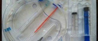
Contrast venography (venography, ascending contrast venography or contrast venography) is an x-ray examination of deep or superficial veins using a contrast agent that provides an image of the blood vessel.
Phlebography determines the patency of the deep veins, the presence of blood clots, valve function and allows for a general assessment of the condition of the deep veins. Phlebography can be used when deep vein thrombosis is suspected, but ultrasound diagnostics cannot accurately exclude it. The study allows us to reliably assess the condition of the iliac veins in obese patients, when it is impossible to conduct a detailed ultrasound scan.
Contrast venography is most often used either during endovascular surgery on the deep veins (angioplasty or installation of a vena cava filter). We use retrograde venography to assess the integrity of venous valves when planning surgery for venous outflow reflux after venous thrombosis.
Ultrasound angioscanning with color mapping (USAS)
After the world's first device for percutaneous flow study was designed in 1960, based on the physical effect of wavelength changes when reflected from moving objects, ultrasound angioscanning began to quickly be introduced into diagnostic testing programs for patients with pathology of the venous system. The developments of medical equipment design engineers made it possible by the mid-70s of the last century to create a fundamentally new class of ultrasonic devices that used the effect of differences in signal reflections from media with different acoustic densities, the so-called B-mode.
The combination of data obtained in one device using both physical effects formed the basis of a class of medical equipment for duplex scanning. Its main disadvantage today is its poor visibility and incomplete information content in clinical use. About 10 years later, by the mid-80s of the last century, medical ultrasound devices were improved with the ability to color code, recorded using the Doppler effect of flows (triplex angioscanning).
The 90s were marked by the active development of computer technologies for processing information (not signals) obtained on the basis of both physical effects involved in the equipment, which today makes it possible not only to calculate, but also to visually assess the state of the venous system using three-dimensional reconstructions of morphological objects and flows, to simulate outcomes of proposed interventions. Since the late 80s of the twentieth century, there has been a clear tendency not only to study the capabilities of the method, but also to gradually replace traditional methods of clinical examination of patients (radiocontrast, plethysmography, clinical).
The method has so firmly gained its position in medicine that special periodical literature on its use has appeared. This was facilitated not only by the high information content and its rapid progress with the development of the method, but also by a significant reduction in the cost of examining angiosurgical and phlebological patients.
USAS: Thrombosis of the venous sinuses of the gastrocnemius muscles (cross section)
UZAS: When compressed by the sensor, all tubular structures (veins) are compressed, except for thrombosed sinuses
This method in this study is of the greatest interest not only because it is highly informative and accessible, but also because ultrasound studies in phlebology began with the diagnosis of thrombosis and thrombophlebitis. To date, the greatest experience has been accumulated on this problem. Even the use of early engineering developments made it possible to increase the specificity and sensitivity of the method in detecting deep phlebothrombosis to 96% and 100%.
In addition to the previously known capabilities of the ultrasound scanning method with color mapping, there are the main ones:
- state of patency of deep veins,
- localization,
- character of thrombomass.
additional:
- the possibility of ultrastructural study of elements of pathologically altered venous valves according to the shape of the reflux flow,
- characterization of the direction and source of phlebohemodynamic disturbances according to the source and direction of reflux,
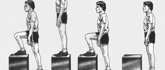
- assessment of the tonic-elastic properties of the walls of deep veins by the reaction of the venous wall to tests that simulate the activation of the patient’s main venous pumps.
In the clinical diagnosis of chronic venous diseases and deep thrombi, the most informative and accumulating information capabilities is the method of ultrasound angioscanning with color mapping. Additional advantages of this method are its absolute safety for the patient and accessibility.
At the stage of making a primary (tentative) diagnosis, it is still advisable to use simple clinical tests that do not require special equipment or additional conditions.
Preparing for diagnosis
- You cannot eat for four hours before the test, and you can only drink water.
- Patients who are allergic (particularly to iodine) or have already had a reaction to contrast should inform their doctor.
- A sedative may be prescribed to calm the patient.
- You may be asked to follow certain dietary restrictions before the test, depending on your specific procedure.
- Tell your doctor about the medications, herbs, or supplements you take. You may be advised to stop taking some of these medications before the test.
How is the diagnosis carried out?
During the procedure, the patient lies on a special X-ray table. The area where the catheter will be inserted is cleared (usually a vein in the arm so that any necessary medications can be administered during the procedure). Sometimes local anesthesia is administered.
A contrast solution is delivered through the catheter. The dye injection causes a warm sensation that can spread throughout the body. The contrast may also cause mild nausea. About 18% of patients experience discomfort from the contrast solution. To fill the deep venous system with dye, a thick tape (or tourniquet) is sometimes placed around the ankle, or the limbs may be angled. The patient is asked to keep his leg still. The doctor watches the movement of the solution through the vein using a fluoroscope. At the same time, a series of photographs are taken.
When the study is completed, saline is injected into the same catheter to clear the veins of contrast, then the catheter is removed and a bandage is applied to the injection site.
Features of phlebography (by location of the veins being examined):
Venography of the lower extremity: the patient is positioned leaning on an inclined X-ray table. The table is tilted so that the legs are raised or lowered. The catheter is inserted into the selected leg or arm. The procedure may take 30 to 45 minutes.
Adrenal venography: the patient lies supine on the X-ray table. The catheter is inserted into the femoral vein. Under the guidance of fluoroscopic visualization, he carefully targets either the renal or suprarenal veins in the abdominal cavity. The procedure takes about 1 hour.
Abdominal venography: the patient lies supine on an x-ray table. The catheter is inserted into the femoral artery. The procedure takes about 1 hour.
1.What is phlebography and indications for it?
Phlebography
is a method of x-ray examination of veins. During the procedure, a special dye (contrast agent) is injected into the veins to more clearly display the processes on x-rays. Phlebography reflects the condition of your veins and the valves in them. Venography is used to examine the veins of the legs, pelvis or arms; veins leading to the heart or leaving the kidneys.
Indications for phlebography
Your doctor may order a venography in one of the following situations:
- Varicose veins.
- Trophic disorders.
- Thrombosis and venous outflow disorders.
- Swelling, pain in the lower leg and thigh of unknown origin.
- Control of the treatment performed.
- Before surgery on veins and arteries (for example, bypass surgery).
- Phlebography allows you to determine the possibility of administering medications into a vein.
After diagnosis
After phlebography, observation in the clinic is necessary for at least 24 hours. Depending on the procedure, you may be advised to rest in bed for a certain period of time.
Patients should drink plenty of fluids to flush any remaining contrast solution from the body. The area where the catheter was inserted may be sore for several days. If you notice swelling, redness, pain, or fever, tell your doctor. In most cases, the patient can resume normal activities the next day.
Possible complications
Phlebography can cause complications such as phlebitis, tissue damage, and deep vein thrombosis in the healthy leg. Complications are quite rare, but they must be taken into account when planning treatment so that the risk of the study does not exceed the risk of the disease for which it is being performed.
A rare side effect (up to 1% of cases) is a serious allergic reaction to the contrast dye. It usually appears 30 minutes after the dye injection and requires medical attention.
Possible risks include blood clot formation in the vein, bleeding, damage to blood vessels, or infection at the site where the catheter was inserted.
Some people may experience an allergic reaction to iodine-based contrast dye. This can cause symptoms such as nausea, sneezing, vomiting, hives, and sometimes a life-threatening reaction called anaphylactic shock (especially in older patients with chronic dehydration or mild kidney failure).
Instrumental examination
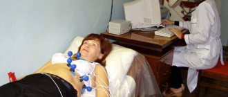
Ultrasound duplex (triplex) angioscanning (UZDAS of veins, duplex, Doppler, Dopplerography, ultrasound of veins, Doppler ultrasound) is currently the most widespread and accessible.
Ultrasound duplex angioscanning
Ultrasound duplex angioscanning is the main diagnostic, non-invasive method of examining a patient with a disease of the body’s venous system. The method allows you to simultaneously visualize the vessel under study, identify intraluminal pathological formations (thrombi), their nature, determine the direction of blood flow in the vein and its parameters, and assess the functional state of the valve system. Availability, quick interpretation of results and absolute safety for the patient under study explain the widespread use of this diagnostic technique at the outpatient stage. The method is perfect for visualizing the superficial and deep vein system in the upper and lower extremities and veins of the neck. At the same time, the use of this technique for diagnosing diseases of the veins of the pelvis, abdominal space and chest is very limited and requires preliminary preparation of the patient and an experienced specialist.
One of the main characteristics of other instrumental methods for examining patients is the need for invasion (puncture) of the vascular bed. Invasive examination methods include: computed and magnetic resonance imaging with a contrast agent, radiocontrast venography, radionuclide venography, intravascular ultrasonography.
The most important criterion for the safety of a diagnostic procedure is the radiation exposure to the patient and medical personnel. Radiation exposure occurs during computed tomography and magnetic resonance imaging, radiopaque phlebography, radionuclide phlebography, ascending and descending phlebography.
Computed tomography (CT venography, computed tomography with contrast)
The essence of the method is to visualize layer-by-layer sections of the body with simultaneous contrasting of the venous bed. Computed tomography does not allow assessing the dynamic state of the body’s venous system, but helps to diagnose occlusive-stenotic lesions of large veins of the pelvis, retroperitoneum and chest.
Magnetic resonance imaging (MRI venography)
The essence of the method is similar to CT venography, but MRI venography can be used if it is impossible to use iodine-containing drugs (individual intolerance to iodine by the patient, the presence of pathological conditions that preclude the use of iodine - pathology of the thyroid gland. An absolute contraindication for this technique is the presence of metal structures in the human body .
X-ray contrast venography
X-ray contrast venography is an invasive procedure and requires hospitalization in a specialized hospital department and preparation for the study. The method allows you to visualize deep and superficial veins and obtain information about morphological changes in the venous system. The essence of the technique is the introduction of an X-ray contrast agent into the venous bed and X-ray visualization of the stained veins of the anatomical area of interest. The indication for the use of radiopaque venography is planning of surgical treatment of patients with occlusion of the iliac, superior and inferior vena cava. The information obtained by contrasting the venous bed makes it possible to present the anatomical and topographic relationships of the veins and surrounding tissues and to identify extravasal compression of the veins.
Radionuclide phlebography
The method allows you to obtain data on the nature and direction of blood flow through the deep, superficial venous system, as well as perforating veins under conditions close to physiological. Radionuclide venography allows you to fully assess blood flow throughout the entire venous system. The method is not readily available in clinical practice due to its high cost and the need for special equipment for implementation.