CT coronary angiography of the heart is a diagnostic method for studying the vessels of the heart, aimed at determining their condition and identifying any anomalies and pathological conditions. It is considered the “gold standard” because it makes it possible to identify pathologies in the early stages of development and see even minor damage to the blood vessels of the heart.
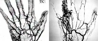
The research method makes it possible to obtain an image of the myocardial network, the pathologies of which are considered the most common initiating factor in cardiovascular diseases. Thanks to it, it is possible to diagnose disturbances in the blood supply to the heart, narrowing and spasms of the coronary vessels, and any other disruptions in the blood supply to the middle muscular layer of the heart.
The procedure involves the use of a contrast agent. In this way, the most accurate three-dimensional image is obtained, after studying which the doctor is able to correctly diagnose and prescribe appropriate treatment.
Control CT coronary angiography after the intervention performed at JSC CELT (term 2-6 months) - 14,500 rubles.
30-45 minutes
(duration of procedure)
Advantages of CT coronary angiography of cardiac vessels in CELT
You can undergo the study at the CELT multidisciplinary clinic. We employ highly qualified specialists with over 15 years of experience. They have perfectly studied the technique of coronary angiography and have everything necessary to carry it out in accordance with international standards.
The procedure is carried out on a modern multislice tomograph, which allows you to obtain the most accurate image and identify any, even the smallest, pathological changes. For contrast, modern safe means are used that minimize any negative impact on the patient’s body.
The price of CT coronary angiography of the heart vessels in Moscow is determined individually, since it depends on the volume of contrast agent, its type and the tasks that need to be solved during the diagnostic study.
The price of coronary angiography of the heart vessels in CELT is presented in the price lists in the corresponding section of our website. You can also find out by calling us at our number. We regularly update the price list data, however, to avoid misunderstandings, we ask you to check the price by phone.
Results of coronary angiography
At the diagnostic center, functional radiologists do not make a final diagnosis, since their responsibilities include only the correct interpretation and description of the scans received. Depending on the clinic’s workload, a report with printed or digital photographs can be received within an hour or the next day. The results of coronary angiography must be taken or sent to the attending doctor, who, based on them, will develop subsequent treatment tactics and decide on the issue of medical or surgical recovery. Treatment methods may include angioplasty, bypass surgery, or a course of medications that help normalize the functions of the vascular network.
Types of coronary angiography, advantages of CT
The purpose of coronary angiography is to visualize the blood vessels of the heart, which is made possible by filling them with a contrast agent. Thus, the doctor gets the opportunity to clearly examine the lumens of the blood arteries and their internal walls. Urografin solution is most often used as a contrast agent.
Experts distinguish three types of coronary angiography, including general, selective and. In the first case, an X-ray machine is involved, and its disadvantage is that the contrast must be injected directly into the vessels of the heart, which significantly complicates the procedure and increases the risk of complications after it.
Selective coronary angiography is a more “advanced” version of the general one. The process focuses on one or more vessels whose condition needs to be determined. As for CT coronary angiography (or, as it is also called, MSCT), it is the most optimal method, since it is safe, effective and allows one to obtain reliable data.
Multislice computed tomography allows you to study not only the condition of the heart vessels, but also its valves. Its cost is quite high, and it can only be carried out in those clinics that have the appropriate equipment - a multislice computed tomograph.
The process uses contrast agents based on iodine compounds, after which scanning and image creation begin. The advantages of this type of coronary angiography are difficult to overestimate:
- minimal invasiveness;
- no need for hospitalization of the patient;
- minimal risk of complications;
- the ability to identify hidden diseases;
- the ability to diagnose pathologies in the early stages of development;
- determination of the type of atherosclerotic changes;
- the ability to evaluate the effectiveness of bypass surgery.
How to prepare for coronary angiography?
Before conducting an X-ray diagnosis of the state of the vascular network or deciding on the upcoming stenting, it is necessary to carry out preparatory manipulations, which include primary research procedures for the composition of the patient’s blood solution. Doctors need to obtain information about blood type, biochemical composition, the presence of severe viral diseases of the immunodeficiency type, hepatitis and other anomalies. After this, classic ECG and EchoCG screenings are carried out.
During the examination, the patient must remain completely calm, motionless, listen and comply with the requests of specialists. It is recommended to prepare yourself emotionally and familiarize yourself in advance with what coronary angiography is and how it is done. There is no need to follow a long-term diet, but it is better to come to the session on an empty stomach, and drink enough fluids a few days before the procedure to prepare the urinary system for the subsequent removal of the contrast agent. The fullness of the gastrointestinal tract does not affect the results of the examination, but if an allergic response to intravenous dye occurs, nausea or vomiting may occur, which is best avoided when carrying out resuscitation or anti-intoxication measures.
Indications and contraindications for CT coronary angiography
Carrying out multislice computed tomography aimed at studying the coronary vessels is advisable in the following cases:
- diagnostic studies as part of preoperative preparation for surgery on the heart or its vessels;
- the development of Kawasaki syndrome in a child, characterized by damage to the blood vessels of the heart with a high risk of blood clots and aneurysms, ruptures of the vessel walls;
- development of a clinical syndrome characterized by a feeling of discomfort in the sternum (angina) during the course of treatment for myocardial infarction;
- suspicions of inflammatory processes of the inner lining of the heart of viral etiology;
- as part of preoperative preparation for any surgical intervention in patients who have suffered a myocardial infarction for an unknown reason;
- lack of the desired effect from conservative treatment of pathological conditions such as angina pectoris, cardiac arrhythmia;
- sudden cardiac arrest for unknown reasons;
- the patient's upcoming organ transplantation;
- recent blunt traumatic injuries to the chest;
- any pathological processes affecting the aorta or coronary blood vessels.
- The procedure has a number of contraindications, since it involves the use of a contrast agent. These include the following:
- pregnancy and lactation in women;
- the patient’s weight is higher than the norm specified in the technical documentation for the tomograph;
- individual intolerance to contrast agents;
- severe form of diabetes mellitus;
- severe liver and kidney failure;
- thyroid diseases;
- any disturbances in the frequency and rhythm of heart contractions - arrhythmia;
- an increase in heart rate from 90 beats per minute - tachycardia.
Where is coronary angiography performed?
The examination is available in any private or government diagnostic institution that has a specially equipped radiography room. You can find a complete list of medical organizations on the website of the single registration center, the telephone number of which is listed at the top of the page.
Choose the name of the procedure, a clinic located in your area, or compare medical institutions by ratings and prices, read the comments and share your own opinion. Book a scan through the portal and receive a special discount from the service in the amount of 1000 rubles for the selected type of examination. If necessary, you can use free transport services to the diagnostic center.
Technique for CT coronary angiography
Since the procedure requires the use of a contrast agent, the patient must undergo an allergic reaction test and diagnosis for diseases, the presence of which is a contraindication to the use of contrast, before undergoing it.
Before the procedure, he will be asked to remove any metal objects from himself, as they may negatively affect its quality. Before scanning begins, the patient lies down on the tomograph table, which smoothly moves into the scanner.
The scan begins after contrast is administered and is accompanied by clicking sounds, which are normal and should not frighten the patient. On average, the procedure lasts from 15 to 20 minutes, during which you need to remain motionless and follow the diagnostician’s commands - for example, hold your breath.
In order to make an appointment with our specialists, fill out and send us the form online or call: +7 (495) 788-33-88.
Coronary angiography (coronary angiography - CG)
Dear patients!
At the National Medical Research Center for Cardiovascular Surgery named after. A.N. Baculeva coronary angiography on an outpatient basis is performed on a paid basis.
Cost 35,000 rubles
For more detailed information, call the multi-line phone from 9:00 to 17:00
Address: 121552, Moscow, Rublevskoye sh. 135
Coronary angiography
(coronary angiography - CG) is the “gold” standard for diagnosing coronary heart disease.
This is an invasive research method. Images of the arteries that supply the heart, the coronary arteries, are achieved by injecting them with a contrast agent through special catheters. Catheters are inserted into the heart vessels through a small puncture in the wrist or, if necessary, through the groin area. Coronary angiography provides the doctor with all the necessary information about the condition of the coronary arteries.
Considering that radial access (through the wrist) is currently used to perform coronary angiography, bed rest after the procedure is not required. The duration of the study itself is 15-20 minutes.
Hospitalization at the Center
The patient arrives at the Center on an empty stomach. After completing the documents, he is placed in the ward and examined by the attending physician. You must have blood tests with you for HIV, hepatitis and syphilis (Wassermann reaction) mdash; no more than 14 days old. In the absence of own results of instrumental examination, an electrocardiogram and ultrasound of the heart (echocardiography) are performed.
Performing coronary angiography
Immediately after this, the patient is transferred to the cath lab to perform coronary angiography. The procedure is performed under local anesthesia, as it does not create serious discomfort during execution. The patient has the opportunity to communicate with the specialist performing the procedure. To perform CG, our Center uses the most modern contrast agents (Visipak, Omnipaque, Xenetics, etc.), which minimizes the risk of allergic reactions. Novocaine is most often used to perform local anesthesia. However, if there is intolerance to any anesthetic, the latter is selected individually.
After the coronary angiography procedure
After the coronary angiography is completed, a pressure bandage is applied to the patient’s wrist, and he is transferred to the ward for rest and observation by a doctor for 2-3 hours. If during this time the patient does not have any complaints, the patient is discharged from the clinic.
Survey results
The patient receives a disc with a digital recording of coronary angiography, as well as consultation with a specialist regarding the need for further examination or treatment. In cases where, during the diagnostic procedure, a life-threatening narrowing of the coronary artery is detected, treatment (transluminal balloon angioplasty with stenting of the affected area of the vessel) is carried out immediately.
Indications:
- identified foci of calcification (calcification) of the valvular apparatus of the heart, cardiac muscle (myocardium) or serous membrane of the heart (pericardium) when performing computed tomography of the chest;
- old post-infarction scars, aneurysms of the great vessels or suspected presence of blood clots in the cavities of the heart;
- high calcium index values when assessing coronary calcification (CT “calcium scoring”);
- suspicion of abnormal development of heart vessels or heart valve insufficiency, for example when performing echocardiography;
- chest pain of unknown etiology, atypical pain;
- the appearance of primary or repeated symptoms of angina pectoris;
- deviation from normal indicators of electrocardiography, stress tests, laboratory tests;
- the need to assess the blood supply to the heart muscle (myocardium) after surgical interventions (coronary artery bypass grafting, coronary artery stenting).
[/td]
The high prevalence of cardiovascular diseases and the first place among the causes of mortality in developed countries makes preventive measures and early diagnosis of heart disease . MSCT of the coronary arteries can be performed as a screening during a preventive (dispensary) examination of a person. Atherosclerosis of the coronary arteries is the main cause of the development of coronary heart disease (coronary heart disease) and myocardial infarction. Modern medicine has a wide range of diagnostic capabilities. And with the advent of high-speed multislice computed tomography (MSCT), a completely new, unique opportunity has emerged for non-invasive assessment of the condition of the coronary arteries, which does not require hospitalization of the patient to a hospital for a complex and unsafe surgical procedure (angiography).
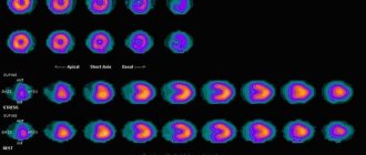
MSCT examination takes only a few minutes, does not require additional preparation, and does not cause pain or emotional anxiety. In addition to assessing the condition of the heart vessels ( coronary “tree” ), MSCT of the coronary arteries provides the doctor with additional equally important information, which allows him to assess the condition of the heart as a whole, and develop tactics for further examination and treatment. MSCT allows you to evaluate the morphological structure of an atherosclerotic plaque , study the valve structures of the heart (calcification of the leaflets, anomalies of valve development), identify myocardial lesions (hypertrophy, scars), and assess the condition of the cavities of the heart and pericardium. Valuable information is provided by determining the function of the left ventricular myocardium with identifying areas of impaired contractility.
Benefits and risks of CT
Advantages
The results of computed tomography allow doctors to make the most accurate, correct diagnosis and timely prescribe treatment. Computed tomography is a non-invasive, painless diagnostic method. Carrying out CT scans very often reduces the required number of studies to a minimum and eliminates the advisability of using invasive techniques.
CT scan:
— allows you to study the state of the bone structure, soft tissues, blood vessels and individual organs layer by layer; - often used in emergency situations, for post-traumatic disorders, for suspected damage to internal organs, bleeding; — can be used if the patient has implanted medical devices of any kind; — used for control during biopsy of organs and pathological areas of the human body. X-rays from a CT scan have no immediate side effects.
Estimated risks and contraindications
The effective dose of radiation from computed tomography is minimal, however, it is always present. The radiation dose is usually specified in the study protocol. In the interests of accurate diagnosis, especially in acute cases, this risk is mitigated. Women are required to inform the referring physician and radiologist about the possibility of pregnancy. Because of the potential risk to the baby, CT scans are not recommended for pregnant women unless there are specific medical indications. It is extremely rare, but allergic reactions to a contrast agent that contains iodine do occur. In such cases, PATERO CLINIC has all the necessary means to stop such reactions.
Manufacturers of intravenous contrast media do not recommend that mothers breastfeed for 24 to 48 hours after receiving or administering contrast media.