MRI procedure
MRI is magnetic resonance imaging, that is, layer-by-layer scanning of the heart. The operation of the tomograph is based on the use of the magnetic resonance effect. During an MRI scan, the machine creates a powerful magnetic field. Cell atoms, when exposed to an RF signal in a magnetic field, begin to vibrate, and this resonance is picked up by a tomograph. The obtained information is translated by an MRI device into three-dimensional images, from which the doctor can determine the presence of the smallest pathologies of the heart muscle and coronary vessels in the patient. The MRI procedure itself is quite simple. The patient is placed on a movable table on his back, the body is fixed with a special coil, and the head is fixed with rollers, after which the table is pushed into a cylindrical chamber. The patient's task is to lie still and calm throughout the magnetic resonance examination. The duration of cardiac MRI is from 25 to 30 minutes. During the procedure, the patient may experience discomfort due to various sounds produced by the device. If a person is concerned about this, they should ask the diagnostician for headphones before the test. This will soften the noise. If the patient begins to feel uncomfortable inside the tomograph tube or experience a panic attack, he always has the opportunity to contact the diagnostician, since the scanner is equipped with two-way communication. After an MRI examination, the patient receives a report from a radiologist and an examination recorded on disk in the form of a series of high-quality images. Waiting for the result can take up to one hour, in rare cases 1-2 days. It all depends on the complexity of the scan and diagnosis. To reduce the waiting time for results, the conclusion in most medical centers in St. Petersburg can be obtained by e-mail.
Previous Next
Ultrasound
Ultrasound is an ultrasound examination of the heart. The operating principle of an ultrasound machine is based on the physical properties of ultrasound. Supersound is not perceived by the human ear, since the vibrations of the sound wave in this case are reproduced at a very high frequency. The ultrasound sensor sends ultrasound into the cavity of the object under study, which, when reflected, creates an echo. It is captured by a computer and converted into images. The anatomical accuracy and level of detail on ultrasound is significantly lower than on cardiac MRI because it is difficult for ultrasound to penetrate the bony barrier of the chest. Ultrasound diagnostics of the heart cannot distinguish between changes ten years ago and changes occurring now. It cannot reveal the severity or depth of the ongoing inflammatory or tumor process. Ultrasound can only indirectly indicate that there are some thrombotic changes in the coronary vessels of the heart.
Why is coronary angiography performed only in a hospital setting?
The main reason that made it mandatory for the patient to be in the hospital when performing this study was the need for puncture (puncture) of the peripheral artery (femoral) and associated complications. The femoral artery is a large vessel located at a depth of 2-4 cm from the skin surface in the groin area. To prevent bleeding, which can be dangerous, after the study the patient is prescribed a limited motor regimen under the supervision of medical personnel. But recently another access has appeared - through the small radial artery, which we palpate on the wrist. An approach called radial, which does not result in serious bleeding. The patient can get up immediately after the examination; bed rest is not necessary. Observation is limited to a few hours. In the CELT clinic, radial (radial) arterial access is used as the main approach. Experience with this technique indicates a lower incidence of local complications, which is 0-0.7%. The possibility of early rehabilitation of the patient after CAG with radial access and the almost complete absence of side effects make it possible to perform CAG on an outpatient basis.
Advantages and disadvantages of ultrasound and MRI of the heart
Today, high-quality cardiac MRI requires very expensive equipment - a tomograph with a magnetic field power of at least 3 Tesla. Research using such an ultra-high-field device will be expensive, so the main disadvantage of cardiac MRI is its high price. In addition, MRI of the coronary vessels and heart cannot measure the speed of blood flow and its actual volume. Cardiac ultrasound can better cope with this task.
What does echocardiography show?
During the procedure, the doctor performs structural imaging and hemodynamic (Doppler) imaging.
Structural visualization includes:
- Visualization of the pericardium (eg, to exclude pericardial effusion);
- Visualization of the left or right ventricle and their cavities (to assess ventricular hypertrophy, wall motion abnormalities, and also to visualize blood clots);
- Valve imaging (mitral stenosis, aortic stenosis, mitral valve prolapse);
- Visualization of the great vessels (aortic dissection);
- Visualization of the atria and septa between the chambers of the heart (congenital heart disease, traumatic injuries).
Dopplerography:
- Visualization of blood flow through heart valves (valvular stenosis and regurgitation)
- Visualization of blood flow through the chambers of the heart (calculation of cardiac output, assessment of diastolic and systolic cardiac function)
Echocardiography for heart failure
- Makes it possible to determine the causes of the development of the chronic form (CHF)
- Left ventricular ejection fraction: reduced (systolic CHF);
- normal (diastolic CHF).
Echocardiography for cardiac arrhythmias
Important! Tachycardia makes it difficult to conduct research and accurately assess the results obtained.
Transthoracic echocardiography and emergency echocardiography are used.
Important parameters:
- Dimensions of the cavities of the heart, especially the atria
- Presence of blood clots
- Ejection fraction estimates are not always reliable
Echocardiography for assessing the condition of cancer patients
Heart damage in cancer patients:
- tumor intoxication;
- cardiotoxicity of chemotherapy drugs;
- cardiotoxicity of radiation therapy.
Indications for specialized echocardiography to determine cardiotoxicity:
- all patients with cancer before starting treatment;
- receiving chemotherapy or radiation therapy (after each course);
- have received chemotherapy or radiation therapy in the past (the frequency depends on the treatment regimen).
When is ultrasound better than cardiac MRI?
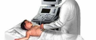
Cardiac ultrasound is well suited for diagnosing both children, from infancy, and adults. An ultrasound examination will clearly show the functional features of the heart:
- efficiency of the heart muscle;
- thickness of the heart walls;
- functional state of four chambers and valves;
- the size of the heart cavities and the pressure in them;
- speed of intracardiac blood flow.
Cardiac ultrasound is better suited than MRI for examining young children, since it does not require immobilization of the patient through anesthesia so that the baby can lie quietly. Ultrasound is an inexpensive way to initially screen the heart if you need to evaluate the condition of the organ due to some primary symptoms of illness or if you want to do an ultrasound of the heart as a preventive measure for cardiovascular diseases. Cardiac MRI is an expert diagnostic method. It is best used when a pathology is identified and it is necessary to verify or clarify the diagnosis.
Holter ECG - if a heart rhythm disorder is suspected!
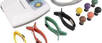
Many patients who go to the doctor report that their heart rate is fast or slow, but the ECG is normal. Then the doctor decides to perform a Holter ECG. Holter is nothing more than an ECG, but its duration is 24 hours and sometimes 48. The patient usually has five electrodes attached to the chest, connected to a special device that contains a memory card on which the daily ECG image is recorded. The device is compact and can be attached to a belt. An ECG recorder is most often performed when cardiac arrhythmia, that is, supraventricular arrhythmia, is suspected:
- atrial fibrillation,
- increased heart rate (tachycardia),
- slow heartbeat (bradycardia),
- heartbeat,
- conductivity blocks.
It is also recommended to perform a holter in case of frequent fainting or loss of consciousness.
This test looks for so-called silent myocardial ischemia in patients with suspected coronary artery disease. This is also a study conducted on patients with a pacemaker to monitor its performance. IMPORTANT! An ECG recorder is not invasive, but there are a few things to keep in mind! After putting on the device, the patient should lead a normal lifestyle. To do this, he must keep a special activity diary in which he records individual actions and the periods of time in which they were performed, for example: climbing stairs, eating, driving, rest and sleep times, as well as symptoms such as dizziness, rapid palpitations, fatigue or shortness of breath, etc. The heart rate monitor also monitors your heart rate at night, giving your doctor valuable information. During the test, remember that the electrodes should not come off, and if you notice this, stick them with adhesive tape. During the examination (24 or 48 hours), you should not swim or shower. After the examination, the card with the record goes to a specialist who, using a special program, downloads the data and analyzes it. Usually, after a few days, the patient receives a description of the examination from a cardiologist, with whom he goes for further consultations.
When is MRI better than cardiac ultrasound?
The level of anatomical detail of the heart area on MRI is higher than on ultrasound. During the scan, the doctor receives up to 1000 images of the mediastinal area. The scanning step of a 3 tesla device can be only 0.8 mm. This means that the doctor can look in detail at the condition of the heart itself and the neighboring coronary vessels. In this case, both planar and three-dimensional data will be provided. MRI best visualizes pathologies and diseases such as:
- inflammatory processes in tissues;
- tumor formations;
- blood clots, vascular malformations, occlusions, aneurysms;
- congenital anomalies of the heart structure;
- heart muscle defects;
- pericardial condition.
Author: Telegina Natalya Dmitrievna
Therapist with 25 years of experience
How is outpatient coronary angiography performed?
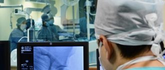
The algorithm for performing coronary angiography on an outpatient basis includes three stages. At the first stage, patients are selected for a diagnostic procedure, and the necessary additional examinations are carried out. Indications for coronary angiography : - identified or suspected ischemic heart disease; - pain in the chest, suspicious for angina pectoris; - myocardial infarction; - planned surgery for heart defects; - heart failure, ventricular arrhythmias. Indications for coronary angiography are determined by the attending physician in accordance with accepted criteria. In preparing the patient for coronary angiography , the necessary tests and studies are performed. In addition to these, additional studies may be prescribed. The second stage of outpatient CAG angiography procedure itself . The patient is admitted to the day hospital ward. After assessing the stability of his condition, premedication is administered and he is transported to the cath lab, where the coronary angiography . After anesthetizing the access area, research begins - a special catheter is passed through the artery of the forearm into the lumen of the coronary arteries . Using a catheter, a radiopaque substance is injected into the blood, thanks to which the lumen of the vessels becomes visible on a special device - an angiograph. During coronary angiography, the degree and size of damage to the coronary vessels is determined, which determines further treatment tactics. This procedure is low-traumatic, which allows it to be performed under local anesthesia without the use of general anesthesia. The duration of the procedure, as a rule, does not exceed 20 minutes. From the operating room, the patient, accompanied by medical personnel, is taken to the day hospital ward. The third stage of outpatient coronary angiography is observation of the patient in a day hospital ward for 4-5 hours after the examination. In the ward, the patient can drink water or juices without restrictions and have lunch. If there are no complications, the patient is sent home. On the day of the outpatient coronary angiography, the patient receives a conclusion with recommendations on further treatment tactics and a disk with the results of coronary angiography . If complications arise during coronary angiography or during the control period, patients are hospitalized in the intensive observation unit of the hospital.
Preparing for the study
There are no special recommendations for preparing for an ultrasound examination. But in order for the results to be most reliable during ultrasound of the heart, it is necessary:
- exclude physical activity the day before;
- a day before, stop drinking coffee, alcohol, and strong tea;
- Avoid eating spicy and fatty foods before eating;
- do not eat for a couple of hours so that the stomach is not full and does not put pressure on the diaphragm;
- do not smoke 2-3 hours before;
- before entering the office, rest a little and calm down in the corridor;
- if there is thick hair on the chest, shave it off if possible;
- Before making an appointment, tell your doctor what medications you are taking and, if necessary, stop taking them for a while;
- lying on the couch, do not worry and remain as calm as possible;
- strictly follow all recommendations of a specialist.
In case of urgent need, echocardiography can be performed without any preparation. When planning a visit to a specialist, you do not need to take anything with you, in particular towels. Everything you need for a cardiac ultrasound is in the treatment room.
