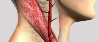
The importance of the carotid arteries, located in the neck and chest, in the human body is difficult to overestimate. The carotid arteries participate in the general blood flow and play a critical role in supplying the brain with oxygen. Any problems related to the carotid arteries can have a negative impact on your overall health. Therefore, today there are quite a lot of examination methods, including ultrasound of the carotid arteries.
Ultrasound of the carotid arteries
Ultrasound of the carotid arteries reliably shows the condition of the blood vessels and is one of the safest and most painless technologies. Ultrasound waves do not harm the body, and the non-invasiveness of the method negates the risk of vessel damage. Therefore, this type of diagnosis of diseases and pathologies of the carotid arteries is the most common. There are practically no obstacles in the path of ultrasonic waves when examining the carotid arteries using ultrasound, which makes it possible to obtain reliable readings about the condition of the vessel. A pleasant advantage of this research method is its low cost.
Indications for cerebral vascular ultrasound
Ultrasound examination is prescribed for patients with angina pectoris, coronary heart disease, osteochondrosis and many other diseases. Diagnostics is indicated if the patient complains of constant headaches and if symptoms indicate the development of pathologies of the blood vessels of the head and neck.
List of indications for ultrasound of blood vessels of the head and neck:
- If you feel dizzy when changing body position.
- Systematic headache.
- Frequent fainting.
- Feeling of lack of air.
- Poor health and weakness.
- Head injuries.
- Vascular dystonia.
- Noise in the head or ears.
- Suspicion of anomalies in the development of blood vessels in the neck or brain.
- Speech impairment.
- Feeling of numbness in the limbs.
- Strong weather dependence.
- Increased cholesterol levels.
- Diabetes.
- Obesity or underweight.
- Diseases of the cervical spine.
For preventive purposes, it is recommended to undergo an ultrasound examination for people over 40 years of age, especially if there is a predisposition to cardiovascular diseases. The study is completely safe, so ultrasound of the head has no contraindications and can be prescribed even to newborns.
Doctors in this field
When is ultrasound scanning of the carotid arteries prescribed?
Duplex ultrasound scanning of the carotid arteries is the general name for ultrasound of the cervical and clavicular regions. Due to vague symptoms, diagnosis of many diseases is difficult, which is why ultrasound of the carotid arteries is prescribed. Indications for ultrasound of the carotid arteries may include headache, dizziness, blurred vision, increased or decreased blood pressure, increased cholesterol levels in the blood, as well as fainting and limb dysfunction.
These alarming symptoms can be signs of many other diseases. Therefore, why to do an ultrasound of the carotid artery is decided by the doctor, based on other signs of a possible disease. Ultrasound of the carotid arteries is also prescribed for vascular damage due to neck injuries, osteochondrosis and spinal injuries.
Indications and contraindications
A number of conditions may serve as indications for ultrasound examination of neck vessels:
- headache;
- arterial hypertension;
- widespread atherosclerosis;
- diabetes;
- unsteadiness of gait;
- dizziness;
- noise in ears;
- spots before the eyes;
- osteochondrosis;
- traumatic brain injuries;
- previous neurosurgical operations or preparation for them;
- consequences of toxic vascular damage;
- syncope (sudden loss of consciousness);
- short-term cerebrovascular accidents.
The use of ultrasonic waves is absolutely safe and has been widely used for decades by healthcare systems around the world. Ultrasound can be performed as often as the medical situation requires. The use of the method may be temporarily limited due to the patient's skin diseases (streptoderma, serious burns).
How is the carotid artery ultrasound procedure performed?
The ultrasound procedure of the carotid arteries does not cause discomfort or pain and is performed with the patient in a supine position. A special pillow is placed under the patient's shoulders so that nothing interferes with the view of the arteries. Throughout the procedure, the sonologist moves the scanner over the skin of the neck, lubricated with gel, and from time to time changes the sensitivity of the device.
There are several methods for ultrasound of the carotid arteries. However, it does not depend on this how ultrasound of the carotid arteries is done - in any case, the sonologist moves the scanner in the neck area, if necessary, telling the patient to turn his head or stretch his neck, examining the subclavian region.
Dopplerography
Using Doppler ultrasound, disturbances in blood flow in the vessels are detected, and ultrasound of the carotid arteries is one of the most effective diagnostic methods. If the blood flow is not visible, the sonologist can apply digital pressure on the artery to obtain more accurate information. In some cases, signals obtained using Doppler ultrasound can be converted into sound, this helps when examining seriously ill patients.
Duplex scanning
Duplex scanning is a combination of Doppler ultrasound, which determines blood flow disturbances, with traditional scanning, which shows the echostructure of the arteries. Duplex scanning of the carotid and vertebral arteries is the most common procedure, since it solves two problems simultaneously.
Triplex scanning
Triplex scanning of the carotid arteries involves research in three modes. The greyscale scanning mode provides information about the structure and shape of the artery, as well as any changes in it. Dopplerography
reveals the quality of blood flow inside the artery. And color Doppler mapping provides a color image that provides comprehensive information about any changes in the structure and blood flow of the artery.
Ultrasound of head and neck vessels
This is a modern ultrasound diagnostic method, which makes it possible to most accurately determine the condition of the neck vessels that supply the brain, shoulder girdle and arms.
The article was prepared by our ultrasound diagnostics doctor Irina Alekseevna Romadova.
When a person is prescribed a study of the brachiocephalic arteries (BCA), he has questions:
— what will he have to deal with?
- How effective is this method?
- isn't it harmful?
— and won’t this procedure cause discomfort?
What is ultrasound of the vessels of the head and neck.
Duplex ultrasound scanning of the brachiocephalic arteries (USD BCA) is a modern ultrasound diagnostic method that makes it possible to most accurately determine the condition of the neck vessels that supply the brain, shoulder girdle and arms.
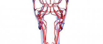
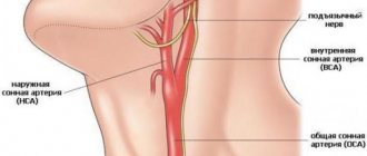
These vessels are called brachycephalic. These include the carotid, vertebral, subclavian arteries and their connection, which forms the brachiocephalic trunk.
The BCA study allows the doctor to determine the presence of a violation of the blood supply to the brain , and to predict the likelihood of worsening the disease in the future.
Types of vascular studies
Vascular examination can be carried out in three ways:
- Ultrasound Dopplerography or Dopplerography . The research is based on the Doppler effect, named after the Austrian scientist Christian Doppler who discovered it.
- Ultrasound scanning or duplex scanning of the vessels of the neck and head is a combination of both the Doppler effect and B-mode (regular gray ultrasound image). That is, the doctor can evaluate the movement and speed of blood flow , as in Dopplerography, as well as the internal walls of blood vessels , deposits on them, the presence of atherosclerotic plaques, thickening, and developmental anomalies. With this method, two functions are examined at once, hence the name duplex. Ultrasound scanning shows a complete picture of the movement of blood through the arteries and veins, which makes it possible to make a more accurate diagnosis.
- Triplex scanning is a type of duplex assessment. However, it provides even more information because it uses the additional technique of color Doppler mapping. A color image of the movement of blood in the vessels makes it possible to more accurately determine changes in blood flow.
Unlike conventional ultrasound, which shows organs in a static state, Doppler ultrasound shows the movement of blood flow and its speed. This is made possible by the difference in speed at which Doppler ultrasound reflects and receives waves.
Undoubtedly, duplex scanning is the most modern research that provides a complete picture of changes in blood vessels. The ultrasound machine in our clinic belongs to the expert class and is equipped with all vascular programs. This makes it possible to most effectively visualize and evaluate pathological changes in the vascular bed.
Doctor's qualifications
We all know that the results of the study directly depend on the qualifications of the doctor performing the study. Our doctor Irina Alekseevna Romadova completed a full course of training in ultrasound angiology (examination of all vessels) at the Northwestern State Medical University named after I.I. Mechnikov, and masters all methods of vascular research. This is undoubtedly a big plus for our patients , because if there is damage to various vessels, then she can evaluate them all, besides, Irina Alekseevna is a former surgeon and is well versed not only in physiology, but also in anatomy .
Safety
Ultrasound examination is completely safe and painless for the patient. The use of this technique has no restrictions or contraindications. Ultrasound scanning can be used repeatedly, both for diagnosis and for monitoring the course of the disease at various stages of treatment.
What pathologies
The most common pathologies diagnosed with ultrasonography are:
- atherosclerotic lesions of the vascular wall
- vascular obstruction
- aneurysms
- deformations and abnormalities of vascular development
- dilation and incompetence of the venous bed
- thrombosis
- damage to the walls of blood vessels
The sensitivity of the method is very high , with ultrasound even minor, initial manifestations of atherosclerosis in the walls of blood vessels are visible, the identification of which will allow timely measures to be taken and prevent the formation of atherosclerotic plaques .
In some situations, it is necessary not only to examine the vascular bed, but also to evaluate the elasticity of the vessels, their elasticity, and ability to respond to changes in blood pressure and oxygen content in the atmosphere ( resistance ). For these purposes, the Carbonik , which is equipped in the ultrasound room at the Smart clinic. It has been clinically proven that with the help of a breath test using the Carbonik device, the most reliable information about changes in the resistivity of the vascular bed is obtained.
This photo shows the process of conducting a study of cerebral vessels with the Carbonik apparatus.
Who is this procedure indicated for?
As a measure to prevent serious complications of cerebrovascular accidents, such as stroke, BCA ultrasonography is recommended to be performed on individuals at risk at least once a year.
- People over 40–45 years of age.
- Patients with diabetes mellitus.
- Persons with a family history of stroke.
- If abnormalities are detected in the lipid profile.
- Patients with pathology of the heart and vascular system.
- Smoking and alcohol abuse.
- Patients with osteochondrosis of the cervical spine.
- Autoimmune vascular disease (hemorrhagic vasculitis)
This study is also indicated for all people with complaints indicating changes in the vessels of the neck.
- constant or recurrent headaches;
- migraine
- dizziness;
- sensation of noise or ringing in the ears;
- absent-minded attention;
- impaired coordination of movement;
- decreased vision;
- memory change;
- blood pressure problems;
- fainting;
- neck injuries;
- condition after a heart attack or stroke.
Preparing for the study
BCA ultrasound does not require , but on the day of the examination it is better to exclude from the diet foods that can change the tone of the vascular wall, such as coffee, strong tea, alcohol, energy drinks, cola. It is advisable to refrain from smoking two hours before the test. If you are taking medications that affect the functioning of the cardiovascular system, be sure to tell your doctor.
Carrying out the procedure
The procedure takes about 30-40 minutes, the patient lies on his back, the chin should be raised up and slightly turned in the direction opposite to the one being examined. The doctor may press a little on the patient’s neck during the examination, but in general this does not cause any discomfort. Our clinic has a “ platinum ” version of the device, including a device for heating the gel , which makes the procedure even more comfortable. The ultrasound scanner will record all the indicators, and the doctor will enter them into the study protocol.
Summary: Duplex ultrasound scanning of the brachiocephalic arteries is accessible and effective diagnostic method. In addition to high resolution, the method is absolutely safe . The Smart Clinic has an expert level ultrasound machine installed, and our doctor, Irina Alekseevna Romadova, is an experienced specialist, which together gives our patients timely and effective diagnosis, and therefore, subsequently, the correct treatment.
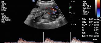
Decoding the results
The interpretation of the diagnosis of ultrasound of the carotid arteries is carried out by the attending physician based on the results obtained. However, a sonologist can also clarify the situation by recording the examination results at the end of the procedure. Typically, a sonologist comments on the visible echostructure of the vessel during scanning and blood flow.
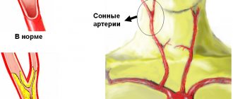
The norm for ultrasound of the carotid arteries is that the blood flow in the internal and external arteries is equal in power, as well as the location of the carotid artery to the left of the aorta. The thickness of the vessel walls should not exceed 1.2 mm, and the pulsation in a healthy artery should be continuous.
What diseases can be detected by ultrasound of the carotid arteries?
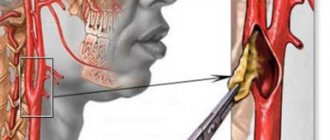
Ultrasound of the carotid arteries allows you to examine not only the brachiocephalic arteries, but also simultaneously assess the condition of the vertebral vessels of the neck. The norm for ultrasound of the carotid arteries implies an internal and external diameter of the walls sufficient for good blood flow. Ultrasound of the carotid arteries most often reveals atherosclerotic plaques and kinks, which cause strokes. Ultrasound of the carotid arteries allows you to see even the structure of plaques, for example, those that have calcium impurities and are especially difficult to treat.
No less dangerous are blood clots and aneurysms, which are clearly visible using ultrasound. Stenosis visible on ultrasound of the carotid arteries may explain ischemic attacks, since blood flow worsened due to narrowing of the artery does not allow the brain to receive enough oxygen. An alarming symptom is a change in the level of the bifurcation of the carotid artery, visible on ultrasound. However, one must understand that ultrasound of the carotid artery can only identify these problems; subsequently, it is recommended to conduct a more in-depth study.
How is the procedure done and why?
Ultrasound of the neck vessels is performed according to standard techniques. The patient lies down on the couch and tilts his head back so that the neck is as open as possible. A gel is applied to the skin to facilitate the passage of ultrasonic waves. The sensor is installed where the vessels pass. The arteries on the right and left are examined alternately. In some cases, functional tests are performed, such as holding your breath, inhaling, pressing on the vessel with your fingers, or hyperventilation.
Ultrasound examination of the arteries and veins of the neck can reveal atherosclerotic plaques, wall dissection, stenosis, and the presence of thrombotic deposits. Pathology of the neck vessels can lead to a lack of oxygen supply to the brain tissue and irreversible consequences for health.
How is ultrasound examination of the vertebral and carotid arteries performed?
Before starting the procedure, the patient will be asked to remove all jewelry from the neck and free it from clothing so that the neck and supraclavicular area are freely accessible to the ultrasound probe. After this, he assumes a lying position on the couch.
The diagnostician applies a gel to the area being examined, ensuring the desired contact of the skin surface with the sensor and eliminating the presence of air bubbles that complicate the examination. It begins with determining the condition of the carotid arteries. The patient is asked to turn his head to the side and first examine the lower part of the right artery, tilting the sensor at different angles.
After this, the diagnostician leads it upward, leading it around the corner of the lower jaw. In this way, he is able to determine the depth and course of the artery, as well as identify the level at which it is divided into external and internal. The final stage is to turn on the three-dimensional color mode, thanks to which you can clearly identify areas of abnormal blood flow. The second carotid artery, as well as all its branches, are examined in the same way. Examination of the vertebral arteries involves a longitudinal placement of the sensor on the neck.
CT scan of neck vessels with contrast
Modern injection systems significantly improve the quality of diagnostics
The introduction of an iodine-based radiopharmaceutical improves the visibility of the smallest structures, which improves the quality of diagnosis. CT angiography of neck vessels with contrast in 95% of cases is not accompanied by extraordinary situations and proceeds without complications. As the drug enters the vein, the patient may experience slight nausea, an unpleasant taste in the mouth, dizziness, and a burning sensation at the injection site. These phenomena disappear on their own within a few seconds to a minute and are considered as normal. A generalized feeling of itching of the skin, difficulty breathing, swelling of the neck, cold sweat, and the appearance of a rash all over the body are signs of a generalized allergic reaction; these symptoms should be reported to the staff immediately via loudspeaker.
Contrast is performed using an injector through an intravenous catheter (the amplifier is injected automatically at certain phases of the study), or the drug is administered simultaneously.
Preparing for the study
No special preparation is required for ultrasound of the neck and head vessels. However, if the patient is taking medications that lower blood pressure or affect blood clotting, the doctor must be informed. He may recommend that you avoid taking medications in some cases.
On the day of the examination, it is recommended to refrain from smoking, drinking alcohol, energy drinks and physical activity. In general, it is not recommended to perform actions that may affect the tone of the vascular wall.
After research
After the examination, the doctor gives you a napkin to wipe off the gel. During this time, a diagnostic protocol is drawn up. If necessary, the protocol can be supplemented by printing an ultrasound image on a printer, although most often a conclusion from an experienced specialist is sufficient. Duplex ultrasound scanning of the neck arteries is a safe and non-invasive method for diagnosing the pathology of the carotid and vertebral arteries and allows us to identify many dangerous diseases. Duplex MAG scanning should be performed in all patients at risk of ischemic stroke or before complex vascular interventions.
Advantages of diagnostics at the Innovative Vascular Center
Our medical center specializes in vascular and endovascular surgery, so it is important for us to obtain detailed information about the condition of the carotid arteries, because ischemic stroke is one of the most dangerous complications in vascular surgery. We perform ultrasound of the carotid arteries on all patients in our hospital and have developed an accurate ultrasound diagnostic algorithm. Prices for research in our center in Moscow are affordable to everyone. An appointment with a vascular surgeon is always accompanied by ultrasound diagnostics.
The advantage of ultrasound of the carotid arteries in our clinic is expert-level ultrasound scanners, evaluation of the results obtained by an ultrasound physician together with the operating vascular surgeon. The accuracy of the results of this study by our specialists is 96%, versus 80% in many other general practice diagnostic centers.