Stagnation of blood in the pelvis is a consequence of various pathologies or poor lifestyle. Blood circulation is disrupted: arterial inflow increases or venous outflow slows down, due to which blood does not flow to all areas adequately: the lower gastrointestinal tract, bladder, elements of the reproductive system of women (uterus, vagina, ovaries) or men (prostate gland, seminal vesicle) ).
Nutrients are transported to organ tissues along with blood. During stagnation, the movement of useful components stops or decreases. Oxygen starvation of all structures begins.
The problem entails the increased development of bacterial, fungal or viral infections: non-circulating or slowly circulating biological fluid is an excellent environment for the spread of pathogenic microorganisms. Since the organs are located anatomically close to each other, the infection quickly spreads through all structures.
Causes
The pathological condition develops more often due to venous obstruction (reflux of the ovarian vein, for example) or varicose veins. Problems arise against the background of impaired hematopoiesis or deformation of the veins, which weaken, become thinner and cannot perform their main function - blood delivery.
The reception is conducted by
Klokov Andrey Nikolaevich
Urologist-andrologist, doctor of the highest category
Make an appointment
Pelvic congestion occurs 2.5 times more often in women. The reason lies in the peculiarities of anatomy and the body’s vivid reaction to even minimal fluctuations in hormones: during menstruation, pregnancy, menopause. Due to changes in the balance of estrogen and progesterone, blood viscosity increases, causing its flow to slow down. Due to hormonal imbalance, women are more likely to develop varicose veins, which also has a detrimental effect on blood circulation.
Pregnancy, if you do not take into account the hormonal factor, entails congestion due to the fact that the growing fetus puts pressure on other organs. Complicated childbirth causes a slowdown in blood flow due to stress on the vessels. At the same time, due to weakened muscle and ligamentous elements, prolapse of organs occurs, which compress the vessels.
Doctors say the most common cause of stagnation is a sedentary, predominantly sedentary lifestyle. A person stays in one position for a long time, for example, when working at a computer or during long car trips, because of this, blood cannot circulate fully.
Blood stasis results from the following problems:
- Decreased elasticity, increased vascular permeability;
- Endocrinological, systemic, autoimmune diseases;
- Inflammatory processes of the genitourinary system, spine, acute or chronic;
- Adhesive processes;
- Developmental anomalies of the pelvic organs;
- Surgical intervention.
Taking hormonal medications, alcohol abuse, and smoking theoretically lead to stagnation of blood circulation. Urologists agree that the practice of interrupted coitus for men causes congestion. For women and men, excessive physical activity on the lumbar region leads to the appearance of foci of inflammation, which often ends in impaired blood flow.
Pelvic venous congestion syndrome
Dear readers, in this article we would like to talk about such a disease as Pelvic Congestion Syndrome (from the English “congestion” - overflow (blood), accumulation) or Pelvic Venous Congestion Syndrome.
This problem is quite common, but is not always well diagnosed.
The true prevalence figures for pelvic congestion syndrome are currently unknown. Due to the variety of manifestations, as will be discussed below, this syndrome is often masked by various diagnoses: endometriosis, inflammatory diseases of the appendages (adnexitis), cystitis (inflammation of the bladder), intestinal diseases, etc. According to some reports, up to 15% of women aged 20 to 50 years have this syndrome, although not all of them experience certain symptoms. However, one should not assume that this is an exclusively female pathology. Thus, the importance of pelvic congestion cannot be underestimated in the development of diseases of the genitourinary system in men, as well as chronic hemorrhoids.
And so, first things first...
The word “syndrome” in medicine means a combination of a certain list (set) of symptoms (disease manifestations), united by a common mechanism of occurrence and development.
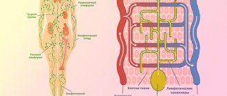
A brief anatomical excursion.
Undoubtedly, you know from the school biology course that the “framework” of our body is bones, which serve not only as a place for muscle attachment, but also as a “receptacle” for a number of human organs. Thus, applicable to the problem under discussion, the pelvic cavity is formed by the connection of four anatomical formations: two pelvic bones, the sacrum and coccyx (Fig. 1), as well as the ligamentous apparatus, muscles, etc. The pelvic cavity contains: the bladder, rectum, uterus with appendages, ovaries and vagina in women, prostate gland in men and many other anatomical formations, such as blood vessels, nerves, etc.
Let us recall that in the human body, the vessels that supply all organs and tissues with blood are arteries and veins. Arterial blood enriched with oxygen and nutrients flows from the heart to organs and tissues through the arteries. Its movement through the vessels is primarily due to the work of the heart - a kind of “pump” that pumps a huge amount of blood per day. In turn, venous blood, coming from the most distant parts of the body to the heart, carries blood poor in oxygen and nutrients, containing large quantities of metabolic products of the body. The movement of venous blood up the vessels is ensured by the joint work of muscles (for example, limbs), which, during their contraction, push venous blood towards the heart, and specific structures of the veins - venous valves (Fig. 2), which do not allow the blood to “submit” to the force gravity and go down.
A feature of the structure of the venous system in the human body is the formation of many plexuses in which the vessels form numerous connections with each other both inside and outside the plexus.
Thus, a large number of plexuses are formed in the pelvic cavity, the largest of which are: vesical, prostatic, rectal, uterine, ovarian, vaginal.
Having somewhat refreshed our knowledge of the structure of the human body, let us dwell on the cause of the development of pelvic congestion syndrome.
The anatomical nature of the development of this syndrome is the weakness of the venous valves and, as a result, incomplete closure of the lumen of the veins when blood returns to the heart against the force of gravity.
The above circumstances cause expansion of the pelvic veins, stagnation of venous blood in the pelvic cavity, and the appearance of corresponding complaints. Causes leading to venous pelvic congestion? First of all, it should be noted that there are primary (not associated with another disease) and secondary, which is a symptom of a disease, pelvic congestion.
The reasons for the development of primary pelvic congestion are still not entirely clear. However, it is believed that its development is genetically predetermined. Thus, its repeatability in a generation is established in 50-70% of cases. First of all, two factors are inherited: the ability of the venous wall to significantly stretch and the insufficient equipment of the veins with valves, with their congenital anatomical inferiority, as mentioned above.
Hormonal influences play a certain role in the development of pelvic congestion, and an increase in the level of estrogen in the blood, which in turn contributes to the expansion of the diameter of the venous vessels of the small pelvis.
Mechanical factors are of no small importance. There is a theory that in the later stages of pregnancy, the pelvic veins are subject to almost 60-fold overload, making them subsequently vulnerable: gradually after childbirth, valve dysfunction and reverse blood flow through the vessel develop (Fig. 3).
Speaking about the secondary nature of pelvic venous congestion, we mean certain structural features of the body and/or diseases that disrupt the outflow of venous blood from the pelvic cavity.
The structural features of the body include the unusual position of the uterus, namely its posterior deviation (retroflexion).
Previous thrombosis (blockage with a blood clot) and/or inflammation of the vein wall (phlebitis) of the vena cava and iliac veins, tumors of the retroperitoneal space, gynecological diseases, which in addition to compression of the veins lead to hormonal imbalance, can be the causes of pelvic venous congestion.
What is the direct cause of pain in pelvic congestion syndrome? The most likely mechanism seems to be that the progressive dilation of the veins and the accumulation of blood in them leads to the release of biologically active substances that have the properties of pain provocateurs (algogens).
The question is also considered that venous congestion itself causes the transmission of a pain impulse to the brain from the periphery. Long-lasting phenomena of stagnation contribute to the development of certain changes in peripheral nerve fibers, which also enhances pathological impulses into the central nervous system, thereby forming a pain component.
How does pelvic venous congestion syndrome manifest?
The manifestations of this syndrome are quite diverse and are represented by the following complaints:
- chronic pelvic pain of a dull, deep, pulling, aching nature, aggravated by postural loads - after long walking, during physical activity and lifting weights, after a long stay in a sitting position. On the contrary, if you lie down, raise the pelvic end, placing a pillow under it, lift legs - pain significantly decreases or disappears;
- feeling of heaviness in the lower abdomen;
- increased pain during and ESPECIALLY after sexual intercourse - this sign is almost obligatory for pelvic congestion syndrome. This circumstance can lead to fear of sexual intercourse (dyspareunia);
- Menstrual irregularities may occur. It is important to emphasize that a characteristic feature of this syndrome is a decrease or complete disappearance of symptoms during menopause (cessation of menstruation);
- in some cases, pain may occur when urinating;
- sometimes excessively heavy menstrual bleeding;
- Depressive and anxiety disorders are very common.
It should be noted that in half of the cases, pelvic venous congestion is combined with varicose veins of the perineum and gluteal region. When the veins of the external genitalia are dilated, a feeling of fullness, itching, pain and burning in the area of the dilated veins is disturbed; swelling of the external genitalia and pain when urinating may appear.
With severe varicose veins, acute thrombosis and inflammation of the vein wall may develop, manifested by intense pain, swelling and redness of the skin. The affected veins are compacted, body temperature is often elevated to 37.5-38.0 ° C. Bleeding from varicose veins of the perineum is not uncommon and can occur on its own, but more often during sexual intercourse or childbirth.
What diagnostic methods are needed for pelvic congestion? The main diagnostic methods are:
- The “gold standard” for diagnosing this disease is pelvic X-ray contrast venography - a method with the introduction of contrast agents into the pelvic veins followed by a series of X-rays, which allows one to obtain a detailed picture of pathologically altered vessels;
- Ultrasound of vessels and pelvic organs:
- traditional - examination through the anterior abdominal wall;
- transvaginal - examination by inserting a device sensor into the vagina.
- CT scan;
- Magnetic resonance imaging is an expensive method, as is computed tomography. Used to exclude/confirm other pathologies of the pelvic organs.
- Laparoscopy is an examination of the abdominal organs and pelvic organs by inserting an endoscope (a device that allows one to assess the condition of organs using vision) through the umbilical ring. Its use is most justified for the simultaneous implementation of therapeutic measures in a number of situations.
How is pelvic venous congestion syndrome treated?
The main recommendation for the treatment of this pathology, as, in general, any other disease, is to avoid self-medication and consult a doctor.
It must be emphasized that pelvic congestion is a chronic disease, with a varied course and individual characteristics for each individual, therefore its treatment is also individual.
There are medical (using drugs) and surgical treatment methods.
If pelvic venous congestion is suspected, treatment begins with medications, physical therapy, compression therapy, and only if such treatment is ineffective (no earlier than 4-6 months) do they decide on surgical treatment.
The most commonly used drugs are the following:
- Phlebotonics - drugs that improve the condition of the vein wall: Detralex, for 2.5-3 months; you can also (Ginkor Fort, Endotelon);
- Agents that improve microcirculation – Pentoxifylline;
- Painkillers - in case of severe pain, use non-steroidal anti-inflammatory drugs: Ibuprofen, Acetylsalicylic acid, Nimesulide. In addition to the analgesic effect, this group of drugs improves blood flow, which also helps to improve the condition;
- Other groups of drugs: antioxidants, drugs for systemic enzyme therapy;
- Antidepressants and anti-anxiety medications – if necessary.
Therapeutic gymnastics and compression treatment, which consists of wearing special therapeutic tights with a high degree of compression, are aimed at improving venous outflow from the pelvic area.
In case of ineffectiveness of drug treatment, therapeutic exercises and compression treatment, surgical treatment is indicated, aimed mainly at “switching off” the affected veins from the blood circulation.
In conclusion, I would like to emphasize that, despite the desire to provide in this article the most complete information about such a disease as pelvic venous congestion syndrome, you should not resort to self-diagnosis and self-medication, since as practice shows, this not only can cause a blow to your health, but also cause a waste of time and material resources.
If you are concerned about your health or the health of those close to you, do not delay visiting a doctor.
Health and prosperity to you and your loved ones!
Complications
Congestion in the pelvis leads to the following problems:
- Reproductive dysfunction;
- Decreased libido;
- Inflammatory processes;
- Haemorrhoids;
- Anorgasmia.
Women cannot become pregnant or bear a healthy fetus and suffer from painful, heavy periods. For men, the pathological condition is the cause of developing impotence and other similar problems.
If the disruption of blood flow turns out to be acute, without medical intervention, tissue necrosis begins, leading to a sharp decrease in organ function. Thrombophlebitis often develops without medical help.
Details about the pelvis
To understand the complexity of stagnation, it is necessary to understand how such a process affects the state of internal organs. Let's go a little deeper into the anatomy.
The small pelvis is the internal space limited by the bony pelvic frame. Women have a naturally wider pelvis than men. The following organs are localized in this space:
- rectum (final section of the gastrointestinal tract);
- bladder.
For women, this space additionally includes:
- uterus - the organ that ensures the bearing of a baby, is localized between the urethra and the rectum;
- vagina - a vaginal “tube” connecting the uterus to the external genital opening;
- ovaries are paired organs that provide the synthesis of sex hormones and regularly produce eggs.
For men, this zone includes:
- prostate gland - an organ that produces secretion for sperm, the gland is located under the bladder;
- seminal vesicle - a small organ located behind the urinary tract, directly above the prostate, it contains seed and, if necessary, throws it out.
All organs are held in anatomically correct places with the help of ligaments. At the same time, they are in close contact with each other. Therefore, any violations in one of them, after some time, affect all the others.
Diagnostics
Ultrasound with Dopplerography or X-ray with a contrast agent (venography) help identify congestion: the machines show that the blood is moving more slowly, the vessels are narrowed or deformed.
Additionally, doctors assess the condition of the organs in the affected area, so they prescribe:
- Ultrasound;
- Radiography;
- MRI;
- CT.
If neoplasms are detected, specialists perform a biopsy followed by microscopy to determine the nature of the tissue.
Laboratory tests help assess the general condition of the body and the functionality of the genitourinary system. Therefore, the patient must pass:
- Blood and urine for general, biochemical analysis;
- Spermogram and prostate juice to determine their quality (men);
- Smear for microflora and cytology (women).
A gynecological examination of women will help determine the condition of the vessels and veins of the vagina.
Treatment of endometritis in women of different ages
Symptoms and treatment of endometritis in women have little to do with age.
After 40 years, acute endometritis usually manifests itself more clearly: this is influenced by hormonal changes due to the upcoming menopause. The transition from acute to chronic at this age occurs much faster, but the format of treatment at 25 and 45 years of age does not differ. Fighting the disease is a difficult but important task, especially for women who have experienced a miscarriage or failed IVF. To make an accurate diagnosis and select individual treatment, MCRM doctors collect a detailed medical history. It is necessary to find out information about menstrual function, the course and outcome of pregnancies, abortions and miscarriages. This will allow you to correctly diagnose the pathology and prepare the endometrium for pregnancy with the help of specially selected drugs.
Complex treatment includes antibiotics with a high ability to penetrate cells, as well as (depending on the type of pathogen) antifungal (antimycotics) and antiviral drugs, local combination drugs, systemic enzyme therapy (use of enzymes) and physiotherapy during rehabilitation. To prevent candidiasis, antifungal drugs are prescribed against the background of antibiotics.
The next stage is hormonal therapy: combined oral contraceptives. In the second phase of the cycle, when planning pregnancy, the doctor prescribes progesterone. Treatment begins on the first day of menstruation.
Before prescribing medications, our clinic undergoes a comprehensive diagnosis:
- ultrasound examination of the pelvic organs;
- hysteroscopy - examination of the inner lining of the uterus (in combination with laparoscopy or alone);
- endometrial examination (obtaining material during hysteroscopy, separate diagnostic curettage or aspiration biopsy);
- immunohistochemical and microbiological examination of the endometrium;
- PCR (femoflor screening): the method will identify pathogenic microflora and the causes of the development of the inflammatory process.
After diagnosis, we select individual treatment. At the first stage, it is necessary to destroy the infectious agent, at the second, to restore the functionality of the endometrium: thanks to treatment, damage is eliminated, regeneration is completed, local hemodynamics (blood movement in the vessels) and the activity of endometrial receptors are restored.
Within the walls of the clinic, we have been developing schemes for many years that help most effectively fight the disease. First of all, the following drugs are prescribed for the entire treatment period (two or three menstrual cycles):
- Monophasic combined oral contraceptives (Yarina, Jess, Zhanin, Femoden, etc.).
- Vitamin therapy: Femibion, Angiovit or Elevit (one capsule per day).
- Immunotherapy: Wobenzym - three capsules three times a day 30 minutes before meals.
Drugs in the first month of treatment:
1. If a pathogen is detected based on the results of femofloral screening, antibacterial or antiviral therapy is prescribed from the first day of the menstrual cycle, depending on the type of pathogen. If a pathogen is not found as a result of femoflor screening, there is no need to take antibiotics!
2. Supporting the normal flora of the gastrointestinal tract and vagina is also important, therefore:
- On the first and last days of taking antibiotics, Diflucan is prescribed: 150 mg orally.
- Drugs that regulate the balance of intestinal microflora, to choose from: Bifiform - one capsule 3 times a day with meals for three weeks,
- Acipol - two capsules 3 times a day for three weeks,
- Enterol - one capsule 2 times a day for 10 days.
- Laktozhinal - one suppository in the vagina at night for two weeks;
Drugs in the second month of treatment:
1. Active immunotherapy: 10 days after menstruation, Genferon is taken - 250 thousand units. twice a day intravaginally.
2. Drugs that activate metabolism in tissues, improve trophism (cell nutrition) and stimulate the regeneration process: Actovegin - one tablet 3 times a day before meals for 30 days.
3. Physiotherapeutic methods have a beneficial effect on the affected tissues, help relieve inflammation, improve the condition of blood vessels and the endometrium.
In the third month of treatment, it is important to consolidate the result: it is best to go to a sanatorium-resort treatment. You can go to the “Baltic Coast” in Zelenogorsk (50 km from the center of St. Petersburg): there are partner doctors who accompany our patients on site. Specialists will know everything about your diagnosis and give timely advice. The cost of an individual program starts from 2060 rubles per day.
Treatment
Gynecologists or urologists work in a group with other specialists. It is important to eliminate not only the consequence, but also the pathology that led to the problem. Endocrinologists, cardiologists, phlebologists, and neurologists are involved in therapy.
Directly to eliminate congestion at the initial stage, doctors use:
- Phlebotropic agents;
- Drugs that ensure normal outflow of venous blood;
- Anti-inflammatory (according to indications);
- Painkillers;
- Vitamin complexes.
Auxiliary ways to reduce congestion are moderate physical activity, special gymnastics (Kegel exercises, for example), and massages.
Physiotherapy, especially electrical stimulation or magnetic stimulation of nerve endings, allows you to restart adequate metabolism.
In difficult situations, specialists choose radical methods of therapy:
- Ovarian vein embolization. Under the influence of special drugs injected into the vein, the walls narrow.
- Sclerotherapy. Also aimed at reducing the lumen of the veins, doctors choose the procedure as an independent method or an addition to embolization.
- Laparoscopy followed by excision of the dilated vein. The operation also allows you to excise adhesions that interfere with normal blood flow.
If the cause of impaired blood flow lies in the prolapse of the pelvic organs, surgeons perform plastic surgery and introduce synthetic implants that hold the pelvic floor.
Treatment of varicose veins of the small pelvis
Effective treatment should be carried out under the supervision of a phlebologist. Therapy includes proper nutrition, exercise, routine, reduced physical activity and compression garments. If necessary, medications are prescribed that improve the condition of the venous walls and normalize their tone, as well as NSAIDs to relieve pain.
If conservative treatment is ineffective, veins are dilated more than ten millimeters, or pain is intractable, surgery is performed.
At the Medical Center you can get qualified help and cure varicose veins using modern methods.
Prevention
Since the onset of a pathological phenomenon is difficult to recognize, it is important to undergo preventive examinations with urologists (gynecologists) and other specialists. Any change in the functioning of the organs of the genitourinary system and other structures should not go unnoticed. Doctors need to be told about all violations.
Prevention measures should also be taken into account:
- To live an active lifestyle. Even when working sedentarily, it is important to take breaks to warm up and exercise. In the evenings, you should give preference to walking, jogging, and swimming.
- Do not abuse alcoholic beverages or smoking.
- Treat acute diseases of the genitourinary system in a timely manner.
- Wear underwear and clothes that fit: they should not be tight.
- Review your diet. Do not indulge in fatty foods or fast food. It is better to focus on plant-based, light foods rich in vitamins.
The information is provided for informational purposes only! If you have questions, we recommend scheduling a consultation with a urologist.
Characteristic symptoms
Blood circulation decreases gradually. Therefore, sudden changes in condition are not typical for pathology. The clinical picture develops gradually and progressively.
The following symptoms are characteristic of blood stagnation in the pelvis:
- Abdominal pain . Discomfort appears in the pelvic area (stomach). It can radiate to the lumbar area, the genital area, the lower limb, and the thigh.
- Increasing pain . Initially, the pain is mild, aching tingling. Over time, the discomfort becomes more intense. In some cases, sudden attacks of pain may appear. They are usually caused by external factors.
- Feeling of pressure . The pain is always accompanied by a feeling of heaviness in the abdomen. Unpleasant pressure increases as discomfort increases.
- Numbness of the lower extremities . Impaired blood flow leads to deterioration of blood circulation in the vascular wall of the legs. Inadequate nutrition underlies tingling, numbness and swelling of the limbs.
- Urinary dysfunction . Stagnation of blood can lead to increased urge to go to the toilet. As the pathology progresses, urinary incontinence gradually develops.
Increased discomfort is observed after playing sports or active physical labor. In women, pain can be triggered by sexual intercourse or menstruation. When lying down, the pain disappears.
Physical therapy
To date, there are no studies examining the role of physical therapy in the treatment of PTS. However, pelvic floor muscle dysfunction and PTS often coexist. Therefore, a person may see a physical therapist who specializes in pelvic dysfunction due to chronic pain or urinary incontinence. The specialist will conduct a thorough assessment and develop a training program based on the patient's impairment. In this sense, physical therapy can be useful because it improves the functioning of the pelvic muscles.