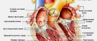
Fetal circulation: mother and child – a single system
In a growing fetus, there are two physiological shunts, i.e. places of communication between circles. Without them, fetal development would be impossible. Blood from the maternal placenta flows through the umbilical vein of the fetus into its inferior vena cava, where, mixing with its venous blood from the lower half of the body, it fills the right atrium. From here the main flow goes through the open oval window, i.e. a hole (defect) in the interatrial septum into the left atrium and left ventricle and further into the large circle. This is the first physiological, natural shunt. From the left ventricle, part of the blood goes to the aorta and vessels of the head and upper half of the body. And that part of the blood that in the right sections passed into the right ventricle through the tricuspid valve, and then into the pulmonary artery, goes into the descending aorta through the second physiological shunt - the patent ductus arteriosus: the normal connection of the pulmonary artery and the aorta.
These are natural shunts, “shunts to save” the growing fetus. Without them, the fetus is not viable, and when they close prematurely, severe congenital defects occur. In the surgery of congenital heart defects, artificial (temporary or permanent) creation of such shunts is one of the widely used treatment methods. But more on that later.
Both physiological shunts close
normally soon after birth, and then both circulation circles begin to function in the mode in which they will work for the rest of their lives. But let’s assume that, in addition to the natural communication between the systemic and pulmonary circulation at the capillary level, something else remains, for example, through a hole in the interventricular or interatrial septum, or in the form of an unclosed arterial duct.
If such a message remains, then the blood flow from any chamber appears in two ways: one is normal, i.e. provided by nature, the second is through a defect or through an open shunt. The blood will partially flow along the second path, since it is easier there, there, in a small circle, there is much less resistance. A shunt is formed “from left to right”: from a large circle to a small one.
Reset from left to right
When a certain volume of blood with each contraction deviates from the normal path and goes from the left to the right, then naturally two problems arise: a lack of blood in the large circle and an overflow of the small circle. The large circle does not suffer: complex compensation mechanisms are quickly activated. But the small circle has a harder time.
a certain amount to live, much less grow.
oxygen, which must be
constantly
, and this delivery is carried out by the heart.
At first, it copes with this, although the conditions in which it must work are far from normal. Its right chambers (depending on the level at which there is communication - atrial, ventricular or great vessels) are filled with blood, increasing in size. The lungs also become filled with blood due to the expansion of their large and small arteries. The left departments also do not remain unaffected, because they perform the work partially in vain. A “vicious circle” arises - an expression that most closely matches our situation. At the same time, the blood entering the systemic circulation is completely saturated with oxygen
, and the color of the child’s skin and mucous membranes is normal.
Right to left shunt and cyanosis
Now let's imagine the opposite situation. Venous, dark blood, which has given oxygen to the tissues, somehow, bypassing the lungs, enters the left side of the heart, the aorta and the arterial system. In other words, in a born child, blood circulates like a fetus, i.e. without a small circle and breathing lungs. But the maternal placenta is no longer there, and with it, there is no source of oxygen. If there are no open paths of communication between the circles, and venous blood is not oxidized anywhere and mixed with arterial blood, then life is impossible, and the child will not be viable. Fortunately, this happens very rarely. But, if there is a message, then through it part of the blood still gets into the small circle and into the lungs, the other part will remain undersaturated. This will be expressed in cyanosis of the skin and mucous membranes. The degree of cyanosis can be very different, as well as the time of its visible manifestation. It can be slightly noticeable or pronounced. Sometimes only others and doctors notice it. The degree of cyanosis depends on the amount of blood that passes through the lungs, and on the degree of its mixing with undersaturated blood in the cavities of the heart, i.e. on the size and level of defects in its septa, as well as on the resistance to blood flow on the way from the heart to the pulmonary arteries and alveoli. The greater this resistance, the less venous blood will enter the small circle and end up in the arteries, and the larger the defect in size, the better the blood will mix in the cavities and the less “cyanosis” will be.
After the birth of a child, the heart, as with defects with a left-to-right reset, works under overload, especially its right sections, and we will talk about this when we describe individual defects. But here we want to emphasize that the very existence of cyanosis can be dangerous, since insufficient oxygen content in arterial blood causes its thickening, an increase in the number of red blood cells and can lead to blockage of small vessels of the body, including the brain, with all the ensuing consequences.
Concept of cross dumping
In some situations, when the septal defects are large enough and the resistance to blood flow is almost the same at the exit of both ventricles, blood may partially flow through the defect in both directions during different phases of the cardiac cycle. That is, at some period of time during one contraction there is a reset from left to right, and in another period during the same cycle, but after a few fractions of a second there is a reset from right to left.
In such cases, they speak of “cross-discharge,” and the degree of oxygen desaturation of the arterial blood will depend on the predominant direction of blood flow. Accordingly, the degree of cyanosis will be visible and pronounced.
Let us say here that defects with such “cross-discharge” most often include very complex, combined defects, including combinations of different disorders of heart development.
Obstructions to blood flow
Congenital obstructions to normal blood flow usually occur due to abnormal development at the junctions of the heart chambers with each other or with the great vessels. Most often this applies to valves. The narrowing is called “stenosis” if it is caused by changes in the valves, and when it concerns the aorta, it is called “coarctation”.
We will discuss this in detail below, but here I would like to note a few points regarding blood flow. Since by the eighth week of intrauterine life of the fetus the heart is basically formed and blood circulation is already occurring, the effect of narrowing and obstruction of normal blood flow affects the early stages of embryo development. If there are no more defects, then the ventricles have to work with increased load, which will result in thickening of the walls, reduction in the size of the cavity, and underdevelopment of the heart chambers. After birth, these phenomena only progress and can become life-threatening already in the first days of a child’s life.
If such obstacles are combined with defects in the septum, then it is easier for the heart to work, because there are other paths for blood in which there is less resistance and the flow chooses such paths of less resistance.
But we have already come close to the classification of vices, i.e. to what defects there are and what happens to the child, whether the heart copes with them and how.
Quoted from the book by G. E. Falkovsky, S. M. Krupyanko. The heart of a child. A book for parents about congenital heart defects
How to get treatment at the Scientific Center named after. A.N. Bakuleva?
Online consultations
- 1. Anatomical and physiological features of the circulatory system. Research methodology
- 2. Blood circulation of the fetus and newborn
- 3. Semiotics of damage to the circulatory system. Research methodology
- 4. Semiotics of the main lesions of the cardiovascular system
- 5. Semiotics and syndromes of damage to the cardiovascular system in children of the first year of life
LECTURE No. 9. The circulatory system of the fetus and newborn. Lesions and methods of studying the organs of the cardiovascular system
1. Anatomical and physiological features of the circulatory system. Research methodology
The weight of the heart in a newborn is 0.8% of body weight, which is more than in adults. The right and left ventricles are approximately equal to each other. The thickness of their walls is about 5 mm. In children, the left ventricle grows more rapidly, the growth of which is stimulated by increasing vascular resistance and blood pressure. In parallel with the growth of the heart, the size of the great vessels also increases. The circumference of the trunk of the pulmonary artery in children is greater than the circumference of the trunk of the ascending aorta. The lumen of the arteries narrows with age relative to the size of the heart and increasing body length.
During the first years of life and adolescence, a series of twists and movements of the heart occur within the chest.
The pulse of newborns is arrhythmic, with unequal duration and unevenness of individual pulse waves and the intervals between them. The pulse in children of all ages is more frequent than in adults, this is explained by a more intense metabolism and the relatively late development of the vagal innervation of the heart.
Children with sufficient physical activity have a lower heart rate than their peers with physical inactivity. The increase in pressure occurs more intensely in the first 2–3 years of life and during puberty.
With age, stroke and minute blood volume increases, and total peripheral resistance decreases.
Methods for studying the cardiovascular system in children
When examined in children with heart disease, various deformations of the chest may be detected in the form of swelling in the area of the heart (heart hump). Circulatory failure is characterized by cyanotic coloration of the distal parts of the extremities: palms, feet, fingertips. When examined, in most children one can see the apical impulse in the form of a weak pulsation in the IV (in older children - in the V) intercostal space, slightly outward from the midclavicular line or on it. Its area does not exceed 1 cm2. By palpation, you can determine the location and assess the extent (localized or diffuse) of the apex beat.
Palmar or digital palpation can determine the symptom of the so-called cat purring, which is observed with stenoses. Palpation of peripheral arteries allows one to judge the characteristics of their pulsation and, to some extent, the condition of the vessel wall. The pulse is examined by palpation in the radial, temporal, carotid, popliteal, posterior tibial, femoral arteries, and on the artery of the dorsum of the foot. Palpation evaluates such properties of the pulse as rhythm, tension, filling, size, uniformity, shape, the number of vibrations of the vascular wall per heart contraction, and a decrease in the pulse during exhalation (paradoxical pulse).
Using chest percussion, the boundaries of the heart are determined.
Auscultation. Listening to the child's heart is carried out in various positions, preferably at the height of inspiration while holding the breath and exhaling completely. Listen with special pediatric flexible stethoscopes with a small bell diameter - no more than 20 mm. Heart sounds in children are distinguished by great sonority and clarity. Heart murmurs in children are distinguished by intensity (loudness), timbre, duration, zone of maximum audibility, and connection with systole or diastole.
To measure blood pressure using the Korotkoff method, cuffs corresponding to the child’s age or shoulder circumference are used. The cuff is placed on the shoulder so that it fits tightly enough to the skin, allowing 1-2 fingers to pass under it; the edge of the cuff should be 2 cm from the ulnar fossa.
The measurement is carried out with the child sitting. The child's hand lies on the table and is turned palm up with complete muscle relaxation. Air is pumped to a pressure value of 30–40 mmHg. Art. exceeding the pressure at which the cessation of arterial pulsation was noted. After this, a stethoscope is installed and they begin to slowly reduce the pressure in the cuff, strictly controlling the moment of the first appearance of tones, and then their complete disappearance. This procedure is repeated three times and the lowest pressure obtained is recorded as the result.
2. Blood circulation of the fetus and newborn
The main blood circulation of the fetus is chorionic, represented by the vessels of the umbilical cord. Chorionic (placental) blood circulation begins to ensure fetal gas exchange already from the end of the 3rd – beginning of the 4th week of intrauterine development. The capillary network of chorionic villi of the placenta merges into the main trunk - the umbilical vein, which runs as part of the umbilical cord and carries oxygenated and nutrient-rich blood. In the fetal body, the umbilical vein goes to the liver and, before entering the liver, through the wide and short ductus venosus (Arantius) gives a significant part of the blood to the inferior vena cava, and then connects with the relatively poorly developed portal vein. After passing through the liver, this blood enters the inferior vena cava through the system of recurrent hepatic veins. The blood mixed in the inferior vena cava enters the right atrium. Purely venous blood from the superior vena cava also flows here, flowing from the cranial areas of the body. At the same time, the structure of this part of the fetal heart is such that complete mixing of the two blood streams does not occur here. Blood from the superior vena cava is directed primarily through the right venous opening into the right ventricle and pulmonary artery, where it bifurcates into two streams, one of which (smaller) passes through the lungs, and the other (larger) through the arterial duct enters the aorta and is distributed between the lower segments of the fetal body.
Blood entering the right atrium from the inferior vena cava enters predominantly into the wide gaping foramen ovale and then into the left atrium, where it mixes with a small amount of venous blood that has passed through the lungs and enters the aorta to the junction of the ductus arteriosus, providing better oxygenation and trophism of the brain, coronary vessels and the entire upper half of the body.
The blood of the descending aorta, which has given up oxygen, returns through the umbilical arteries to the capillary network of the chorionic villi of the placenta. In this way, the circulatory system functions, which is a vicious circle, separate from the mother’s circulatory system, and operating exclusively due to the contractility of the fetal heart. The viability of the fetus depends on its supply of oxygen and the removal of carbon dioxide through the placenta into the maternal circulation. The umbilical vein carries oxygenated blood only to the inferior vena cava and portal veins. All fetal organs receive only mixed blood. Blood circulation of a newborn
At birth, a restructuring of blood circulation occurs, which is extremely acute. The most significant points are the following:
1) cessation of placental circulation;
2) closure of the main fetal vascular communications (venous and arterial duct, oval window);
3) switching the pumps of the right and left hearts from parallel to sequentially connected;
4) inclusion in full of the vascular bed of the pulmonary circulation with its high resistance and tendency to vasoconstriction;
5) increased oxygen demand, increased cardiac output and systemic vascular pressure.
With the onset of pulmonary respiration, blood flow through the lungs increases almost 5 times, and vascular resistance in the pulmonary circulation decreases 5-10 times. The entire volume of cardiac output flows through the lungs, while in the prenatal period only 10% of this volume passed through them. Due to a decrease in resistance in the pulmonary bed, an increase in blood flow into the left atrium, and a decrease in pressure in the inferior vena cava, a redistribution of pressure in the atria occurs, and the shunt through the oval window ceases to function.
Immediately after the first breath, under the influence of the partial pressure of oxygen, a spasm of the ductus arteriosus occurs. However, the duct, which is functionally closed after the first respiratory movements, can open again if the efficiency of breathing is impaired. Anatomical closure of the ductus arteriosus occurs later (in 90% of children by the 2nd month of life). Due to the cessation of blood circulation, blood flow through the venous duct also stops, which becomes obliterated. The small (pulmonary) and systemic circulations begin to function.
3. Semiotics of damage to the circulatory system. Research methodology
To assess the state of the cardiovascular system, an ECG study is used. The technique of taking an ECG, the lead system and the theoretical basis of the method are common to all ages. However, the interpretation of ECG results in children is more complex due to age-related differences in individual ECG indicators.
The P wave reflects the spread of excitation in the atrial myocardium. The first half of the wave up to its apex corresponds to the excitation of the right atrium, the second - to the left. The duration of the P wave in healthy children does not exceed 0.1 s.
In standard lead III, the wave can be negative, biphasic or smoothed.
The P – Q or P – R interval includes the P wave and the iso-electric line from the end of the P to the Q, or R wave. The interval changes with the pulse rate, and its proper normal values are estimated from the tables.
The Q wave is the most variable element of the pediatric ECG. Often in healthy children there is a deep Q wave in lead III.
The R wave is always directed upward (except in cases of congenital dextracardia).
Newborns are characterized by fluctuations in tooth height within the same lead (electrical alternation).
The S wave is an intermittent negative wave. At an early age it is often deep in standard lead I.
The ventricular QRS complex, reflecting the spread of excitation in the ventricular myocardium (depolarization) and the extinction of this excitation (repolarization), has a total duration in children that does not exceed 0.35-0.40 s and is closely related to the heart rate.
Electrical axis of the heart. It is determined by the degree of unilateral dominance of the electrical activity of the ventricles and the position of the heart in the chest.
In the chest leads, the ratio of the R and S waves changes significantly with age, this is due to the rotation of the heart that occurs in the first years of life and changes in the degree of adherence of the right ventricle to the surface of the chest.
A phonocardiogram allows you to objectively evaluate heart sounds and identify additional murmurs. In the structure of the first tone there are three components, in the structure of the second tone there are two. The first component of the first tone is muscular and characterizes atrial contraction; the second component is caused by the tension of the closed valves, the atrioventricular valves, and the final low-amplitude component is caused by the vibration of the ventricular myocardium, the walls of the aorta and the pulmonary artery. A feature of FCG in children is the relatively high frequency of registration of the third heart sound, which is recorded at low frequencies with a predominant detection at the apex of the heart (in 60–70% of children). IV (atrial) heart sound, also low-frequency, is often recorded.
It is detected mainly in the third intercostal space to the left of the sternum in 1/3 of children. The V sound is recorded in 6% of children above the apex of the heart. A special feature is also the high frequency of detection of small, or functional, noises.
In preschool children, systolic murmurs are the most common.
An x-ray allows you to evaluate the size and shape of the heart.
Vectorcardiography is the recording of the electrical field of the heart on the screen of a cathode ray tube.
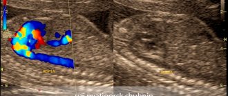
Echocardiography is an ultrasound examination of the heart, which allows you to obtain data on the size of the cavities of the heart, the thickness of its walls and septa, the size of the aorta, pulmonary artery, observe the movement of the valve leaflets, their shape, see the sagging of the valve leaflets, etc. Also, using this study, you can identify inflammatory effusion in the pericardial cavity and intracardiac tumors or thrombi.
Sphygmography is the recording of the movement of the arterial wall that occurs under the pressure of a blood wave with each contraction of the heart.
Rheocardiography is a method of studying blood circulation, which is based on recording pulse fluctuations in the resistance of the human body to alternating electric current of sound frequency.
Functional tests of the cardiovascular system. To assess the functional state of the cardiovascular system, dosed physical activity is used, which makes it possible to judge the adequacy of the cardiovascular system’s response to each of them.
4. Semiotics of the main lesions of the cardiovascular system
Myocarditis is an inflammatory lesion of the heart muscle. Most often, myocarditis occurs with rheumatism, as well as with infectious diseases (infectious-allergic myocarditis).
Myocarditis occurs most severely in children during the neonatal period and the first weeks of life. The incidence of myocarditis increases at school age.
When examining the heart, a significant expansion of the boundaries of relative cardiac dullness, muffling of heart sounds, especially the first tone, and increased heart rate (tachycardia) are recorded.
A systolic murmur is heard at the apex of the heart. ECG abnormalities occur: a decrease in T wave voltage and a decrease in the S-T interval, conduction disturbances are detected - sinoauricular, ventricular, intraventricular blockade, and there may be extrasystole.
Myocarditis can leave behind disturbances in myocardial contractility or persistent forms of arrhythmias.
Endocarditis is inflammation of the inner lining of the heart.
This group also includes inflammatory lesions of the valves - valvular endocarditis, or valvulitis. The most common forms of endocardial damage are rheumatic and infectious (bacterial or septic) endocarditis. Rheumatic endocarditis occurs after a child has suffered an acute streptococcal infection (tonsillitis) or an exacerbation of chronic tonsillitis.
Pericarditis is relatively rarely observed as an isolated inflammatory lesion of the pericardium. More often, pericarditis accompanies other inflammatory lesions of the heart - myocarditis or endomyocarditis. Depending on the amount of effusion in the pericardial cavity, it can be dry or exudative. The latter, according to the nature of the effusion, are divided into serous, hemorrhagic and purulent. On auscultation, the main symptom is weakening of tones and pericardial friction noise.
The ECG reveals a sharp decrease in voltage and a shift of the S-T interval up from the isoline, characteristic of pericarditis.
Bicuspid valve insufficiency. A weakening of the tone is heard at the apex of the heart, an increase in the second tone at the pulmonary artery and a systolic murmur at the apex of the heart or at the V point.
Narrowing of the left venous orifice. The apical impulse is weakened, and a “cat’s purr” is palpated. A loud and short (popping) first sound and diastolic murmur at the apex of the heart are heard. The emphasis of the second tone on the pulmonary artery is determined, often its bifurcation or splitting.
Combined mitral disease is characterized by the dominance of clinical and instrumental signs of one type of defect, usually mitral regurgitation, in combination with less pronounced manifestations of orifice stenosis, in particular the presence of a typical murmur of mitral stenosis above the apex of the heart.
Aortic valve insufficiency. The apical impulse upon palpation is strengthened and shifted outward and downward. The borders of the heart are expanded to the left.
At the apex, a weakening of the first tone is heard, at the base, or in the III–IV intercostal spaces to the left of the sternum, a proto-diastolic murmur is heard.
The noise is often quiet, gentle, flowing, and is better heard in a standing position with the body tilted forward.
Tricuspid valve insufficiency. Severe shortness of breath and weakness, upon examination - cyanosis of the lips, face, limbs, pulsation of the neck veins, epigastric pulsation, expansion of the right border of the heart. A systolic murmur is heard over the lower part of the sternum.
In case of heart lesions, the ECG can reveal the following rhythm changes: sinus tachy- and bradycardia, atrioventricular rhythm, coronary sinus rhythm, migration of the rhythm source, linked rhythm, sinoauricular block, intraatrial block, atrioventricular block, complete and incomplete atrial and ventricular block, peduncle block His bundle, extrasystole, paroxysmal tachycardia, atrial fibrillation.
Also, an ECG can be used to diagnose hypertrophy of the right and left atria and hypertrophy of the right and left ventricles.
5. Semiotics and syndromes of damage to the cardiovascular system in children of the first year of life
A ventricular septal defect is characterized by a rough systolic murmur along the left edge of the sternum with a maximum in the 4th intercostal space, at the left edge of the sternum, but usually not carried into the left axillary region, with a wide area of irradiation in the region of the heart. Percussion reveals an increase in the size of the heart to the right and left; X-ray - enlargement of the right and left ventricles of the heart, increased pulmonary pattern due to overflow of the pulmonary circulation; ECG - hypertrophy of both ventricles of the heart.
A patent ductus arteriosus is manifested by a continuous systolic-diastolic murmur in the 2nd–3rd intercostal spaces at the left edge of the sternum.
In newborns, only the systolic component of the noise and an intensified second sound on the pulmonary artery are heard. X-rays show an increase in the size of the heart mainly due to its left parts, bulging of the pulmonary artery arch and overflow of the pulmonary vessels; on the ECG - hypertrophy of the left parts of the heart or both ventricles.
Atrial septal defect is characterized by moderate systolic murmur in the 2nd intercostal space to the left of the sternum, accentuated 2nd tone on the pulmonary artery. Percussion reveals expansion of the boundaries of the right atrium and ventricle, overflow of the vessels of the pulmonary circulation, and on the ECG - deviation of the electrical axis to the right, hypertrophy of the right ventricle.
Tetralogy of Fallot is manifested by cyanosis, dyspnea-cyanotic attacks, systolic murmur along the left edge of the sternum. The intensity of the noise is inversely proportional to the severity of the defect; the second tone on the pulmonary artery is weakened. X-rays show a depleted pulmonary pattern, a small heart in the shape of a boot, a sunken pulmonary artery arch, and an ECG showing hypertrophy of the right ventricle.
Transposition of the great vessels is characterized by general cyanosis from birth; There may be no murmur, or the systolic murmur of a concomitant septal defect or pulmonary stenosis may be heard. X-rays show enlargement of the right parts of the heart, often the heart is in the form of an egg lying on its side, narrowing of the vascular bundle in a direct projection, and ECG shows hypertrophy of the right ventricle.
Hypoplasia of the left ventricle of the heart is usually diagnosed in the neonatal period, since patients rarely live more than one month. It is characterized by severe shortness of breath with a frequency of up to 100 breaths per minute, gray skin tone, and acrocyanosis.
The pulse is sharply weakened, the heartbeat is sharply increased, murmurs over the heart area may not be audible.
X-ray shows an overflow of the pulmonary circulation due to the venous bed, a huge shadow of the heart due to the right sections, and an ECG shows hypertrophy of the right ventricle.
Pulmonary artery stenosis is manifested by a rough systolic murmur with a maximum in the 2nd–3rd intercostal spaces along the left edge of the sternum, a sharp weakening or absence of the 2nd tone in the pulmonary artery. Attacks of cyanosis may occur due to the discharge of blood from the right side through the oval window. Percussion and x-ray shows a significant enlargement of the heart due to the right parts, and the ECG shows hypertrophy of the right ventricle and right atrium.
Aortic stenosis is characterized by pallor of the skin, weak peripheral pulse, systolic murmur in the second intercostal space to the right of the sternum, amplified by the apical impulse. Percussion and radiological examination indicate an enlargement of the heart due to the left ventricle, and an ECG shows hypertrophy of the left ventricle.
Coarctation of the aorta is manifested by severe shortness of breath and an abundance of moist rales in the lungs, characteristic of this defect in young children. The murmur over the heart area may not be heard or may be heard on the left or right of the sternum in the 2nd intercostal space. Sometimes this noise is heard only on the back. The main signs of the defect are a sharp weakening of the pulse and a decrease in blood pressure in the legs.
The pulse in the arms is full and high, the blood pressure in the arms is normal or elevated. X-rays show an increase in the size of the heart due to the left or right parts, an increase in the pulmonary pattern, and an ECG shows hypertrophy of one left or both ventricles of the heart.
Table of contents