The heart is a muscular pump that ensures continuous movement of blood through the vessels. Together, the heart and blood vessels make up the cardiovascular system. This system consists of the systemic and pulmonary circulation. From the left side of the heart, blood first moves through the aorta, then through large and small arteries, arterioles, and capillaries. In the capillaries, oxygen and other substances necessary for the body enter the organs and tissues, and from there carbon dioxide, metabolic products, are removed. After this, the blood turns from arterial to venous and again begins to move towards the heart. First along the venules, then through smaller and larger veins. Through the inferior and superior vena cava, blood again enters the heart, only this time into the right atrium. A large circle of blood circulation is formed.
Venous blood from the right side of the heart is sent through the pulmonary arteries to the lungs, where it is enriched with oxygen, and returns to the heart again - this is the pulmonary circulation.
Inside, the heart is divided by partitions into four chambers. The two atria are divided by the interatrial septum into the left and right atria. The left and right ventricles of the heart are separated by the interventricular septum. Normally, the left and right parts of the heart are completely separate. The atria and ventricles have different functions. The atria store blood that flows into the heart. When the volume of this blood is sufficient, it is pushed into the ventricles. And the ventricles push blood into the arteries, through which it moves throughout the body. The ventricles have to do more hard work, so the muscle layer in the ventricles is much thicker than in the atria. The atria and ventricles on each side of the heart are connected by the atrioventricular orifice. Blood moves through the heart in only one direction. In the systemic circle of blood circulation from the left side of the heart (left atrium and left ventricle) to the right, and in the small circle from the right to the left.
The correct direction of blood flow is ensured by the valve apparatus of the heart:
Valves:
- tricuspid
- pulmonary
- mitral
- aortic
They open at the right time and close, preventing blood flow in the opposite direction.
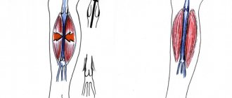
Aortic valve
Closes the entrance to the aorta. It also consists of three valves, which look like crescents. Opens when the left ventricle contracts. In this case, blood enters the aorta. When the left ventricle relaxes, it closes. Thus, venous blood (poor in oxygen) from the superior and inferior vena cava enters the right atrium. When the right atrium contracts, it moves through the tricuspid valve into the right ventricle. Contracting, the right ventricle ejects blood through the pulmonary valve into the pulmonary arteries (pulmonary circulation). Enriched with oxygen in the lungs, the blood turns into arterial blood and moves through the pulmonary veins to the left atrium, then to the left ventricle. When the left ventricle contracts, arterial blood enters the aorta through the aortic valve under high pressure and spreads throughout the body (systemic circulation).
The heart muscle is called the myocardium
There are contractile and conductive myocardium. The contractile myocardium is the actual muscle that contracts and produces the work of the heart. In order for the heart to contract in a certain rhythm, it has a unique conduction system. The electrical impulse to contract the heart muscle occurs in the sinoatrial node, which is located in the upper part of the right atrium and spreads through the conduction system of the heart, reaching every muscle fiber
First, both atria contract, then both ventricles, thereby ensuring the flow of blood to all organs and tissues of the body. The heart muscle has two membranes (external and internal). The inner lining of the heart is called the endocardium. The outer lining of the heart is called the pericardium.
Circulation circles
From previous articles you already know the composition of blood and the structure of the heart. It is obvious that the blood performs all functions only thanks to its constant circulation, which is carried out thanks to the work of the heart. The work of the heart resembles a pump that pumps blood into the vessels through which blood flows to the internal organs and tissues.
The circulatory system consists of a large and small (pulmonary) circulation, which we will discuss in detail. They were described by William Harvey, an English physician, in 1628.
Systemic circulation (BCC)
This circulatory system serves to deliver oxygen and nutrients to all organs. It begins with the aorta, the largest vessel, emerging from the left ventricle, which sequentially branches into arteries, arterioles and capillaries. The famous English scientist, physician William Harvey discovered the BCC and understood the meaning of the blood circulation.
The capillary wall is single-layered, so gas exchange occurs through it with surrounding tissues, which also receive nutrients through it. Respiration occurs in the tissues, during which proteins, fats, and carbohydrates are oxidized. As a result, carbon dioxide and metabolic products (urea) are formed in the cells, which are also released into the capillaries.
Venous blood collects through venules into veins, returning to the heart through the largest - the superior and inferior vena cava, which flow into the right atrium. Thus, BCC begins in the left ventricle and ends in the right atrium.
Blood passes through the BCC in 23-27 seconds. Arterial blood flows through the arteries of the BCC, and venous blood flows through the veins. The main function of this circulation is to provide oxygen and nutrients to all organs and tissues of the body. In the vessels of the BCC there is high blood pressure (relative to the pulmonary circulation).
Pulmonary circulation (pulmonary)
Let me remind you that the BCC ends in the right atrium, which contains venous blood. The pulmonary circulation (PCC) begins in the next chamber of the heart - the right ventricle. From here, venous blood enters the pulmonary trunk, which divides into two pulmonary arteries.
The right and left pulmonary arteries with venous blood are directed to the corresponding lungs, where they branch to capillaries intertwining the alveoli. Gas exchange occurs in the capillaries, as a result of which oxygen enters the blood and combines with hemoglobin, and carbon dioxide diffuses into the alveolar air.
Oxygenated arterial blood collects in venules, which then drain into the pulmonary veins. The pulmonary veins with arterial blood flow into the left atrium, where the ICC ends. From the left atrium, blood flows into the left ventricle, the site of origin of BCC. Thus, two circles of blood circulation are closed.
ICC blood passes through in 4-5 seconds. Its main function is to saturate venous blood with oxygen, as a result of which it becomes arterial blood rich in oxygen. As you noticed, venous blood flows through the arteries in the ICC, and arterial blood flows through the veins. Blood pressure here is lower than BKK.
Interesting Facts
On average, a person’s heart pumps about 5 liters every minute, and over 70 years of life - 220 million liters of blood. In one day, the human heart makes approximately 100 thousand beats, over a lifetime - 2.5 billion beats.
© Bellevich Yuri Sergeevich 2018-2021
This article was written by Yuri Sergeevich Bellevich and is his intellectual property. Copying, distribution (including by copying to other sites and resources on the Internet) or any other use of information and objects without the prior consent of the copyright holder is punishable by law. To obtain article materials and permission to use them, please contact Yuri Bellevich
.
Conduction system of the heart
The heart, like any organ, has its own nervous system. The nervous system of the heart has several levels. The first and main pacemaker of the heart is the sinus node, located in the right atrium. It is subject to the atrioventricular node, which is located on the border between the atria and ventricles and quite often slows down the heart rate set by the sinus node. Then the nerve impulse goes to the ventricles of the heart along the branches of the Hiss bundle, which are divided into the smallest nerve endings - Purkinje fibers.
Pulmonary hypertension
Pulmonary hypertension is a rare disease that affects the lungs. The pulmonary arteries are involved in this condition. If left untreated, this disease can lead to heart failure.
Features of the pulmonary arteries?
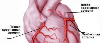
The group of arteries that supply blood to the lungs are called pulmonary arteries. These arteries are located on the right side of the heart. These large blood vessels carry blood from the right side of the heart to the lungs. When the blood pressure within these arteries is high, it results in pulmonary hypertension.
What is pulmonary hypertension?
Increased pressure in the pulmonary artery leads to pulmonary hypertension. The heart consists of an upper and lower chamber and two sides on the left and right, and septa on the muscular wall. The lower right chamber is called the right ventricle, which helps pump blood to the pulmonary arteries. These arteries carry blood to the lungs, where it becomes rich in oxygen. Likewise, the left atrium receives oxygen-rich blood from the lungs. This blood enters the lower left chamber, the left ventricle.
Now, when the walls of the pulmonary arteries are tightened, narrowed or blocked due to the presence of blood clots, it becomes very difficult for the heart to pump blood to the lungs.
The difficulty in pumping blood is the increase in pressure in the arteries. This leads to the development of pulmonary hypertension.
Causes of pulmonary hypertension
As mentioned above, blood flows through the arteries from the right side of the heart to the lungs. When certain changes occur in the arteries, it leads to an increase in pressure. These changes include narrowing, hardening of the wall, or blockage of the pulmonary artery.
The cause of these changes is mainly idiopathic. This means that the exact cause or factor leading to pulmonary hypertension is unknown. In this case, it is called idiopathic pulmonary hypertension. It is believed that there is a gene involved in this disease that predisposes some people to pulmonary hypertension. Idiopathic pulmonary hypertension is a rare disease.
Secondary pulmonary hypertension is a disease caused by concomitant diseases. This is the most common type of pulmonary hypertension.
Medical conditions that may lead to secondary pulmonary hypertension:
- Pulmonary embolism
- Scleroderma
- Lupus
- Congenital heart defects
- Sickle cell anemia
- Schistosomiasis
- Hemolytic anemia
- Systolic or diastolic dysfunction
- Valve diseases
- Chronic obstructive pulmonary disease (COPD)
- Collagen lung disease
- People living in mountainous areas for a long time
- Sleep disorders such as sleep apnea
- Gaucher disease
- Thyroid diseases
- Chronic renal failure
- Splenectomy, etc.
Symptoms of pulmonary hypertension
Signs and symptoms of pulmonary hypertension are not observed during the initial stages of the disease. Symptoms usually occur as the condition worsens.
Some symptoms include:
- Fatigue
- Dyspnea
- Angina pectoris
- Swelling around the ankles and feet
- Fainting
- Cough
- Cyanosis
- Heartbeat feeling
Complications of pulmonary hypertension.
Pulmonary hypertension can lead to various complications.
These include:
- Internal bleeding in the lungs. You should immediately consult a doctor if you notice coughing or vomiting blood, as this can lead to the death of the patient.
- Rapid heartbeat, dizziness, fainting are signs of arrhythmia. These are other complications of pulmonary hypertension.
- Formation of blood clots in the arteries, leading to increased pressure in the pulmonary artery.
- Enlargement of the right ventricle as it becomes harder to work to pump blood to the lungs. Soon, the right chamber becomes deformed and this leads to right-sided heart failure.
It becomes harder for the heart to pump blood as pressure increases. This leads to hardening of the affected arteries, which further leads to fibrosis. This leads to right-sided heart failure (cor pulmonale). Over time, blood flow to the lungs decreases. This results in a decrease in the oxygen-carrying capacity of the blood. Thus, patients usually suffer from shortness of breath and fainting, due to insufficient oxygen supply to the body. With intense physical exertion, this can lead to a heart attack, which often leads to death.
Diagnosis of pulmonary hypertension
It is very difficult to diagnose pulmonary hypertension in the initial stages. Only when a patient begins to experience symptoms that indicate heart or lung disease does the doctor perform tests to rule out pulmonary hypertension.
First, physical examination helps in pointing out the characteristic signs of pulmonary hypertension. These include cardiac sound abnormalities, pulmonary regurgitation, increased jugular venous pressure, peripheral edema, abdominal edema, etc.
Diagnostic tests to confirm pulmonary hypertension include:
- Blood tests to rule out autoimmune diseases, HIV, or liver disease.
- Electrocardiography
- Arterial blood gas measurements
- Pulmonary function test Pulmonary function test
- Breast x-ray
- CT scan
- Transesophageal echocardiography
- Right heart catheterization
- Lung biopsy
Treatment of pulmonary hypertension
Pulmonary hypertension has no specific cure, but treatment aims to control symptoms and slow the progression of the disease. Treatment depends on the severity of the individual condition.
It includes:
- Medicines like diuretics, anticoagulants, vasodilators, calcium channel blockers, etc. Medicines like tadafil and sildenafil help in the treatment of pulmonary hypertension.
- Surgery is indicated in some cases of severe pulmonary hypertension. Atrial septostomy helps in opening the chambers of the heart and reducing pressure on the heart.
Lifestyle changes for pulmonary hypertension
You can control symptoms by making lifestyle changes.
These changes include
- Avoid strenuous physical activity and get plenty of rest. This will reduce fatigue and episodes of shortness of breath.
- Quit smoking as it puts extra pressure on the heart and lungs.
- Eat healthy foods and reduce salt intake in your diet.
- Yoga and meditation can help you stay calm, reduce stress and improve your quality of life.
- Avoid driving to places at high altitudes. Those living in such areas should try to move to regions with lower atmospheric pressure.
Pulmonary hypertension, if left untreated, can cause death within 3 years of diagnosis. However, today, with improved treatment options, quality of life has increased dramatically. Women of childbearing age with pulmonary hypertension are asked to avoid pregnancy. This is because they have a high mortality rate and it also affects the life of their fetus. Thus, such women should follow the advice of their doctor before planning a pregnancy.
Pulmonary hypertension is easy to control today. Medicines and other treatments help slow the progression of the disease. Talk to your doctor for more details.
Arteries
Not a single cell can survive without nutrients and oxygen. They are delivered by arteries. They carry oxygen-rich blood throughout the body. When you breathe, oxygen enters your lungs. where the delivery of oxygen throughout the body begins. First to the heart, then through the systemic circulation to all parts of the body. There, the blood exchanges oxygen for carbon dioxide and then returns to the heart. The heart pumps it back to the lungs, which take in carbon dioxide and give out oxygen, and so on endlessly. There are also pulmonary arteries of the pulmonary circulation, they are located in the lungs and through them blood, poor in oxygen and rich in carbon dioxide, enters the lungs, where gas exchange occurs. This blood then returns to the heart through the pulmonary veins.
Cellular structure of blood
Blood consists of two components: plasma (50-60%) and suspended formed elements (40-50%).
The second category includes:
· Erythrocytes (red blood cells) are the most numerous of the formed elements. According to official studies, one drop of blood contains about 5 million red blood cells. Red blood cells are responsible for transporting gases - oxygen and carbon dioxide. They contain the protein hemoglobin, which binds oxygen molecules in the lungs. Red blood cells deliver oxygen to all tissues and organs, after which they absorb carbon dioxide and carry it to the lungs. It is removed from the body during respiration.
· Leukocytes (white blood cells) - elements that protect our body from foreign bodies and compounds, are part of the immune system. White blood cells recognize and attack pathogens through the production of antibodies and macrophages. When an infection enters the body, the production of leukocytes increases significantly. Normally, their quantity is inferior to the concentration in the blood of other formed elements.
· Platelets (blood platelets) are cells that provide coagulation (clotting) of blood flowing from a damaged vessel and protect the body from heavy blood loss. They stick to the hole in the damaged vessel, forming a “sealing” plug to stop bleeding. It is the platelets that can stick together and form pathological blood clots inside the vessels, called thrombi.
All formed elements are synthesized by the bone marrow and distributed through plasma, the liquid part of the blood.
Blood vessels
Blood vessels have different shapes, structures and volumes, depending on their role in the body.
1. Arteries are the strongest vessels in the human body. Their walls are dense and elastic, consisting of three layers - endothelium, smooth muscle fibers and fibrous tissue. The task of the arteries is to saturate all organs and tissues with blood enriched with oxygen and nutrients. An exception is the arteries of the pulmonary circulation, through which venous blood flows from the heart to the lungs. The largest arterial vessel is the aorta.
2. Veins perform the function of transporting waste blood, saturated with carbon dioxide, back to the heart. This vein fluid is obtained from capillaries. Like an artery, a vein consists of several layers - endothelial, soft connective, dense connective and muscle. Venous walls are several times thinner and more vulnerable than arterial walls. For this reason, as you move away from the heart, the movement of venous blood may be disrupted - the pressure in the capillaries is almost equal to atmospheric pressure, and a normal flow is not created. Therefore, in hemodynamics, the vessels are assisted by the venous valves and the venous pulse.
3. Capillaries are the thinnest vessels, similar in volume to human hair. They are branches of large peripheral arteries. It is through them that tissues and organs are supplied with oxygen and nutrients. They also communicate with the veins to carry cellular waste. Consequently, these tiny vessels are at the same time the breadwinners and caretakers of our body.
Normal blood circulation within the vascular system is ensured by blood pressure.