Many specialists, including neurologists and cardiologists at MedicCity, are confident that MRI scanning of cerebral vessels should certainly be included in the program of mandatory medical examination of the population.
MRI diagnostics allows you to detect pathological changes in blood vessels at the earliest stages. This means that we are able to prevent the development of the most dangerous conditions, such as atherosclerosis, stroke, heart attack, and tumor processes. MRI angiography of cerebral vessels can save your life and the lives of your loved ones!
Diagnostics based on the phenomenon of magnetic nuclear resonance are not only extremely informative and accurate, but also do not have a harmful effect on the body and can be carried out many times.
The study of the condition of cerebral vessels makes it possible today to identify and treat diseases that for many years have robbed people of their mental and physical health at an unacceptably early age.
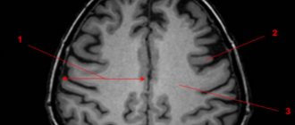
1 MRI of cerebral vessels
2 MRI of cerebral vessels
3 MRI of cerebral vessels
What is the difference between MRI of cerebral vessels, MRI angiography of cerebral vessels and MRI of the brain
Patients often wonder what kind of test they were prescribed and whether different types of diagnostics duplicate each other.
First of all, let's define the terms. When they talk about MRI of cerebral vessels and magnetic resonance angiography of cerebral vessels, we are talking about the same study.
At one time, angiography was quite invasive and required local anesthesia. During the procedure, the patient had a puncture of the carotid artery and a contrast agent was injected into the blood through a special needle. X-rays were then used to take a series of pictures of the area of interest.
The invention of MRI made it possible to improve the method and reduce the number of unpleasant sensations and contraindications. Now the patient does not experience pain, he is not exposed to radiation, and the scan itself has become much more informative.
Due to the fact that MRI angiography provides excellent visualization of the area under study, the study can be performed without a contrast agent. The use of contrast is resorted to in difficult cases, for example, in cancer diagnostics.
1 MRI of the brain
2 MRI of the brain
3 MRI of the brain
MRI angiography of cerebral vessels determines the intensity of blood flow in the arteries, veins and capillaries, helps to identify painful changes at an early stage, confirm or refute an alarming diagnosis, clarify the scope of the upcoming surgical intervention, track the dynamics of recovery after a stroke, etc.
This diagnosis differs from MRI of the brain in the nature of the visualization:
MRI of the brain is not suitable for observing blood vessels, but it makes it possible to see the structure of nervous tissue and brain matter.
MRI angiography depicts the vascular structures of the brain, but not the substance of the brain.
To obtain the most complete picture of structural changes, the techniques can be used simultaneously, complementing each other.
CT angiography: what is it?
Multislice computed tomography is a modern method of radiological diagnosis of the pathology of organs and their systems. During the study, several processes take place simultaneously:
- Forward movement of the table with the patient on it;
- Movement of the X-ray tube in a spiral around the subject being examined;
- Registration of an X-ray image by a sensor located on the opposite side and transferring it to a computer screen.
As a result, the doctor receives a series of layer-by-layer images of the examined area of the body, after which the information is processed by a computer and a two- or three-dimensional image is built. It often happens that after a standard study, contrast enhancement of the vessels of the pathological focus is necessary to determine the nature of its blood circulation - then CT angiography of the vessels appears in the diagnostic plan. This study is aimed at obtaining reliable data on the patency of blood vessels, the direction of their course and disturbances in blood flow.
The medical diagnostic center “Medicine of the Northern Capital” uses a modern multislice tomograph Siemens Somatom installed in 2016.
What does MRI of cerebral vessels show?
With this research you can:
- track post-traumatic changes (possible contusions, hemorrhages, hematomas, etc.);
- detect signs/monitor the consequences of a stroke;
- detect signs of multiple sclerosis;
- identify changes associated with vascular atherosclerosis;
- identify cerebral circulation disorders;
- identify developmental anomalies;
- detect the danger of blood clots in the vessels of the head and neck;
- identify the presence of infection, inflammation, neoplasm, and many others.
1 MRI of cerebral vessels
2 MRI of cerebral vessels
3 MRI of cerebral vessels
How the study is performed
Cerebral angiography is performed in the operating room of the cath lab department. The patient's head is gently fixed to the table with a special tape passing through the frontal area. This is necessary to eliminate head movements during the examination and obtain a high-quality “unblurred” image.
In the department, before transportation to the operating room, premedication is carried out using a mild sedative and an antiallergic drug.
The puncture site in the area of the femoral artery through which the examination is carried out must first be shaved the day before and treated with an antiseptic in the operating room. The examination is performed under local anesthesia. Through a small incision with a diameter of 2.0-3.0 mm, a catheter is inserted into the mouth of the arteries of the brain, through which contrast is injected, and the image is recorded on a computer. If there is pathology of the cerebral vessels, this will be reflected in the resulting image. After completion of the study, a pressure aseptic bandage is applied to the puncture site. For 12 hours after the examination, it is necessary to remain in bed in a position with the lower limb extended on the side where the intervention was performed. After completing the study, it is recommended to drink at least 0.5-0.7 liters of clean water to ensure rapid removal of the contrast from the body.
For whom is MRI of cerebral vessels recommended?
MRI angiography of cerebral vessels is usually required by such conditions and diseases as:
- previous head injuries (TBI, SHM);
- strokes, micro-strokes;
- frequent headaches of unknown origin;
- frequent dizziness;
- episodes of loss of consciousness;
- coordination problems;
- visual or auditory dysfunction;
- epilepsy;
- rapidly developing diabetes mellitus;
- suspicion of a tumor process;
- tracking the results of conservative or surgical treatment, etc.
Contraindications to MRI of cerebral vessels
This diagnostic procedure has a fairly narrow list of contraindications. Among them:
- patient weight over 120 kg;
- severe form of claustrophobia;
- inability to lie in one position for a long time and motionless;
- early stages of pregnancy;
- the presence in the body of metal implants, prostheses, insulin pumps, vascular clips and devices (pacemakers, defibrillators, etc.).
1 MRI of the brain
2 MRI of the brain
3 MRI of the brain
How to prepare for the test
Before the study, be sure to inform your doctor about the presence of:
- allergies to strawberries, shellfish, seafood or iodine-containing substances;
- history of bleeding of unknown origin;
- an allergic reaction to a contrast agent in the past;
- pregnancy.
Before the study, it is necessary to exclude food and liquid intake for 4-8 hours. Before the examination, you need to remove all jewelry, decorations and costume jewelry to prevent their contours from interfering with the resulting image.
How to prepare for MR angiography
There is no special preparation for this examination.
Before lying on the tomograph table, the patient must get rid of electronic gadgets and remove all metal objects and jewelry.
You can take earplugs with you for comfort - the equipment creates quite intense noise during operation.
The procedure may take 30-60 minutes, so you need to be patient.
There is no need to be afraid - if the patient experiences any unpleasant sensations, the procedure can be interrupted at any time (although this is usually not required; the examination can be tolerated without problems even by children).
Once the diagnosis is complete, the patient is released and the MRI physician begins to interpret the results.
What is it for?
Angiographic research is used to clarify the diagnosis and prescribe the most effective treatment. Also, such an examination is carried out in preparation for surgery, making it possible to determine the location and condition of the vessels. Angiography is combined with endovascular interventions: angioplasty and stenting. Sometimes the decision about the need to implant a stent into a vessel is made immediately during an angiographic examination - in this case, stenting is carried out simultaneously with angiography.
Stages of MRI of neck vessels and MRI of cerebral arteries
The procedure is very simple and easy. Let's take a closer look at what will happen during an appointment with a specialist:
- The patient enters the office with a tomograph installed.
- Fits on a mobile couch.
- The doctor can fix the limbs with special fasteners.
- The patient is placed in the scanner corridor.
- The average time spent in the device's chamber is 60 minutes.
The equipment may produce characteristic noises during operation, so in order to get rid of loud sounds, you are allowed to wear earplugs. Also, during the session, the light in the tunnel is always on and there is a two-way connection, through which the patient can contact the doctor if he feels discomfort. If a scan is scheduled with the introduction of a contrast agent, then before immersion in the tomograph, the nurse injects the drug into the body intravenously.
Interpretation of the received report after MRI of the cerebral arteries
The information is processed by a technologist after scanning. Typically, report generation takes up to 60 minutes. In the photographs, the attending physician will be able to see various pathologies, cracks and various modifications. All deviations from the norm will be recorded by the device and displayed on the computer monitor. With the information received, it is worth making an appointment with the doctor with whom you are being treated so that he can prescribe you a timely course of therapy or refer you to a more qualified specialist.
Indications
- Presence of headaches of unknown origin;
- Detection of atherosclerosis, in order to identify the risk of stroke;
- Presence of head injuries;
- If other diagnostic methods do not provide convincing information regarding the diagnosis;
- Systemic and cerebral vasculitis;
- Risk of hemorrhage;
- To assess the patient’s condition after surgery;
- The presence of malignant and benign neoplasms in blood vessels;
- Any abnormalities in vascular development.
After research
Tell your doctor or nurses if you feel:
- Itching, dry throat
- Nausea
- Chest discomfort
- Vision problems
- Weakness in one or more limbs
At the end of the study, you will be transferred to a ward. It is necessary to remain in bed for several hours. The doctor will periodically examine the access site and assess the general condition. If there are no problems, then discharge can be made the next day.
In what situations should the examination not be carried out?
Earlier we talked about the most important advantages of this scan, but despite their large number, there are also contraindications for MRI. There are absolute and partial bans. Let's consider when you should never do an MRI of the vessels of the neck. So:
- If the body contains inserts, implants or other devices consisting of metal.
- If the patient is overweight, standard tomographs are designed for people weighing up to 130 kg and waist size up to 150 cm.
Doctors include the following as relative indicators:
- Heart failure.
- First trimester of pregnancy.
- Mental and psychological disorders.
- Fear of closed spaces.
- Severe conditions of a person in which he is unconscious (coma, connection to a ventilator, etc.).
Also, it is worth saying that relative restrictions can in some cases be eliminated if scanning is done under anesthesia. Quite often, doctors use clinical sleep so that the patient can remain completely still for some time. This method is used for patients who are unable to control their movements, children and people with mental disorders.
Operating principle of an MRI machine
The essence of the operation of the resonant device used for MRI of the neck and other parts of the human body is to produce electromagnetic waves during the procedure. After entering the body, they collide with hydrogen atoms, where they are activated. As a result, a resonance occurs between the chemical element and the radiation. After the process is completed, hydrogen returns to its original state. The received data is transferred to a special computer, which is connected to the device. On the screen, specialists can see a detailed image and characteristics of various pathologies inside that cannot be noticed during a routine examination or using other diagnostic methods. At the end of the appointment, the patient receives several images printed on wide-format film or the results are recorded on a removable drive. Using pictures of the required area, the doctor can identify the level of progression of the disease and the clear location of the inflammatory process.
The search service “Unified Recording Center” offers its professional services for individual selection of the most suitable option for undergoing MRI testing of the neck and other areas. With our help, you can find out: information about the nearest clinics specializing in such tests, the cost of the procedure, the number of available places for the current and upcoming ones, as well as many other useful information. In addition, using our search system, you can independently make an appointment with any specialist you like. To do this, you will need to enter some information and wait a couple of seconds, the system itself will select acceptable options for you. If you have any additional questions or difficulties, please contact our operators at the hotline number, which you will find at the top of the site.
Indications for prescribing angiography
Despite the significant information content when examining vessels of a particular location, strict indications are needed in order to perform CT angiography. All indications for angiography can be divided into:
- General, which determine the choice of research method;
- Particular ones, characteristic for the study of a certain vascular bed, which we will discuss below.
General indications for angiography include the need to:
- determining the exact location of the pathology;
- clarification of its nature: narrowing, blockage, compression of the vessel, aneurysm;
- calculating the effective lumen of the vessel;
- selection of the best treatment in this particular case: surgical or medicinal;
- assessing the effectiveness of the blood flow bypass.
Contraindications for angiography
Contraindications to manipulation are divided into relative and absolute.
However, if manipulation is necessary for health reasons, almost any contraindication can be circumvented. Limitations to diagnosis are associated with the presence of chronic or acute pathology, which may be aggravated by the administration of a contrast agent. Before angiography, you should tell your doctors if you know you have the following diseases or conditions:
- Acute or chronic kidney pathology;
- Increased thyroid function;
- Pregnancy at any stage;
- Weight more than 150 kg;
- Compounded allergic history;
- Pathology of blood clotting;
- Neurological diseases that do not allow a person to remain motionless while an angiography procedure is performed.
Contraindications for CT scanning
Computed tomography is prohibited at any stage of pregnancy, since X-rays can cause abnormalities in the development of the fetus.
Also, diagnostics are not recommended for people with a body weight exceeding 200 kg.
Since CT is based on the use of contrast, it has a number of additional limitations:
- Since iodine is used as a contrast agent, it can cause allergic reactions in patients. This is why the procedure is not prescribed to people allergic to iodine;
- If the patient has previously been diagnosed with renal failure, this is a contraindication for CT scanning, as it can lead to serious consequences;
- Since the contrast penetrates into milk, the procedure is not prescribed to women during breastfeeding;
- If the patient has severe injuries or shock, computed tomography with contrast is also not recommended.