COGNITIVE IMPAIRMENTS IN PATIENTS WITH ISCHEMIC STROKE IN THE VERTEBROBASILAR BASIS.
Kameneva N.N.
FGKU "416 VG" of the Russian Ministry of Defense,
Russia, Voronezh
Annotation.
Every year, stroke affects about 6 million people in the world, and in Russia - more than 450,000, i.e. Every 1.5 minutes, one of our compatriots suffers a stroke for the first time [1]. Along with the “classic” manifestations of stroke, such as motor and sensory disorders
and speech disorders, cognitive dysfunction is of great importance, which in a quarter of patients a year after a stroke reaches the degree of dementia (DM) [2,3,4]. The development of cognitive impairment (CI) not only increases the disability of patients and complicates their rehabilitation, but also
increases the risk of recurrent stroke and death [3,5,6,7]. Numerous studies have been carried out to study CI in ischemic stroke (IS) in the carotid region. However, it should be taken into account that about a third of all strokes and more than half of transient ischemic attacks occur in the vertebrobasilar region (VBB). [2,3,8,9,10,11]. Based on this, the study of post-stroke cognitive impairment (PSCI) in patients with ischemic stroke in the VSB is very relevant.
Keywords:
cognitive impairment, ischemic stroke, post-stroke cognitive impairment, infarction in the vertebrobasilar region.
Determination of cognitive functions.
Cognitive (cognitive) functions should be understood as the most complex functions of the brain (GM), with the help of which the process of rational cognition of the world is carried out, as well as purposeful interaction with it. Cognitive functions include [11]:
• Gnosis - perception of information, the ability to recognize information coming from the senses and combine elementary sensory sensations into holistic images.
• Praxis is the ability to acquire, retain, and use a variety of motor skills that are based on learned and automated sequences of movements.
• Memory - the ability to imprint, store and repeatedly reproduce received information.
• Intelligence - the ability to analyze information, identify similarities and differences, general and particular, main and secondary, the ability to abstract, solve problems, build logical conclusions.
• Speech - the ability to understand spoken speech and express one's thoughts in words.
• Attention - the ability to maintain a level of mental activity that is optimal for mental activity [11,12,13].
Classification of cognitive impairment.
KN is usually divided into mild, moderate and severe.
Thus, mild cognitive impairment is a syndrome of cognitive decline of various types caused by age-related and/or pathological changes in the brain that do not significantly affect daily activities [11,12].
In foreign literature, such cognitive impairments are called “subjective” cognitive disorders, since they do not impede normal professional and social activities, but they can be identified based on the patient’s subjective assessment [13].
Moderate cognitive impairment is cognitive impairment that goes beyond the age norm, does not lead to loss of independence and independence in everyday life, but causes difficulties in carrying out complex activities, acquiring new skills and learning.
Severe cognitive impairment refers to persistent
or transient cognitive impairment of various etiologies that make it impossible to carry out everyday, professional and social activities (dementia, delirium, depressive pseudodementia, isolated amnesia, aphasia, apraxia) [10,11,12,13].
DM is a polyetiological syndrome that develops in various diseases of the brain. There are about 100 different diseases that can be accompanied by dementia. The leaders in the list of causes of dementia in old age are Alzheimer's disease (AD), cerebrovascular diseases, the so-called mixed dementia (AD in combination with cerebrovascular disorders) [11,12,14].
Previously, it was traditional to divide DM into cortical and subcortical. However, at present this division seems outdated. In chronic and acute neurological diseases, isolated damage to cortical or subcortical structures almost never occurs [11,12,13].
Post-stroke cognitive impairment
.
For a long time, cognitive impairment as a consequence of stroke has not received enough attention, despite the importance of cognitive function for quality of life [7,10,11,13,15].
PICN should be understood as any cognitive impairment that is detected in the first 3 months after a stroke or at a later date, but usually no later than a year after the stroke. If CIs develop after a year or more, as a rule, other causes should be excluded, while stroke can be considered as one of the factors in the development of cognitive deficit [4,11,10].
It is important to note that in the first 6 months after a stroke, CIs occur in 40–60% of elderly people [1,11,16]. At the same time, the prevalence of dementia in the first 3-6 months after a stroke ranges from 5 to 32%, and after 12 months - from 8 to 26%, and vascular dementia in the first 5 years after a stroke occurs in 42% of patients [1 ,4,16].
Based on the degree and prevalence of cognitive deficit, three variants of PCI can be distinguished:
• focal (monofunctional) CIs associated with focal lesions of the brain and involving only one cognitive function (aphasia, amnesia, apraxia, agnosia); • multiple CIs that do not reach the level of dementia (post-stroke moderate cognitive impairment): • multiple CIs that cause a violation of social adaptation (regardless of the existing motor or other focal neurological deficit), allowing the diagnosis of dementia (post-stroke MD) [4].
PICNs are heterogeneous in their origin [4,10]. In some cases, the main contribution to the development of DM is made by latently developing neurodegenerative diseases (primarily AD) and/or progressive microvascular pathology, accompanied by diffuse damage to the white matter and multiple lacunar infarcts [12]. In such situations, a stroke acts as the “last straw”, leading to the manifestation of an already formed cognitive deficit. Thus, based on the mechanism of development of DM, the following forms of PICN can be distinguished:
• PCI caused by a single infarction (affecting the “strategic zone”, see below); • PCI caused by a multi-infarct condition (in such patients a single clinical episode of stroke is possible, but CT or MRI reveals multiple “silent” infarcts, lacunar or territorial); • PICN due to single or multiple infarctions arising against the background of leukoencephalopathy; • PICN associated with worsening of previously manifested asthma or its clinical debut; • PICN caused by single or multiple intracranial hemorrhages [4]. There are so-called “strategic zones”-structures of the brain, which are especially closely related to the regulation of cognitive activity. Most often, the zones included in the basin of the anterior and posterior cerebral arteries (prefrontal cortex, medial temporal lobes, thalamus), basal ganglia (primarily the caudate nucleus, to a lesser extent the globus pallidus), adjacent white matter, and also the area at the junction of the occipital, temporal and parietal cortices (especially the angular gyrus). The clinical picture when each of these “strategic zones” is affected is relatively specific [4,17].
Cognitive impairment in ischemic stroke in the VBB.
Thus, a meta-analysis of works devoted to the study of PCI in patients with infarction in the VBB shows the following.
When the diencephalon and midbrain are damaged, pronounced CIs develop within the framework of the so-called mesencephalothalamic syndrome, which develops gradually. Initially, there are transient episodes of confusion, which are combined with neuropsychological disorders in the form of anxiety. After some time, apathy, restriction of daily activity, including failure to comply with personal hygiene rules, disorientation, and increased drowsiness develop. This is accompanied by amnesia and confabulations, which may resemble Korsakoff's syndrome [3,10,12,18].
Damage to the junction of the occipital, parietal and temporal lobes of the brain, predominantly the dominant hemisphere, leads to maladaptation of the patient in everyday life. Developing:
•visuospatial agnosia (difficulty in forming an idea of the spatial relationships between objects),
•simultaneous agnosia (impaired ability to simultaneously perceive several objects or an entire situation),
•constructive apraxia (difficulty placing objects in two-dimensional and three-dimensional spaces, while the patient cannot put together a whole from parts),
•acalculia (numbing disorder),
•semantic aphasia (impaired understanding of logical-grammatical speech structures) [12].
Work has been carried out to study PCI in cerebellar infarction. Thus, it was found that the severity of cognitive disorders in cerebellar IS depends on the location of the lesion: strongly expressed CIs are observed with an infarction in the territory of the superior cerebellar artery, and mild ones are observed with an infarction in the territory of the posterior inferior cerebellar artery. At the same time, in patients with an infarction in the area of the posterior inferior cerebellar artery, switching of attention is predominantly impaired; in patients with an infarction in the area of the superior cerebellar artery, constructive abilities and spatial representation are predominantly impaired [19].
It should be said that a very small number of studies have been conducted aimed at studying PCI in patients with ischemic stroke in the VBD, which makes this issue open for further study.
Literature.
1. Gusev E.I., Skvortsova V.I., Platonova I.A. Therapy of ischemic stroke. //Consilium Medicum, 2003. Special. issue pp. 18-25. 2. Schaefer PW et al. Diffusion-Weighted MR Imaging of the Brain. // Radiology-2000.-3.
3. Ulyanova O.V., Kutashov V.A. On the issue of cardiogenic risk factors for ischemic stroke in young people / Ulyanova O.V., Kutashov V.A. // Cardiovascular therapy and prevention. 2015. T. 14. pp. 62-64.
4. Levin O.S., Usoltseva N.I., Yunishchenko N.A. Post-stroke cognitive impairment: mechanisms of development and approaches to treatment./ Levin O.S., Usoltseva N.I., Yunishchenko N.A. // Difficult Patient 2007;5(8):29-36.
5. Gusev E.I. Skvortsova V.I. Cerebral ischemia. M.: Medicine, -2001.-P.328l.217-P.331-345.
6. Kutashov V.A., Sakharov I.E., Kutashova L.A., Chemordakov I.A. Neurology in clinical examples / Kutashov V.A., Sakharov I.E., Kutashova L.A., Chemordakov I.A. // Under the general editorship of A.V. Kutashova. Moscow, 2021. Ser. Medicine in clinical examples.
7. Surzhko G.V., Kutashov V.A., Khabarova T.B., Ulyanova O.V. Psychocorrection of anxiety and depressive disorders in patients with stroke in the early recovery period / Surzhko G.V., Kutashov V.A., Khabarova T.B., Ulyanova O.V. //Voronezh-2017
8. Budnevsky A.V., Kutashov V.A., Kravchenko A.Ya. Clinical manifestations of antiphospholipid syndrome with damage to the central nervous system / Budnevsky A.V., Kutashov V.A., Kravchenko A.Ya. //Clinical medicine. 2021. T. 94. No. 5. P. 391-394.
9. Polyanskaya O.V., Kutashov V.A. Clinical case of treatment of post-stroke depression with sleep disorders and patient refusal to eat / Polyanskaya O.V., Kutashov V.A. // Applied information aspects of medicine. 2021. T. 20. No. 2. P. 127-138. 10. Kutashov V.A., Budnevsky A.V., Priputnevich D.N., Surzhko G.V. Psychological characteristics of patients with consequences of acute cerebrovascular accident that impede social adaptation. / Kutashov V.A., Budnevsky A.V., Priputnevich D.N., Surzhko G.V. // Bulletin of neurology, psychiatry and neurosurgery. 2014. No. 8. P. 8-13.
11. V.V. Zakharov, N.N. Yakhno. Cognitive disorders in old and senile age / Manual for doctors. - 2005. - pp. 4-5, 10-16.
12. Dementia: a guide for doctors / N.N. Yakhno, V.V. Zakharov, A.B. Lokshina, N.N., Koberskaya, E.A. Mkhitaryan. – 3rd ed. – M.: MEDpress-inform, 2011 pp.-8-10., pp. 12-13, p. 80-81.
13. Storandt M, Grant EA, Miller F. et al. Longitudinal course and neuropathologic outcomes in original revised vs MCI and pre-MCI // Neurology. - 2006. - Vol. 67. — P. 467–474
14. Zakharov V.V., Yakhno N.N. Memory impairment. - M.: GeotarMed, 2003. - pp. 110–111 Dementia: a guide for doctors / N.N. Yakhno, V.V. Zakharov, A.B. Lokshina, N.N. Koberskaya, E.A. Mkhitaryan. – 3rd ed. – M.: MEDpress-inform, 2011 p.-5.
15. Stroke Recovery & Rehabilitation. J. Stein, R. L. Harvey, R. F. Macko et al. (eds). NY: Demos Medical Publishing, 2009.
16. Kispaeva T.T., Skvortsova V.I. Early criteria for diagnosing cognitive dysfunction in patients with first cerebral stroke. Journal Neurol. and psychiatrist named after. S.S. Korsakova (Stroke App) 2008;23:7–9.
17. Leys D., Henon H., Mackowiak-Cordoliani MA, Pasquier F. Poststroke dementia // Lancet Neurol 2005; 752-759
18. “Ischemic thalamic infarctions” V.A. Yavorskaya, O.B. Bondar, E. L. Ibragimova, V.M. Krivchun, Kharkov Medical Academy of Postgraduate Education, City Clinical Hospital No. 7, Kharkov (International Medical Journal, No. 1, 2009
19. V.V.
Goldobin. Cerebellar strokes. Dynamics of clinical indicators and cognitive functions in the acute and recovery periods / V.V. Goldobin // dissertation work, - 2006. KOGNITIVNYYe NARUSHENIYA U PATSIYENTOV S ISHEMICHESKIM INSUL'TOM V VERTEBRO-BAZILYARNOM BASSEYNE
Kameneva
N.N.
FGUU "416 VG" of the Ministry of Defense of Russia,
Russia, Voronezh
Annotation.
Annually in the world, stroke affects about 6 million people, and in Russia - more than 450,000, i.e. every 1.5 minutes someone from our compatriots for the first time has a stroke [1]. Along with the “classic” manifestations of stroke, such as motor, sensitive disorders and speech disorders, cognitive dysfunction is of great importance, which in a quarter of patients after a stroke has reached the degree of dementia (DM) [2,3, 4]. The development of cognitive impairment (CN) not only increases the disability of patients, makes it difficult to rehabilitate them, but also increases the risk of recurrent stroke and death [3,5,6,7].
Numerous studies have been carried out to study CN in ischemic stroke (AI) in the carotid basin. However, it should be borne in mind that about a third of all strokes and more than half of the transient ischemic attacks occur in the vertebral-basilar basin (WBB). [2,3,8,9,10,11]. On this basis, the study of post-stroke cognitive impairment (PICP), in patients with ischemic stroke in WBB, is very relevant.
Key words
: cognitive impairment, ischemic stroke, post-stroke cognitive impairment, infarction in the vertebro-basilar basin.
References:
1. Gusev EI, Skvortsova VI, Platonova IA Therapy of ischemic stroke. // Consilium Medicum, 2003. Spec. no. pp. 18-25.
2. Schaefer PW et al. Diffusion-Weighted MR Imaging of the Brain. // Radiology-2000.-3.
3. Ulyanova OV, Kutashov VA On the issue of cardiogenic risk factors for ischemic stroke in young people / Ulyanova OV, Kutashov VA // Cardiovascular therapy and prevention. 2015. P. 14. P. 62-64.
4. Levin OS, Usoltseva NI, Yunishchenko NA Post-stroke cognitive impairment: mechanisms of development and approaches to treatment. / Levin OS, Usoltseva NI, Yunishchenko NA .// The difficult patient 2007, 5 (8): 29-36.
5. Gusev EI Skvortsova VI Ischemia of the brain. M: Medicine, -2001.-P.328l.217-P.331-345.
6. Kutashov VA, Sakharov IE, Kutashova LA, Chemordakov IA. Neurology in clinical examples / Kutashov VA, Sakharov IE, Kutashova LA, Chemordakov IA // Under the general editorship of VA Kutashov. Moscow, 2021. Ser. Medicine in clinical examples.
7. Surzhko GV, Kutashov VA, Khabarova TB, Ulyanova OV Psychocorrection of anxiety-depressive disorders in patients with stroke in the early recovery period / Surzhko GV, Kutashov VA, Khabarova TB, Ulyanova OV // Voronezh-2017g.
8. Budnevsky A.V., Kutashov V.A., Kravchenko A.Ya. Clinical manifestations of antiphospholipid syndrome in central nervous system damage / Budnevsky AV, Kutashov VA, Kravchenko A.Ya. //Clinical medicine. 2021. T. 94. No. 5. P. 391-394.
9. Polyanskaya OV, Kutashov VA Clinical case of treatment of post-stroke depression with sleep disorders and patient's refusal to eat / Polyanskaya OV, Kutashov VA // Applied information aspects of medicine. 2021. T. 20. No. 2. P. 127-138.
10. Kutashov VA, Budnevsky AV, Priputnevich DN, Surzhko GV Psychological features of patients with acute cerebral circulation impairment, complicating social adaptation. / Kutashov VA, Budnevsky AV, Priputnevich DN, Surzhko GV // Bulletin of Neurology, Psychiatry and Neurosurgery. 2014. No. 8. P. 8-13.
11. V.V.Zakharov, N.N. Yakhno. Cognitive disorders in elderly and senile age / Manual for Doctors.-2005.-p.4-5, 10-16.
12. Dementia: a guide for doctors / NN Yakhno, VV Zakharov, AB . Lokshina, NN, Koberskaya, EA Mkhitaryan. — 3rd ed. - M.: MEDPress-Inform, 2011 pp.-8-10., P.12-13, p. 80-81.
13. Storandt M, Grant EA, Miller F. et al. MCI and pre-MCI // Neurology. - 2006. - Vol. 67. - P. 467-474
14. Zakharov VV, Yakhno NN Memory impairment. - M.: GeotarMed, 2003. - P. 110-111 Dementia: a guide for doctors / NN Yakhno, VV Zakharov, A. Lokshina, NN Koberskaya, EA Mkhitaryan. — 3rd ed. — M.: MEDPress-Inform, 2011 p.-5.
15. Stroke Recovery & Rehabilitation. J. Stein, R. L. Harvey, R. F. Macko et al. (eds). NY: Demos Medical Publishing, 2009.
16. Kispaeva TT, Skvortsova VI Early criteria for diagnosis of cognitive dysfunction in patients with the first cerebral stroke. Jour. The neurologist. and a psychiatrist. SS Korsakov (appendix Insult) 2008; 23:7-9.
17. Leys D., Nennon H., Mackowiak-Cordoliani MA, Pasquier F. Poststroke dementia // Lancet Neurol 2005; 752-759
18. “Ischemic thalamic infarcts” V.A. Yavorskaya, O.B. Bondar, EL Ibragimova, VM Krivchun, Kharkiv Medical Academy of Postgraduate Education, City Clinical Hospital No. 7, Kharkov (International Medical Journal, No. 1, 2009
19. VV Goldobin. Strokes of the cerebellum. Dynamics of clinical indicators and cognitive functions in acute and recovery periods / VV Goldobin // dissertational work, -2006.
Causes and mechanisms
Cerebral veins can be divided into 2 subtypes: superficial and deep. The veins that are located in the soft membrane (superficial) are designed to drain blood from the cerebral cortex, and those that are located in the central parts of the hemispheres (deep veins) serve to drain blood from the white matter.
This rather simplified description of the complex route of blood outflow allows us to understand why for such a long time doctors cannot determine the true causes of cerebrovascular accidents.
At the moment, doctors have learned that cerebral venous discirculation occurs due to pathological processes in the cavity between the membranes of the brain or in the cervical and spinal plexus. In 75% of cases, these pathological processes are cervical osteochondrosis or atherosclerotic plaques.
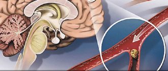
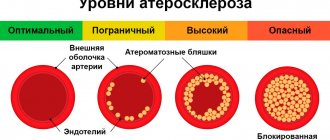
Factors limiting blood flow in the VBS (circulatory failure): atherosclerotic stenosis (narrowing), thrombosis, embolism; post-traumatic dissection of the vertebral arteries; extravasal (extravascular) compression due to pathology of the spine or neck muscles, as well as scar tissue;
deformation of arteries with constant or periodic disruption of their patency. It is assumed that, in addition to the mechanical influence, extravascular factors may cause spasm of the arteries when their periarterial plexus is irritated. In almost two thirds of cases, these changes concern the extracranial part of the vertebral arteries and are often accessible for elimination. Therefore, identifying extravascular factors is of particular importance.
Excessive outflow of blood to other areas of the vascular system occurs through the steal mechanism (creating ischemia in the VBS).
Also important are an increase in hematocrit, viscosity, fibrinogen, platelet aggregation and adhesion and a decrease in the deformability of erythrocytes in aggravating on.
Most often, the clinical manifestations of circulatory failure in the VBS are the interactions of a number of listed causes and mechanisms. At the same time, modern methods of clinical and instrumental research make it possible to identify the main really significant, and sometimes the only, pathogenetic factor and justify ways to eliminate it.
Causes of obstruction of blood flow from the brain
It is quite difficult to determine exactly what exactly provoked the disruption of the normal outflow of blood from the brain, because more than one year may pass after the event that provoked the blockage. The main causes of venous discirculation may be:
- pulmonary and heart failure;
- compression of extracranial veins;
- jugular vein thrombosis;
- brain tumors;
- traumatic brain injury;
- cerebral edema;
- systemic diseases (lupus erythematosus, Wegener's granulomatosis, Behcet's syndrome).
Discirculation can be provoked by either one disease or a complex of several unpleasant symptoms. For example, a mutation in the prothrombin protein in combination with the use of contraceptives in the form of pills increases the risk of developing dysgemia (another name for venous discirculation).
What does ignoring the problem lead to?
Ignoring symptoms for a long time results in oxygen and glucose not reaching the brain. This can lead to neurological problems. Lack of treatment can provoke more severe conditions.
Stroke
If any tumor blocks the flow of blood in the carotid artery, a heart attack or stroke may occur. As a result, some brain tissue may die. The death of even a small amount of tissue can affect speech, coordination, and memory. The severity of the consequences of a stroke depends on how much tissue has died and how quickly the outflow of venous blood has been restored. Some patients are able to fully recover, but most of those affected suffer irreversible changes.
Forecasts for venous discirculation
The prognosis and speed of recovery will depend on several factors.
For example, the survival prognosis for dysgemia may be quite negative if the patient has had a stroke or thrombosis. But if the cause of the disease is hypertension or diabetes, the prognosis will be much better.
Presence of hypoxia
The prognosis will be poor if venous discirculation previously led to hypoxia. Even after eliminating dysgemia, sudden loss of consciousness or problems with the musculoskeletal system are possible.
Most of all, the result of treatment will depend on the age and general health of the patient. Young people with good immunity have the best prognosis for a full recovery.
Symptoms and signs of the disease
In neurology, manifestations of clinical signs of vertebrobasilar syndrome include:
- frequent headaches in the back of the head, throbbing or pressing;
- attacks of dizziness, which as the disease progresses lead to fainting, worsen after prolonged neck immobility (sleep, sedentary work), and are sometimes accompanied by nausea;
- clearly manifested discomfort, a feeling of numbness in the neck;
- the appearance of tinnitus, which becomes constant as the disease progresses;
- visual disturbances: blurred contours, fog or spots before the eyes;
- imbalance, unsteadiness when walking.
To prevent acquired vertebrobasilar syndrome from spoiling the quality of life, the general recommendation for each person is to adhere to a physical activity regime and use a special warm-up. Gymnastics for the prevention of VBI disease is performed smoothly, without tension. It is good to do exercises in the morning after sleep, slowly warming up. Periodically take courses in relaxing massage and physiotherapy.
The most dangerous poses for gymnastics for patients with vertebrobasilar syndrome are the head thrown back, lying on the stomach (and you can’t sleep like that with this disease) and circular rotations of the head with a large amplitude. Effective exercises to reduce the manifestations of the disease are warm-ups with bending and stretching the head. Each exercise must be repeated 10 times, then move on to the next one. This is the complex:
- tilting your head forward, reach your chin to your chest, after a few seconds return to the original position;
- tilt your head to the right and left, reaching your shoulder;
- slowly rotate your head in a circle, first in one direction, then in the other direction;
- pull your head forward slowly, then return to the starting position;
- Pull your head up, fix the position of your head for a few seconds, relax.
After this, use the complex to warm up the whole body, also repeating the exercises 10 times:
- turn your body to the right and left, stretching in the direction of the turn;
- standing straight, raise your arms with your palms together and hold in this position for a short time;
- stand on one leg for as long as possible, change legs.
Risk factors
In addition to the above diseases, disruption of venous blood flow can provoke an unhealthy lifestyle. If you find yourself with at least one of the risk factors listed below, you need to make an appointment with a neurologist to discuss measures to prevent dysgemia.
High blood pressure and a sedentary lifestyle are the first step to dysgemia.
The following deviations should alert you:
- presence of diabetes mellitus;
- high blood pressure;
- obesity degree 2 or higher;
- high cholesterol;
- high triglyceride levels;
- passive lifestyle.
How to treat venous discirculation?
The doctor may recommend several different treatment methods, depending on the identified causes of the disease. But most patients will be advised to make changes to their daily lifestyle, namely:
- stop smoking and drinking alcohol;
- perform simple physical exercises daily;
- follow a diet to lower cholesterol levels;
- Monitor your blood sugar and blood pressure daily.
As for the drug treatment of patients with venous discirculation, specific therapy is prescribed, which includes taking anticoagulants or thrombolytics (depending on the medical history). But the use of systemic anticoagulation as primary treatment is recommended for all patients without exception (even for a child and in the presence of intracranial hemorrhage).
Drug therapy is the most effective method of treating dysgemia
Most often, drugs containing heparin are prescribed. When administered intravenously, its action begins immediately, which is very important for patients with acute dyshemia.
Enoxaparin sodium is a low molecular weight heparin and is prescribed if it is necessary to restore venous outflow to patients suffering from allergic reactions, or for prophylaxis. The main advantage of enoxaparin is the possibility of intermittent administration of the drug, which allows the patient not to go to the hospital, but to take advantage of outpatient treatment.
Warfarin is prescribed to patients with bleeding disorders for whom heparin and enoxaparin are strictly contraindicated. The drug has a slight effect on coagulation activity, but the therapeutic effect can only be seen after a few days. Therefore, such treatment is not prescribed in the acute stages of dyscirculation.
The dose of the administered drug must be carefully monitored by a doctor, so use at home is excluded. Higher doses are administered at the beginning of treatment in order to speed up the time to restore normal outflow, but at the same time, this tactic leads to an increased risk of bleeding. Treatment with warfarin should be continued for 3-6 months to obtain lasting results.
Surgical intervention to get rid of dyscirculation is prescribed in extreme cases
If the disturbances in the venous system are too severe, the doctor may recommend surgery to quickly improve the flow of blood from the brain. But surgery is prescribed only if medical methods do not work.
Types of surgical operations recommended for dysgemia:
- endarterectomy (removal of the inner lining of the affected artery);
- bypass surgery: a new blood vessel is placed near the narrowing of the vein to create a new route for blood flow;
- angioplasty: a balloon catheter is inserted into a narrow section of the artery to widen the walls and improve blood flow.
Treatment of vertebrobasilar insufficiency
Treatment is prescribed by a neurologist.
The main directions of therapy for VBI are determined by the nature of vascular damage. Regular (daily) monitoring of blood pressure and mandatory diet correction (restriction of table salt, fatty foods), avoidance of alcohol consumption and smoking, and dosed physical activity are required.
If there is no positive effect for 3-6 months, drug therapy should be carried out. Reducing blood pressure can begin with any of the following groups:
- diuretics (indapamide, Hypothiazide),
- ACE inhibitors (lisinopril, enalapril),
- calcium channel blockers (amlodipine, felodipine),
- beta blockers (carvedilol, metoprolol, bisoprolol),
- sartans (valsartan, azilsartan, olmesartan).
It is preferable to use complex therapy of 2-3 drugs in low doses rather than increasing one drug to the maximum (diuretic + ACE inhibitor, diuretic + b-blocker, beta blocker + calcium channel blocker). If necessary (lack of effect from treatment, poor tolerability of drugs), the drug is replaced with a drug from a different pharmacological group.
In those patients whose cause of vertebrobasilar insufficiency is a tendency to thrombus formation, increased blood viscosity, an effective way to prevent attacks is to restore the properties of the blood and prevent the formation of blood clots. The most common drug with an antithrombotic effect is acetylsalicylic acid. It is currently believed that the optimal therapeutic dose is to take the drug 0.5-1.0 mg/kg body weight per day (the patient should receive 50-100 mg of acetylsalicylic acid daily). The inability to use acetylsalicylic acid requires the use of other drugs, in particular clopidogrel .
To improve the condition of the vascular wall, dipyridamole is indicated. The daily dose can vary from 75 to 225 mg (25 to 75 mg 3 times a day), in some cases the volume of the drug is increased to 450 mg. Dipyridamole is taken 1 hour before meals, the tablet is not chewed, and must be washed down with a full glass of water. The duration of the course of using dipyridamole is usually 2-3 months. Cancellation is carried out gradually, the dose is reduced over 1-2 weeks. The drug is contraindicated in acute myocardial infarction, rest angina, severe congestive heart failure, and heart rhythm disorders.
Modern antithrombotic drugs are clopidogrel, ticlopidine, rivaroxaban, apixaban. Nicergoline can help improve cerebral circulation. The maintenance dose of nicergoline is set individually and is 5-10 mg 3 times a day. Cinnarizine has proven itself well. Treatment begins with minimal dosages (12.5 mg 3 times a day) with a gradual increase in dose (25-50 mg 3 times a day after meals). Also prescribed are piracetam 0.8 g 3 times a day for 2 months, Cerebrolysin 5-10 ml intravenously 5-10 injections per course of therapy.
A very convenient combination is the drug Phezam, containing 25 mg of cinnarizine and 400 mg of piracetam. The undoubted advantage of the drug is its ease of dosing. The effect is observed when taking 1-2 capsules 3 times a day. The duration of treatment is determined individually and depends on the nature and severity of the neurological deficit. The average course duration is 1-3 months. Carnitine hydrochloride is administered intravenously, 5-10 ml of a 20% solution per 300-400 ml of physiological (isotonic) solution, the course of treatment is 8-12 injections. A prerequisite is a speed of no higher than 60 drops per minute.
Betahistine has proven itself to be effective in eliminating attacks of dizziness in vertebrobasilar insufficiency. The drug is used at a dose of 8-16-24 mg 2-3 times a day. It is advisable to start treatment with small doses, gradually increasing them if necessary. The course of treatment is long (2-3 months). In order to reduce the intensity of episodes of dizziness and accompanying symptoms (nausea, vomiting), especially those provoked by movement, meclozine is prescribed. The daily dose is variable and ranges from 25 to 100 mg.
Massage occupies a special place in treatment. The effect on muscles, ligaments and tendons helps to increase vascular tone and their adequate response to changes in blood pressure.
Diagnostic methods
Recently, the information capabilities of instrumental diagnostic methods in clinical practice have increased. Ultrasound methods for studying the vascular system of the brain have become the most accessible and safe. Doppler ultrasound allows you to obtain data on the patency of the vertebral arteries, linear speed and direction of blood flow in them.
Compression-functional tests make it possible to assess the condition and resources of collateral circulation, blood flow in the carotid, temporal, supratrochlear and other arteries. Duplex scanning demonstrates the condition of the arterial wall, the nature and structure of stenotic formations. Transcranial Doppler ultrasound with pharmacological tests is important for determining cerebral hemodynamic reserve.
Doppler ultrasound (USDG) - detection of signals in the arteries gives an idea of the intensity of microembolic flow in them, cardiogenic or vascular embologenic potential. Data on the condition of the main arteries of the head obtained by MRI angiography are extremely valuable.
When deciding on thrombolytic therapy or surgical intervention on the vertebral arteries, contrast X-ray panangography becomes of decisive importance. Indirect data on the vertebrogenic effect on the vertebral arteries can also be obtained from conventional radiography performed with functional tests.
Otoneurological research occupies a special place, especially if it is supported by computer electronystagmographic and electrophysiological data on auditory evoked potentials characterizing the state of brain stem structures, as well as their MRI.
The sequence of application of the listed instrumental research methods is determined by the peculiarities of determining the clinical diagnosis.
If the patient complains of several of the above symptoms, then all the doctor’s efforts will be aimed at identifying and treating the cause of the dyscirculation. This involves a physical examination and a medical history. To confirm the violation of venous outflow, several studies are prescribed with visualization of veins in the brain and vertebrobasilar region.
Complete blood count
It is prescribed to detect antinuclear antibodies and determine the erythrocyte sedimentation rate. If the results of the analysis confirm the presence of antibodies and a reduced ESR, then an additional study is prescribed to determine complement components and the level of antibodies to anti-deoxyribonucleic acid. The results of the above tests will reveal that the cause of dysgemia is systemic lupus erythematosus or Wegener's granulomatosis.
An electroencephalogram with impaired outflow of venous blood may be normal. But this study is strongly recommended after unilateral thalamic infarction. A slowdown in the basic alpha rhythm indirectly indicates coordination abnormalities and problems with blood outflow.
An EEG can help a doctor identify venous dyscirculation
CT is an important imaging modality and is often indicated for the initial diagnosis of dysgemia. In the tomograph image, you can see whether any neoplasm or thrombosis is the cause of dyshemia.
CT angiography
CT angiography is also prescribed to visualize the cerebral venous system. Only angiography can indicate the absence of flow in the venous channels.
Contrast magnetic resonance imaging is an excellent method for visualizing blood flow in the great cerebral veins. It is prescribed if angiography does not reveal any disturbances in the outflow of venous blood into the VBB.

