Pulmonologist
Prokhina
Maria Egorovna
Experience 38 years
Pulmonologist
Make an appointment
Pulmonary edema is a pathological, very serious condition, which is characterized by the release of transudate into the lung tissue. As a result, gas exchange is disrupted, which leads to serious consequences, including death.
Emergency care for pulmonary edema is the only thing that can increase the patient’s risks of survival and recovery. A person in such a situation requires immediate medical attention.
Pulmonary edema itself is most often a complication that accompanies serious problems of organs and body systems, for example, the cardiovascular system, gastrointestinal tract, etc.
Causes
In fact, there are many causes of pulmonary edema - they are different for different diseases. Let us name, for example, a few general prerequisites:
- cardiosclerosis after a heart attack, acute myocardial infarction;
- hypertension, arrhythmia;
- heart failure;
- congenital or acquired heart defects;
- chronic bronchitis, lobar pneumonia, bronchial asthma;
- complications due to ARVI, measles, influenza, scarlet fever, whooping cough and other diseases;
- prematurity in newborns;
- serious kidney problems;
- traumatic brain injury, brain surgery, etc.;
- inhalation of toxic substances.
These and many other reasons are not direct factors contributing to the development of pulmonary edema. But against the background of such conditions, it can develop, which is necessarily taken into account during hospitalization with all of the above.
Surgical methods
In a situation of rapid increase in effusion with the development of symptoms of cardiac tamponade, a thoracic or cardiovascular surgeon performs puncture of the pericardium with drainage of the contents. The procedure is performed after local anesthesia and under the control of ECHO-CG.
- Pericardioscopy is a method of visualizing the pericardial cavity with targeted biopsy (sampling a piece of tissue) to determine the nature and cause of the lesion.
- Surgery to form a pericardial “window” is an intervention to create an opening from the pericardial space into the pleural space for permanent drainage and prevention of cardiac tamponade. The method is used for frequent massive effusions, which occur more often in diseases caused by tumor processes.
- Pericardectomy is a radical method of treating constrictive pericarditis with the removal of both layers of the pericardium. The operation is performed on an open heart through a sternal approach.
Varieties
There are different types of pulmonary edema:
- fulminant. It develops extremely quickly, in a few minutes - the outcome in this case is only fatal;
- spicy. Symptoms increase over four hours and the risk of death is very high. Such swelling often occurs with heart attack, suffocation, and traumatic brain injury;
- subacute. The development of symptoms alternates between active and quieter stages. Occurs in liver failure;
- protracted. It can develop within twelve hours, even several days, and not have a clear manifestation. It manifests itself in heart failure, as well as chronic lung diseases.
Obviously, each option requires different actions. If the patient can still be saved, speed of response will be a key factor.
Symptoms
It is possible to describe the symptoms of pulmonary edema only in general terms, since certain types of pathology occur with blurred characteristics. Signs include the following:
- severe weakness;
- shallow, very rapid breathing;
- dry cough;
- dry wheezing;
- severe shortness of breath;
- puffiness of the face and neck;
- bubbling breathing and moist wheezing;
- foam at the mouth with a pink tint;
- lethargy, confusion;
- shallow breathing;
- thready pulse.
Some signs of pulmonary edema contradict each other for the reason that everything can start with one condition and end with another. For example, rapid breathing occurs for several minutes or hours, and then it weakens. In the fastest and most dangerous forms of edema, the patient's death occurs from suffocation (asphyxia).
Are you experiencing symptoms of pulmonary edema?
Only a doctor can accurately diagnose the disease. Don't delay your consultation - call
Kinds
Pericarditis is classified according to many characteristics. The most important among them is the cause of occurrence. On this basis, infectious pericarditis and non-infectious pericarditis are distinguished.
Infectious:
- viral pericarditis is the most common in occurrence; its causative agents include enteroviruses, herpes viruses, adenoviruses, and parvovirus;
- parasitic and bacterial, including tuberculous pericarditis (the most common in developing countries - up to 60%), pneumococcal, meningococcal, gonococcal, streptococcal, staphylococcal, mycoplasma and others;
- fungal - an extremely rare option.
Non-infectious:
- autoimmune pericarditis, develops more often with systemic lupus erythematosus, rheumatoid arthritis, Sjogren's syndrome, scleroderma, systemic vasculitis, sarcoidosis, Still's disease;
- neoplastic, the most common cause is malignant neoplasm of the lung, breast, lymphoma;
- metabolic, uremic pericarditis is more common (impaired excretion of protein metabolic products by the kidneys), also develops with myxedema (an extreme form of hypothyroidism - insufficiency of thyroid hormones), anorexia;
- traumatic pericarditis, occurs due to chest injuries, esophageal perforation, radiation (radiation pericarditis), percutaneous coronary intervention;
- medicinal, due to long-term use of various groups of drugs.
It often happens that the cause of inflammation of the pericardial sac remains unknown; such pericarditis is called idiopathic.
In the correct formulation of the diagnosis, there is another important classification of the disease - syndromic:
Acute pericarditis
- Acute pericarditis is characterized by rapid development and lasts no more than 1 week. Based on the substrate in the cavity, fibrinous (dry) pericarditis is distinguished, when a lot of fibrin (a protein from the blood plasma) and little fluid is deposited, and effusion pericarditis (or exudative) with the formation of mostly fluid.
Subacute
- The subacute version of inflammation is not so active and lasts from 1 week to 3 months. There are constrictive, constrictive-effusion. Constriction occurs due to thickening of the pericardial layers, the formation of calcifications and hard adhesions between them, which greatly reduces the possibility of normal movement of the heart inside the bag during its contractions. In this case, the function of relaxation of the ventricles and their filling with blood suffers most.
Recurrent
- Recurrent pericarditis is a disease that is characterized by the return of clinical symptoms some time after stopping treatment - an intermittent option, and permanent, in case of immediate recovery of symptoms after discontinuation of drugs. Constriction and cardiac tamponade are rare phenomena in the recurrent course of the disease.
Chronic
- Chronic pericarditis is inflammation of the pericardium lasting more than 3 months. Patients often suffer for years or even until the end of their lives. It manifests itself as a constrictive variant, in the form of effusion (exudative pericarditis); it is also distinguished as adhesive or adhesive, in which the pericardial layers are soldered together, but softer in structure than in the constrictive variant. The adhesive form is formed after exposure to radiation. In addition, mixed forms can be observed, for example adhesive-exudative pericarditis.?
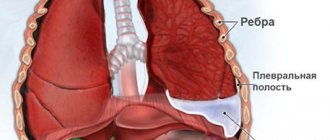
Pericardial effusion
- Effusion in the pericardial cavity is formed in acute (develops quickly) and chronic (accumulates for a long time) variants of the disease. In addition, effusion is classified according to the following criteria: distribution in the cavity - localized (between adhesions) or surrounding (throughout the entire cavity);
- influence on hemodynamics - no effect. cardiac tamponade or constriction;
- composition of the contents - exudate (for infectious inflammation), transudate (for non-infectious nature), blood, less often air or gas (for certain types of bacterial damage);
- size based on echocardiographic (ultrasound of the heart) assessment - small <10 mm, moderate 10-20 mm, pronounced >20 mm.
Cardiac tamponade
- Cardiac tamponade is a severe life-threatening condition. It consists of the accumulation of fluid, blood, pus, gas in the pericardial cavity with the development of compression of the heart in the absence of treatment. As a result, it cannot supply the remaining organs with blood in the required volume. As a result, their deficiency develops.
Diagnostics
If the symptoms of pulmonary edema are not pronounced, additional studies are required in parallel with emergency care:
- biochemical screening. This is a blood test;
- study of blood gases;
- ECG, ultrasound of the heart;
- X-ray of the chest area;
- pulmonary artery catheterization.
In many cases, diagnosis of pulmonary edema is possible immediately - only based on the signs that appear visually in the patient and without additional examination.
How to Diagnose Fluid Around the Heart
To accurately diagnose the cause of fluid accumulation around a person’s heart, the following steps must be taken:
- collecting a complete medical history
- conducting a medical examination
- ordering diagnostic tests
- use of medical imaging
- analysis of pericardial fluid samples
Medical imaging is an effective way to determine if someone has fluid around the heart. When a doctor suspects a person has this disease, researchers recommend they use an echocardiogram as their first diagnostic tool. This test can also show doctors the amount and location of fluid.
Other tests include:
- chest x-ray
- CT scan
- MRI of the heart
Pericardiocentesis is another important tool that doctors use to determine what is causing pericardial effusion. This procedure involves using a needle to take a sample of fluid around the heart for testing. Sometimes it is necessary to leave a drain in the pericardial space to remove all the fluid from the sac.
The consistency of the fluid , which may be watery or high in protein, helps doctors determine what is causing the fluid buildup and how best to treat it.
The researchers report that although the procedure is risky, it could be a potentially life-saving technique for people with cardiac tamponade.
When people don't have cardiac swabs, doctors usually don't use pericardiocentesis to diagnose fluid around the heart chambers unless the medical team is concerned about a bacterial infection.
Treatment
There is not and cannot be a single program for the treatment of pulmonary edema. Emergency care for a patient includes measures to reduce venous return to the heart and supply humidified oxygen. Often the patient is transferred to mechanical ventilation, and a tracheostomy may be performed.
Various drugs are also additionally administered: analgesics, diuretics, drugs that reduce pressure in the pulmonary circulation, drugs for the heart, antibacterial agents and much more. Further, treatment of pulmonary edema, if the attack is removed, comes down to treating the underlying disease that caused this pathology.
The prognosis for edema is very serious. Depending on the causes of pulmonary edema, mortality can range from 20 to 90%. The sooner the problem is identified, the higher the chances of recovery. But in many cases, the patient can only be saved by timely treatment of those diseases that contribute to the occurrence of pathology - such prevention of pulmonary edema, for example, is the only way to reduce the risks of the fulminant form of the disease, which in itself is already fatal and cannot be treated.
Questions and answers
What causes pulmonary edema?
There are dozens of factors that contribute to the development of this pathology. They are associated with other diseases of different body systems. Clinical recommendations for pulmonary edema are largely determined by the situation.
Can pulmonary edema lead to death?
Yes, the risk of death in the case of such a pathology is very high, and for the fulminant form it is the only possible outcome of events. This is an extremely dangerous condition that requires immediate professional help.
How to treat pulmonary edema?
This is done exclusively in medical institutions, most often in intensive care. Only doctors know everything you need to know about the symptoms and treatment of pulmonary edema in adults or children - the pathology cannot be eliminated without special knowledge and skills.
What are the features of pulmonary edema in children?
Unlike adults, this pathology occurs much less frequently in children due to other diseases. The underlying cause is usually allergies or exposure to toxins. Or the cause may be congenital anomalies of various body systems.